Review
. 2022 Nov 3;23(21):13457.
doi: 10.3390/ijms232113457.

The IL-33/ST2 Pathway in Cerebral Malaria

Affiliations
Review

The IL-33/ST2 Pathway in Cerebral Malaria

Corine Glineur et al. Int J Mol Sci. .

Abstract

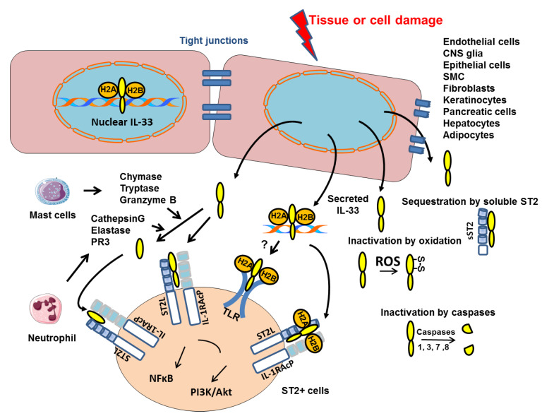
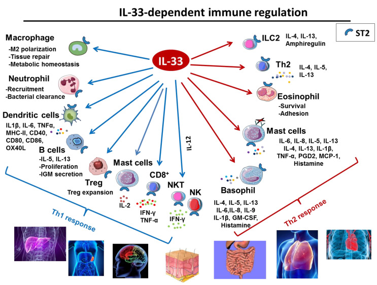
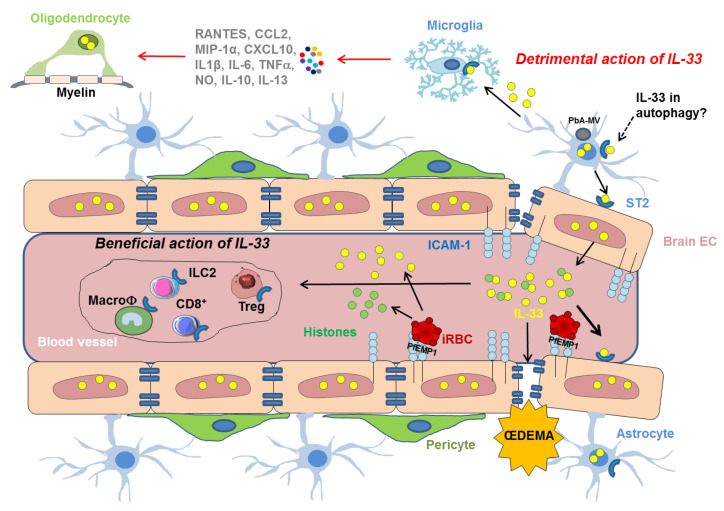
Interleukin-33 (IL-33) is an immunomodulatory cytokine which plays critical roles in tissue function and immune-mediated diseases. IL-33 is abundant within the brain and spinal cord tissues where it acts as a key cytokine to coordinate the exchange between the immune and central nervous system (CNS). In this review, we report the recent advances to our knowledge regarding the role of IL-33 and of its receptor ST2 in cerebral malaria, and in particular, we highlight the pivotal role that IL-33/ST2 signaling pathway could play in brain and cerebrospinal barriers permeability. IL-33 serum levels are significantly higher in children with severe Plasmodium falciparum malaria than children without complications or noninfected children. IL-33 levels are correlated with parasite load and strongly decrease with parasite clearance. We postulate that sequestration of infected erythrocytes or merozoites liberation from schizonts could amplify IL-33 production in endothelial cells, contributing either to malaria pathogenesis or recovery.

Keywords: IL-33; Plasmodium; ST2; astrocytes; blood–brain barrier; central nervous system; cerebral malaria; endothelial; inflammation; red blood cells.


Conflict of interest statement

The authors declare no conflict of interest.

Figures

Figure 1
Figure 1
Mechanisms of intracellular IL-33 action and secretion. IL-33 is constitutively present in the nucleus of structural cells where it binds to chromatin by docking to the pockets of the histone H2A–H2B dimer. Upon cell or tissue damage, the IL-33 alarmin, is released extracellularly as a chromatin complex and is able to bind to receptors such as ST2 complexed to the coreceptor IL-1 receptor accessory protein (IL-1AcP) or supposedly TLR. IL-33 processing by inflammatory proteases produced by mast cells or neutrophils can generate mature forms with increased activity (up to 30-fold). Conversely, cleavage of the IL-1-like domain of IL-33 by caspases 1, 3, 7 or 8 could be an important mechanism of IL-33 inactivation during apoptosis. Extracellular IL-33 may also be sequestrated by soluble ST2 (sST2) or oxidized (formation of S-S disulfide bridges). All these inactivation mechanisms limit the range and duration of the ST2-dependent responses in vivo.
Figure 2
Figure 2
Role of IL-33 in immune response. IL-33 is a potent multi-organ pro-inflammatory cytokine acting on cells involved in Th1 orTh2 responses.
Figure 3
Figure 3
Mechanistic insights on the cellular and molecular players involved in the effect of IL-33/ST2 pathway, following the sequestration of iRBC in the vasculature of the BBB. IRBCs bind to ICAM-1 at the surface of endothelial cells via the Plasmodium erythrocyte membrane protein 1 (PfEMP1). IRBCs may also release IL-33 (yellow circles) that can activate ICAM-1 expression on EC promoting the binding of iRBC. Histones (green circles) released by iRBC may synergize with IL-33. IL-33 drives the expansion of ILC2 cells, leading to the polarization of the anti-inflammatory M2 macrophages, which in turn expand Tregs giving a beneficial role of the IL-33/ST2 pathway in ECM. IL-33 may also have detrimental activities by inducing vascular permeablity responsible for œdema and targeting cells involved in the integrity of BBB, such as microglia which can then produce IL-1β resulting in neuroinflammation and IL-33 overexpression in oligodendrocytes. Autophagy pathways participate in the neuropathophysiological mechanisms of CM in driving the transfer of PbA-microvesicles (PbA-MVs-grey circle) and the induction the proinflammatory response in astrocytes. This pathway could also involve the IL-33/ST2 activity.

Similar articles

Cited by

References

    1. Idro R., Marsh K., John C.C., Newton C.R.J. Cerebral Malaria: Mechanisms of Brain Injury and Strategies for Improved Neurocognitive Outcome. Pediatr. Res. 2010;68:267–274. doi: 10.1203/PDR.0b013e3181eee738. - DOI - PMC - PubMed
    1. Hunt N.H., Golenser J., Chan-Ling T., Parekh S., Rae C., Potter S., Medana I.M., Miu J., Ball H.J. Immunopathogenesis of Cerebral Malaria. Int. J. Parasitol. 2006;36:569–582. doi: 10.1016/j.ijpara.2006.02.016. - DOI - PubMed
    1. Medana I.M., Turner G.D.H. Human Cerebral Malaria and the Blood-Brain Barrier. Int. J. Parasitol. 2006;36:555–568. doi: 10.1016/j.ijpara.2006.02.004. - DOI - PubMed
    1. van der Heyde H.C., Nolan J., Combes V., Gramaglia I., Grau G.E. A Unified Hypothesis for the Genesis of Cerebral Malaria: Sequestration, Inflammation and Hemostasis Leading to Microcirculatory Dysfunction. Trends Parasitol. 2006;22:503–508. doi: 10.1016/j.pt.2006.09.002. - DOI - PubMed
    1. Bagot S., Nogueira F., Collette A., do Rosario V., Lemonier F., Cazenave P.-A., Pied S. Comparative Study of Brain CD8+ T Cells Induced by Sporozoites and Those Induced by Blood-Stage Plasmodium Berghei ANKA Involved in the Development of Cerebral Malaria. Infect. Immun. 2004;72:2817–2826. doi: 10.1128/IAI.72.5.2817-2826.2004. - DOI - PMC - PubMed

LinkOut - more resources