. 2022 Nov 4;23(21):13537.
doi: 10.3390/ijms232113537.

Neurodegenerative Disorder Risk in Krabbe Disease Carriers

Affiliations

Neurodegenerative Disorder Risk in Krabbe Disease Carriers

Lorenza Vantaggiato et al. Int J Mol Sci. .

Abstract

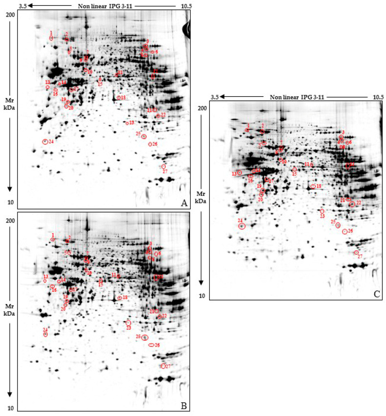
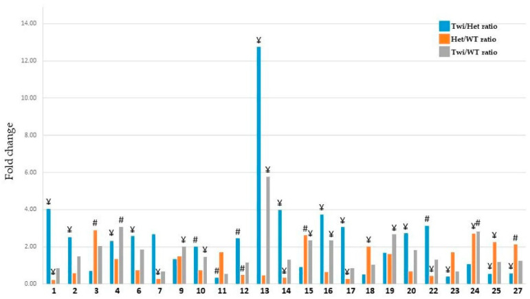
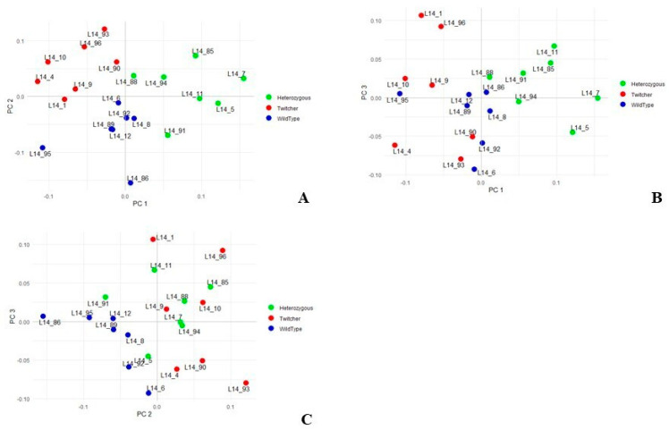
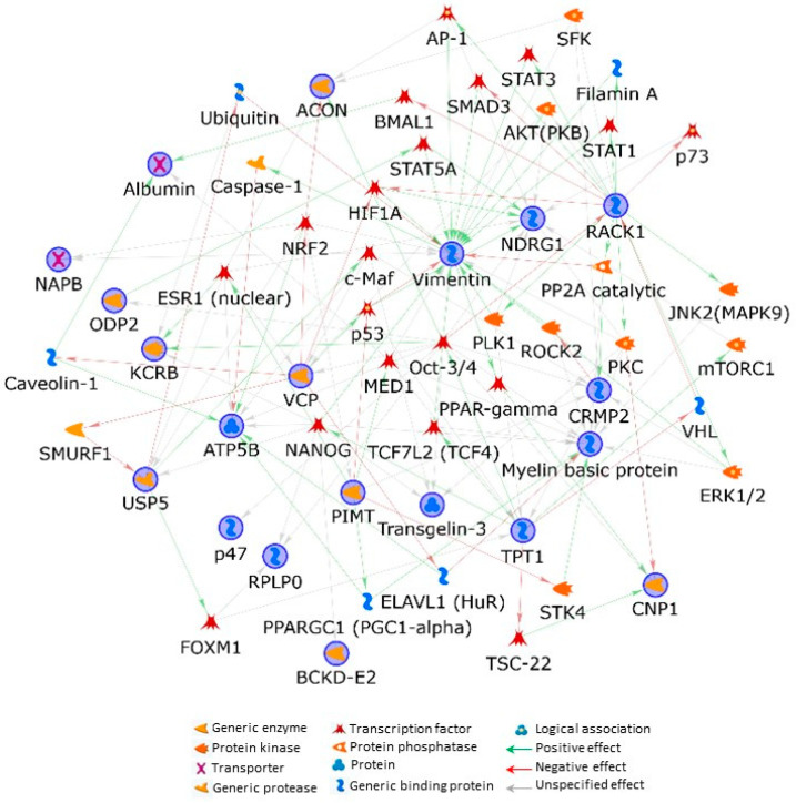
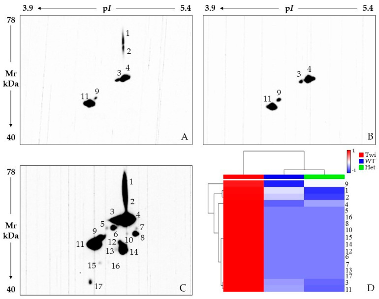
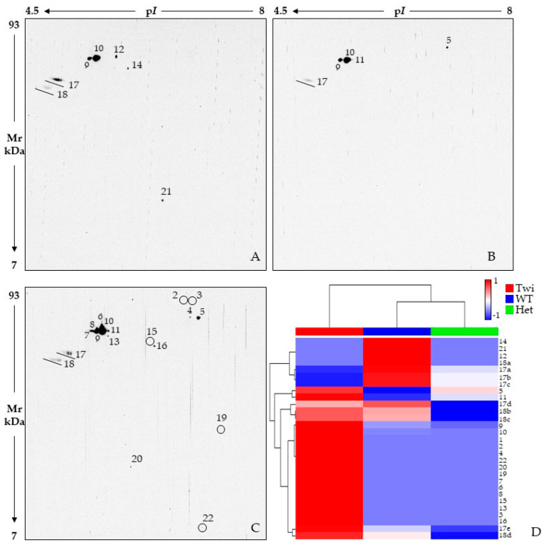
Krabbe disease (KD) is a rare autosomal recessive disorder caused by mutations in the galactocerebrosidase gene (GALC). Defective GALC causes aberrant metabolism of galactolipids present almost exclusively in myelin, with consequent demyelinization and neurodegeneration of the central and peripheral nervous system (NS). KD shares some similar features with other neuropathies and heterozygous carriers of GALC mutations are emerging with an increased risk in developing NS disorders. In this work, we set out to identify possible variations in the proteomic profile of KD-carrier brain to identify altered pathways that may imbalance its homeostasis and that may be associated with neurological disorders. The differential analysis performed on whole brains from 33-day-old twitcher (galc -/-), heterozygous (galc +/-), and wild-type mice highlighted the dysregulation of several multifunctional factors in both heterozygous and twitcher mice. Notably, the KD-carrier mouse, despite its normal phenotype, presents the deregulation of vimentin, receptor of activated protein C kinase 1 (RACK1), myelin basic protein (MBP), 2',3'-cyclic-nucleotide 3'-phosphodiesterase (CNP), transitional endoplasmic reticulum ATPase (VCP), and N-myc downstream regulated gene 1 protein (NDRG1) as well as changes in the ubiquitinated-protein pattern. Our findings suggest the carrier may be affected by dysfunctions classically associated with neurodegeneration: (i) alteration of (mechano) signaling and intracellular trafficking, (ii) a generalized affection of proteostasis and lipid metabolism, with possible defects in myelin composition and turnover, and (iii) mitochondrion and energy supply dysfunctions.

Keywords: Alzheimer disease; Lewy body; Parkinson disease; demyelination; globoid cell leukodystrophy; multiple sclerosis; neurodegeneration; neuroinflammation; ubiquitin dependent degradation.


Conflict of interest statement

The authors declare that they have no current or potential conflict of interest regarding this article.

Figures

Figure 1
Figure 1
Reference whole brain lysate protein patterns of 33-day-old wild-type (WT) (A), heterozygous galc +/− (Het) (B) and Twitcher galc −/− (Twi) (C) mice. Red circles and numbers point out differentially abundant protein spots detected among the three sample classes. Numbers matched those listed in Table 1 and Figure 2.
Figure 2
Figure 2
Histogram representing the %Vol ratios of Twi vs. Het (blue bar), Het vs. WT (orange bar), and Twi vs. WT (grey bar) as reported in Table 1. The numbers in the x axis correspond to those of differentially abundant protein spots identified by mass spectrometry, for more clarity: 1: Ubiquitin carboxyl-terminal hydrolase 5; 2: Transitional endoplasmic reticulum ATPase; 3: Aconitate hydratase, mitochondrial; 4: Aconitate hydratase, mitochondrial; 6: Aconitate hydratase, mitochondrial; 7: Albumin; 9: Dihydrolipoyllysine-residue acetyltransferase component of pyruvate dehydrogenase complex, mitochondrial; 10: Dihydropyrimidinase-related protein 2; 11: Lipoamide acyltransferase component of branched-chain alpha-keto acid dehydrogenase complex, mitochondrial; 12: 2′,3′-cyclic-nucleotide 3′-phosphodiesterase; 13: Vimentin; 14: ATP synthase subunit beta, mitochondrial; 15: Protein NDRG1; 16: NSFL1 cofactor p47; 17: Tropomodulin-2; 18: 60S acidic ribosomal protein P0; 19: Beta-soluble NSF attachment protein; 20: Creatine kinase B-type; 22: Receptor of activated protein C kinase 1; 23: Protein-L-isoaspartate(D-aspartate) O-methyltransferase; 24: Translationally-controlled tumor protein; 25: Transgelin-3; 27: Myelin basic protein. # and ¥ indicate statistical relevance, i.e. p ≤ 0.01 and 0.01 < p ≤ 0.05, respectively.
Figure 3
Figure 3
Principal component analysis (PCA) performed on %Vols of spots matched among WT, Het, and Twi mouse samples. Plots highlight spatial distribution of the 21 whole brain analyzed samples—7 from WT mice (blue symbols); 7 from Het mice (green symbols); and 7 from Twi mice (red symbol)—along the PC1 and PC2 (A), PC1 and PC3 (B), and PC2 and PC3 (C).
Figure 4
Figure 4
MetaCore protein network built by processing significant protein differences occurring among Twi, Het, and wild-type mice. Experimental proteins, circled in blue, were cross-linked by using the shortest-path-network (SPN) building tool. This generates hypothetical networks by cross-linking experimental factors and expanding protein interactions to other proteins, not present in the processed experimental list but supported by the MetaCore database, that are needed to functionally correlate user up-loaded proteins, which do not directly interact. Except for tropomodulin-2, all the processed proteins entered into the SPN.
Figure 5
Figure 5
2D Western blot analysis with anti-vimentin antibody on 33-day-old wild-type (WT) (A), heterozygous galc +/− (Het) (B) and twitcher galc −/− (Twi) (C) mice. Heatmap analysis (D) scaled values of spot intensity. Color changes from blue to red indicate less or higher signal intensity, respectively. Each row corresponds to a VIME protein species while each column corresponds to one of the three tested conditions, as highlighted by the colored bar from the horizontal dendrogram: Twi = red, WT = blue, and Het = green. Row spot numbers match those in (AC).
Figure 6
Figure 6
2D Western blot analysis by using anti-ubiquitin antibody on 33-day-old wild-type (WT) (A), heterozygous galc +/− (Het) (B), and Twitcher (Twi) (C) mice. Small signals are circled to facilitate their viewing. Circles are used to improve the visualization of weaker immunoreactive signals (C). Heatmap analysis (D) performed on scaled values of spot intensity. Color changes from blue to red indicate less or higher signal intensity, respectively. Each row corresponds to a ubiquitinated protein species while each column corresponds to one of the three tested conditions, as highlighted by the colored bar from the horizontal dendrogram: Twi = red, WT = blue, and Het = green. Row spot numbers match those in (AC). Spots from the same isoelectric series (i.e., 17 and 18 lines) are named in alphabetical order, from the most acid pI value to the most basic one.

References

    1. Komatsuzaki S., Zielonka M., Mountford W.K., Kölker S., Hoffmann G.F., Garbade S.F., Ries M. Clinical Characteristics of 248 Patients with Krabbe Disease: Quantitative Natural History Modeling Based on Published Cases. Genet. Med. 2019;21:2208–2215. doi: 10.1038/s41436-019-0480-7. - DOI - PubMed
    1. Suzuki K. Globoid Cell Leukodystrophy (Krabbe’s Disease): Update. J. Child Neurol. 2003;18:595–603. doi: 10.1177/08830738030180090201. - DOI - PubMed
    1. Ryckman A.E., Brockhausen I., Walia J.S. Metabolism of Glycosphingolipids and Their Role in the Pathophysiology of Lysosomal Storage Disorders. Int. J. Mol. Sci. 2020;21:6881. doi: 10.3390/ijms21186881. - DOI - PMC - PubMed
    1. Schmitt S., Castelvetri L.C., Simons M. Metabolism and Functions of Lipids in Myelin. Biochim. Biophys. Acta. 2015;1851:999–1005. doi: 10.1016/j.bbalip.2014.12.016. - DOI - PubMed
    1. Giussani P., Prinetti A., Tringali C. The Role of Sphingolipids in Myelination and Myelin Stability and Their Involvement in Childhood and Adult Demyelinating Disorders. J. Neurochem. 2021;156:403–414. doi: 10.1111/jnc.15133. - DOI - PubMed

Substances