. 2022 Nov 5;23(21):13578.
doi: 10.3390/ijms232113578.

SIRT1 Promotes Host Protective Immunity against Toxoplasma gondii by Controlling the FoxO-Autophagy Axis via the AMPK and PI3K/AKT Signalling Pathways

Affiliations

SIRT1 Promotes Host Protective Immunity against Toxoplasma gondii by Controlling the FoxO-Autophagy Axis via the AMPK and PI3K/AKT Signalling Pathways

Jina Lee et al. Int J Mol Sci. .

Abstract

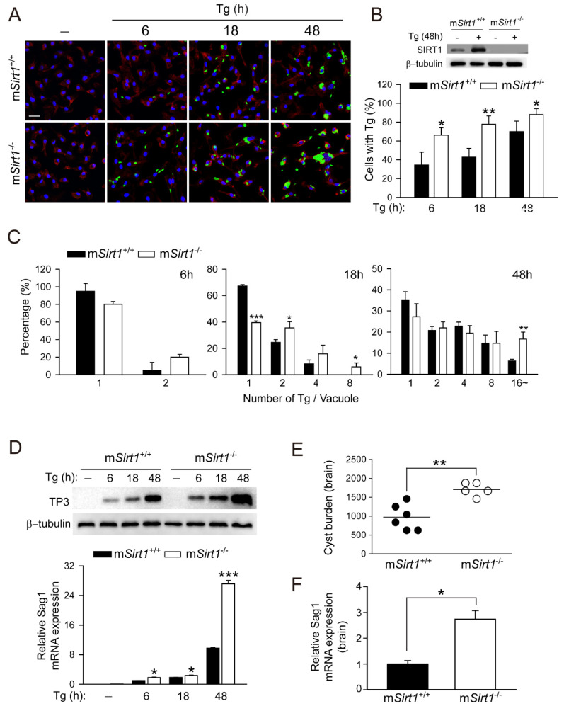
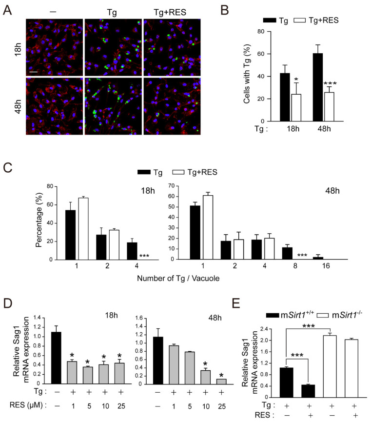
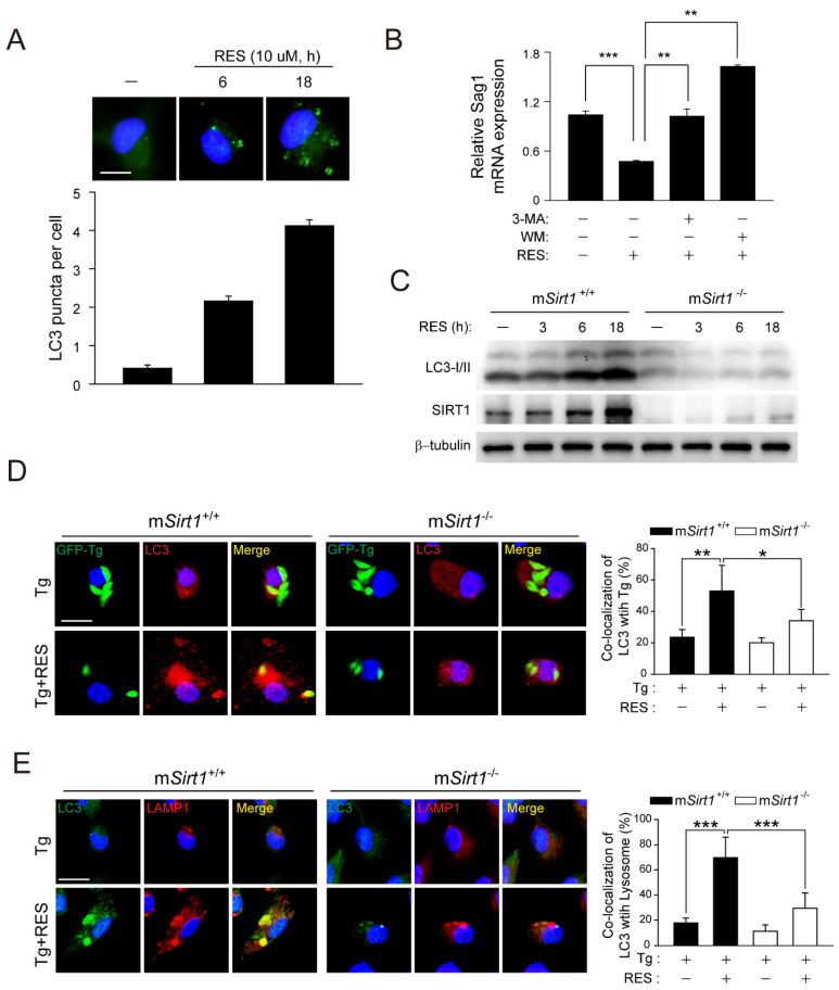
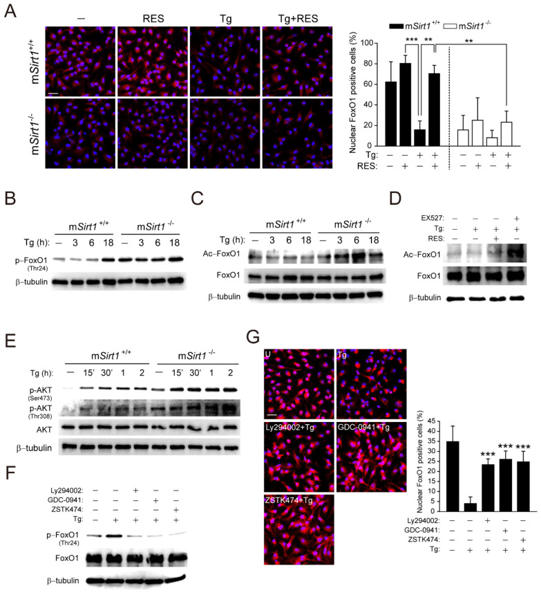
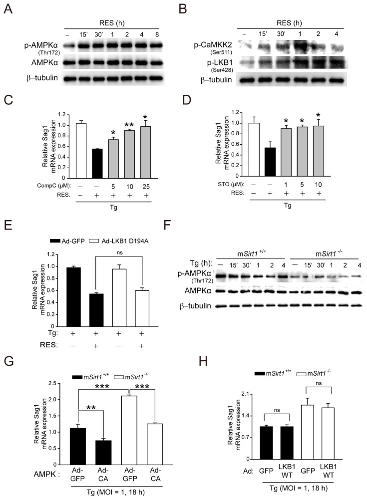
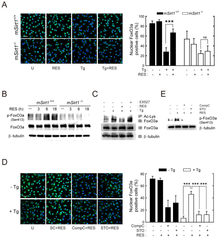
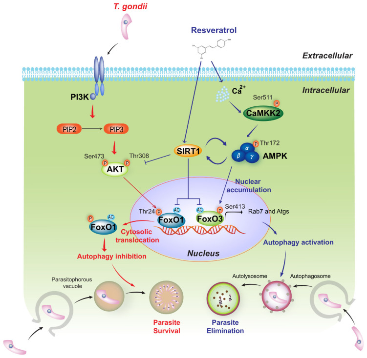
Sirtuin 1 (SIRT1) regulates cellular processes by deacetylating non-histone targets, including transcription factors and intracellular signalling mediators; thus, its abnormal activation is closely linked to the pathophysiology of several diseases. However, its function in Toxoplasma gondii infection is unclear. We found that SIRT1 contributes to autophagy activation via the AMP-activated protein kinase (AMPK) and PI3K/AKT signalling pathways, promoting anti-Toxoplasma responses. Myeloid-specific Sirt1-/- mice exhibited an increased cyst burden in brain tissue compared to wild-type mice following infection with the avirulent ME49 strain. Consistently, the intracellular survival of T. gondii was markedly increased in Sirt1-deficient bone-marrow-derived macrophages (BMDMs). In contrast, the activation of SIRT1 by resveratrol resulted in not only the induction of autophagy but also a significantly increased anti-Toxoplasma effect. Notably, SIRT1 regulates the FoxO-autophagy axis in several human diseases. Importantly, the T. gondii-induced phosphorylation, acetylation, and cytosolic translocation of FoxO1 was enhanced in Sirt1-deficient BMDMs and the pharmacological inhibition of PI3K/AKT signalling reduced the cytosolic translocation of FoxO1 in BMDMs infected with T. gondii. Further, the CaMKK2-dependent AMPK signalling pathway is responsible for the effect of SIRT1 on the FoxO3a-autophagy axis and for its anti-Toxoplasma activity. Collectively, our findings reveal a previously unappreciated role for SIRT1 in Toxoplasma infection.

Keywords: AMP-activated protein kinase; Class O of forkhead box transcription factors; PI3K/AKT signalling pathway; Sirtuin 1; Toxoplasma gondii; autophagy; bone-marrow-derived macrophages.


Conflict of interest statement

The authors declare no conflict of interest.

Figures

Figure 1
Figure 1
Myeloid SIRT1 is essential for host protection against T. gondii infection in primary macrophages and mice. (AC) BMDMs from mSirt1+/+ and mSirt1−/− mice were infected with GFP-conjugated T. gondii RH strain (MOI = 1) for the indicated periods. Cells were fixed and stained with Texas Red®-X phalloidin for F-actin in the cytoskeleton (red) and DAPI for nuclei (blue), respectively. (A) Fluorescent images showing the number of intracellular T. gondii. (B,C) Number of T. gondii RH-infected cells (for B) and T. gondii RH per vacuole (for C) were quantified. Scale bar = 25 µm (D) BMDMs from mSirt1+/+ and mSirt1−/− mice were infected with T. gondii RH strain (MOI = 1) for the indicated time periods. TP3 protein expression (top) and Sag1 mRNA expression (bottom) was examined by immunoblot and qPCR analysis, respectively. (E,F) mSirt1+/+ and mSirt1−/− mice (n = 5 per group) were infected with 40 cysts of the ME49 strain (i.p. injection) for 3 weeks. (E) The number of cysts in the brain was counted under a microscope. (F) Sag1 mRNA expression in brain homogenates was evaluated by real-time qPCR analysis. Data are representative of three independent experiments and are presented as means ± SD (for BD) or ± SEM (for F). * p < 0.05, ** p < 0.01, *** p < 0.001, compared with control cultures or control mice infected with T. gondii (two-tailed Student’s t-test). Tg, Toxoplasma gondii.
Figure 2
Figure 2
RES treatment promotes the elimination of intracellular T. gondii in primary murine macrophages. (AC) BMDMs were infected with a GFP-conjugated RH strain (MOI = 1) for 2 h, followed by the further treatment of RES (10 µM) for the indicated times. Cells were stained with Texas Red®-X phalloidin for F-actin in the cytoskeleton (red) and DAPI for nuclei (blue), respectively. (A) Fluorescent images showing the number of intracellular T. gondii. (B,C) Number of T. gondii RH-infected cells (for B) and T. gondii RH per vacuole were quantified. Scale bar = 25 µm (D,E) qPCR analysis for evaluating Sag1 mRNA expression. (D) T. gondii-infected BMDMs were stimulated with increasing concentrations of RES (1, 5, 10, or 25 µM) for 18 h (for left) or 48 h (for right). (E) BMDMs isolated from mSirt1+/+ and mSirt1−/− mice were infected with RH strain of T. gondii (MOI = 1) for 2 h and then further stimulated with RES (10 µM) for 48 h. Data are representative of three independent experiments and are presented as means ± SD. * p < 0.05 and *** p < 0.001, compared with control cells (two-tailed Student’s t-test). Tg, Toxoplasma gondii; RES, resveratrol.
Figure 3
Figure 3
RES induces autophagy activation in a SIRT1-dependent manner. (A) BMDMs were stimulated with RES (10 µM) for various times and subjected to immunofluorescence microscopy for analysing LC3 puncta formation (top). Quantification of LC3 punctate foci per cell (bottom). Each experiment included a minimum of 100 cells scored in 5 random fields. Scale bar = 10 µm. (B) BMDMs were pre-treated with 3-MA (10 µM, 2 h) or WM (100 nM, 2 h) and further incubated with RES for 18 h. The mRNA expression of the Sag1 gene was evaluated by real-time qPCR analysis. (C) BMDMs from mSirt1+/+ and mSirt1−/− mice were stimulated with RES for the indicated time periods and subjected to immunoblot analysis of LC3 and β-tubulin. (D,E) BMDMs from mSirt1+/+ and mSirt1−/− mice were infected with a GFP-conjugated RH strain (for D) or a T. gondii RH strain (for E) for 2 h, followed by further treatment of RES (10 µM) for 18 h. (D) Cells were immunostained with an anti-LC3 antibody (Alexa Fluor 594-conjugated goat anti-rabbit IgG, red) and the level of colocalisation T. gondii with LC3 was quantified. Scale bar = 10 µm. (E) LC3 (Alexa Fluor 488-conjugated goat anti-rabbit IgG, green), Alexa Fluor 594-conjugated LAMP1 (red), and DAPI (blue) were detected by confocal microscopy. Immunofluorescence microscopy images were obtained from one representative of 3 independent samples, with each experiment containing a minimum of 50 cells scored in 7 random fields. Scale bar = 10 µm. Data are representative of three independent experiments and are presented as means ± SD. * p < 0.05, ** p < 0.01, and *** p < 0.001, compared with control cells (two-tailed Student’s t-test). Tg, Toxoplasma gondii; RES, resveratrol; 3-MA, 3-methyladenine; WM, wortmannin.
Figure 4
Figure 4
SIRT1 deficiency leads to the excessive activation of PI3K/AKT-mediated FoxO1 upon T. gondii infection. (A) BMDMs from mSirt1+/+ and mSirt1−/− mice were infected with a RH strain (MOI = 1, for 2 h) and then further stimulated with RES (10 µM) for 18 h. Cells were immunostained using an anti-FoxO1 antibody (Alexa Fluor 568-conjugated goat anti-rabbit IgG, red) and DAPI for nuclei (blue). Cells were then subjected to immunofluorescence microscopy. Representative images (left) and quantitative data (right) showing the cytoplasmic translocation of FoxO1 are from one representative of 3 independent samples, with each experiment containing a minimum of 50 cells scored in 3 random fields. Scale bar = 25 µm. (B,C,E) BMDMs from mSirt1+/+ and mSirt1−/− mice were infected with T. gondii for the indicated time periods. The phosphorylation (for B) and acetylation (for C) of FoxO1 or AKT phosphorylation (Ser473 and Thr308, for E) were determined by immunoblot analysis. (D) BMDMs were infected with T. gondii for 18 h in the presence of RES (10 µM) or EX527 (4 µM). The level of FoxO1 acetylation was evaluated by immunoblot analysis. (F,G) BMDMs were infected with T. gondii for 18 h in the presence of LY294002 (10 µM), GDC0941 (250 nM), or ZSTK474 (10 nM). (F) The level of FoxO1 phosphorylation was evaluated by immunoblot analysis, and total protein was determined by monitoring β-tubulin (BF), AKT (E), or FoxO1 (C,D,F) as a loading control. (G) Immunofluorescence microscopy analysis was assessed to determine the nuclear and cytoplasmic localisation of FOXO1, as described in Figure 4A. Scale bar = 25 µm. Data are representative of three independent experiments and are presented as means ± SD. ** p < 0.01 and *** p < 0.001, compared with mSirt1+/+ cells (for A) or control cells (for G) (two-tailed Student’s t-test). Tg, Toxoplasma gondii; RES, resveratrol.
Figure 5
Figure 5
CaMKK2-dependent AMPK signalling is crucial for the activation of SIRT1-mediated antiparasitic responses against Toxoplasma infection in primary murine macrophages. (A,B) BMDMs were stimulated with RES (10 µM) for the various time periods, and then immunoblot analysis was assessed to evaluate the phosphorylation of AMPKα (for A) or CaMKK2 and LKB1 (for B). (C,D) BMDMs were pretreated with increasing concentrations of CompC (5, 10, or 25 μM, for C) or STO (1, 5, or 10 μM, for D) for 45 min, then infected with T. gondii RH strain (MOI = 1) for 18 h in the presence or absence of RES. Sag1 mRNA expression was examined by qPCR analysis. (E) BMDMs transduced with Ad-GFP, Ad-LKB1 WT, or Ad-LKB1 D194A for 36 h (MOI of 10), respectively, were infected with a T. gondii RH strain (MOI = 1) for 2 h, then further incubated with RES (10 µM) for 22 h. Sag1 mRNA expression was evaluated by qPCR analysis. (F) BMDMs from mSirt1+/+ and mSirt1−/− mice were infected with T. gondii for various time periods. Cell lysates were subjected to immunoblot analysis using antibodies against p-AMPKα (Thr172), AMPKα, and β-tubulin. (G,H) BMDMs from mSirt1+/+ and mSirt1−/− mice were transduced for 36 h with adenovirus-expressing GFP (Ad-GFP, for G,H), constitutively active AMPK (Ad-CA, for G), or WT LKB1 (Ad-LKB1 WT, for H), at a MOI of 10, and each cell then was infected with a T. gondii RH strain (MOI = 1) for 18 h. The mRNA expression of the Sag1 gene was evaluated by real-time qPCR analysis. Data are representative of three independent experiments and are presented as means ± SD. * p < 0.05, ** p < 0.01, *** p < 0.001 (two-tailed Student’s t-test). Tg, Toxoplasma gondii; RES, resveratrol; CompC, Compound C; STO, STO-609; adenovirus-expressing GFP, Ad-GFP; adenovirus-expressing WT LKB1, Ad-LKB1 WT; adenovirus-expressing LKB1 D194A mutant, Ad-LKB1 D194A; ns, not significant.
Figure 6
Figure 6
RES treatment promotes the activation of FoxO3a, which is mediated by SIRT1 and AMPK. (A) BMDMs from mSirt1+/+ and mSirt1−/− mice were infected with the RH strain (MOI = 1, for 2 h) and then further stimulated with RES (10 µM) for 18 h. Cells were fixed and immunostained with an anti-FoxO3 antibody (Alexa Fluor 488-conjugated goat anti-rabbit IgG, green) and DAPI for nuclei (blue). Cells were then subjected to immunofluorescence microscopy. Representative images (left) and quantitative data (right) showing the cytoplasmic translocation of FoxO3a were obtained from one representative of 3 independent samples, with each experiment containing a minimum of 50 cells scored in 7 random fields. Scale bar = 25 µm. (B) BMDMs from mSirt1+/+ and mSirt1−/− mice were stimulated with RES (10 µM) for the indicated time periods. The phosphorylation of FoxO3 (Ser413) was determined by immunoblot analysis. (C) BMDMs were infected with T. gondii for 18 h in the presence of RES (10 µM) or EX527 (4 µM). Cell lysates were subjected to immunoprecipitation analysis using an anti-acetylated-lysine antibody (Ac-Lys), and then the immunoprecipitates were analysed by Western blot using an anti-FoxO3a antibody. Cell lysates were harvested and subjected to Western blot analysis for FoxO3a and β-tubulin, as loading controls. (D) BMDMs were pretreated with Compound C (25 µM) or STO-609 (10 µM) for 45 min, then infected with RH strain (MOI = 1) for 18 h in the presence or absence of RES (10 µM). Cells were fixed, immunostained, and quantified as described in Figure 6A. Representative images (left) and quantitative data (right) showing the cytoplasmic translocation of FoxO3a. Scale bar = 25 µm. (E) BMDMs were pretreated with Compound C (25 µM) or STO-609 (10 µM, 45 min) and then stimulated with RES (10 µM, 18 h). The phosphorylation of FoxO3 (Ser413) was determined by immunoblot analysis. Data are representative of three independent experiments and are presented as means ± SD. *** p < 0.001 (two-tailed Student’s t-test). Tg, Toxoplasma gondii; RES, resveratrol; CompC, Compound C; STO, STO-609; ns, not significant.
Figure 7
Figure 7
Essential function and molecular mechanism of SIRT1 in modulating the host immune response to T. gondii infection. In macrophages, myeloid-specific SIRT1 increases autophagy activity during T. gondii infection, thereby eliminating intracellular T. gondii by inducing the colocalisation of parasitophorous vacuoles with autophagosomes/lysosomes. As a key regulatory mechanism of autophagy, SIRT1 attenuates the acetylation of the transcription factor FoxO1 and its PI3K/AKT-dependent phosphorylation (Thr24) in response to T. gondii, thus regulating FoxO1 transactivation activity by preventing its nuclear exclusion. Moreover, SIRT1 promotes FoxO3a deacetylation and its CaMKK2/AMPK-dependent phosphorylation (Ser413), leading to nuclear accumulation and the transactivation of FoxO3a. Pharmacological activation of SIRT1 by RES induces antiprotozoal autophagy by regulating FoxO1 and FoxO3a activity via the PI3K/AKT and CaMKK2/AMPK signalling pathways, respectively.

References

    1. Elmore S.A., Jones J.L., Conrad P.A., Patton S., Lindsay D.S., Dubey J.P. Toxoplasma gondii: Epidemiology, feline clinical aspects, and prevention. Trends Parasitol. 2010;26:190–196. doi: 10.1016/j.pt.2010.01.009. - DOI - PubMed
    1. Harker K.S., Ueno N., Lodoen M.B. Toxoplasma gondii dissemination: A parasite’s journey through the infected host. Parasite Immunol. 2015;37:141–149. doi: 10.1111/pim.12163. - DOI - PubMed
    1. Montoya J.G., Liesenfeld O. Toxoplasmosis. Lancet. 2004;363:1965–1976. doi: 10.1016/S0140-6736(04)16412-X. - DOI - PubMed
    1. Sasai M., Pradipta A., Yamamoto M. Host immune responses to Toxoplasma gondii. Int. Immunol. 2018;30:113–119. doi: 10.1093/intimm/dxy004. - DOI - PubMed
    1. Luft B.J., Hafner R., Korzun A.H., Leport C., Antoniskis D., Bosler E.M., Bourland D.D., 3rd, Uttamchandani R., Fuhrer J., Jacobson J., et al. Toxoplasmic encephalitis in patients with the acquired immunodeficiency syndrome. Members of the ACTG 077p/ANRS 009 Study Team. N. Engl. J. Med. 1993;329:995–1000. doi: 10.1056/NEJM199309303291403. - DOI - PubMed

MeSH terms

Substances