. 2022 Nov 6;23(21):13606.
doi: 10.3390/ijms232113606.

L-Type Ca2+ Channel Inhibition Rescues the LPS-Induced Neuroinflammatory Response and Impairments in Spatial Memory and Dendritic Spine Formation

Affiliations

L-Type Ca2+ Channel Inhibition Rescues the LPS-Induced Neuroinflammatory Response and Impairments in Spatial Memory and Dendritic Spine Formation

Jieun Kim et al. Int J Mol Sci. .

Abstract

Ca2+ signaling is implicated in the transition between microglial surveillance and activation. Several L-type Ca2+ channel blockers (CCBs) have been shown to ameliorate neuroinflammation by modulating microglial activity. In this study, we examined the effects of the L-type CCB felodipine on LPS-mediated proinflammatory responses. We found that felodipine treatment significantly diminished LPS-evoked proinflammatory cytokine levels in BV2 microglial cells in an L-type Ca2+ channel-dependent manner. In addition, felodipine leads to the inhibition of TLR4/AKT/STAT3 signaling in BV2 microglial cells. We further examined the effects of felodipine on LPS-stimulated neuroinflammation in vivo and found that daily administration (3 or 7 days, i.p.) significantly reduced LPS-mediated gliosis and COX-2 and IL-1β levels in C57BL/6 (wild-type) mice. Moreover, felodipine administration significantly reduced chronic neuroinflammation-induced spatial memory impairment, dendritic spine number, and microgliosis in C57BL/6 mice. Taken together, our results suggest that the L-type CCB felodipine could be repurposed for the treatment of neuroinflammation/cognitive function-associated diseases.

Keywords: Ca2+ channel blocker; LPS; felodipine; gliosis; neuroinflammation; spatial memory.


Conflict of interest statement

The authors declare no conflict of interest.

Figures

Figure 1
Figure 1
The L-type Ca2+ channel blocker felodipine suppresses LPS-induced proinflammatory cytokine levels in BV2 microglial cells. (a) Structure of felodipine. (b) MTT assay of the effect of felodipine on cell viability (felodipine: n = 7/group). (c) BV2 microglial cells were treated with LPS, followed by felodipine (1, 2.5, or 5 μM) as shown, and real-time PCR was performed (n = 8/group). (d) BV2 microglial cells were treated with LPS, followed by 5 μM felodipine as shown, and RT-PCR was conducted (n = 8/group). (e) ELISA analysis of the effect of felodipine post-treatment on secreted COX-2 levels (n = 8/group). (f) BV2 microglial cells were pre-treated with felodipine (1, 2.5, or 5 μM), followed by LPS as shown, and real-time PCR was performed (n = 8/group). (g) BV2 microglial cells were pre-treated with 5 μM felodipine, followed by LPS as shown, and RT-PCR was conducted (n = 16/group). (h) ELISA analysis of the effect of felodipine pre-treatment on secreted COX-2 and IL-1β levels (n = 8/group). * p < 0.05, ** p < 0.01, *** p < 0.001. V: vehicle; L: LPS; F or Felodi: felodipine.
Figure 2
Figure 2
Felodipine downregulates LPS-stimulated proinflammatory responses through the L-type Ca2+ channel and TLR4 signaling in BV2 microglial cells. (a,d) Real-time PCR analysis of cacna1d gene expression in cells treated with 1 μM felodipine (a) or 5 μM felodipine (d), followed by LPS as shown (n = 8/group). (b,c,e,f) Real-time PCR analysis of cox-2 and il-1β mRNA levels in cells transfected with cacna1d siRNA (60 nM) or scramble (control) siRNA for 24 h and subsequently treated with 1 μM felodipine (b,c) or 5 μM felodipine (e,f) and with LPS as shown (n = 8/group). (g,h) RT-PCR and real-time PCR analyses of proinflammatory cytokine cox-2 expression in cells treated with TLR4 inhibitor (TAK-242), 5 μM felodipine, and LPS (felodipine: n = 22/group). * p < 0.05, ** p < 0.01, *** p < 0.001. V: vehicle; L: LPS; F or Felodi: felodipine.
Figure 3
Figure 3
Felodipine inhibits LPS-stimulated AKT and STAT3 phosphorylation in BV2 microglial cells. (a) Western blot analysis of AKT phosphorylation in cells treated with 5 μM felodipine or vehicle (1% DMSO) for 45 min, followed by LPS (200 ng/mL) or PBS for 45 min (n = 6/group). (b) Immunocytochemical staining of p-AKTS473 in cells treated with 5 μM felodipine or vehicle (1% DMSO) for 45 min, followed by LPS (200 ng/mL) or PBS for 45 min (Veh, n = 443; LPS, n = 578; LPS+Felodi, n = 918). (c,d) Expression of the proinflammatory cytokine cox-2 in cells treated as shown with an AKT inhibitor (MK-2206), 5 μM felodipine, and LPS and analyzed via RT-PCR (c, n = 4/group) or real-time PCR (d, n = 8/group). (e) Nuclear fractionation and Western blotting analysis of LPS-mediated nuclear p-STAT3S727 levels in cells treated with 5 μM felodipine or vehicle (1% DMSO) for 30 min, followed by LPS (200 ng/mL) or PBS for 5.5 h (n = 6/group). (f) Immunocytochemical staining analysis of LPS-induced nuclear STAT3 phosphorylation in cells treated with 5 μM felodipine or vehicle (1% DMSO) for 30 min, followed by LPS (200 ng/mL) or PBS for 5.5 h (Veh, n = 414; LPS, n = 326; Felodi+LPS, n = 308). * p < 0.05, ** p < 0.01, *** p < 0.001, scale bar: 20 μm. V: vehicle; L: LPS; F or Felodi: felodipine, M: MK-2206.
Figure 4
Figure 4
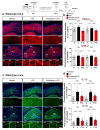
Daily administration of felodipine for 3 days decreases LPS-induced microgliosis in the cortex and hippocampus of C57BL/6 mice. (a) Experimental scheme of felodipine treatment in vivo. (b) Coordinates and specific locations of the hippocampus used for immunofluorescence staining. (c) C57BL/6 mice were injected with vehicle (5% Tween20 + 5% PEG300 in saline) or felodipine (5 mg/kg, i.p.) daily for 3 days. On day 3, LPS (10 mg/kg, i.v.) or PBS was injected, and immunofluorescence staining was conducted with an anti-Iba-1 antibody. (d) Quantification of data from (c) (n = 5 mice/group. Vehicle, n = 20 brain slices; LPS, n = 20 brain slices; felodipine+LPS, n = 16 brain slices. * p < 0.05, ** p < 0.01, *** p < 0.001, scale bar = 100 μM. V: vehicle; L: LPS; F or Felodi: felodipine.
Figure 5
Figure 5
Daily administration of felodipine for 3 days diminishes LPS-mediated proinflammatory cytokine COX-2 and IL-1β levels in the brain in C57BL/6 mice. (a,c) Immunofluorescence staining of proinflammatory cytokine COX-2 and IL-1β expression in brain slices from C57BL/6 mice injected daily with vehicle (5% Tween-20 + 5% PEG300 in saline) or felodipine (5 mg/kg, i.p.) for 3 days, followed by LPS (10 mg/kg, i.v.) or PBS on day 3. (b,d) Quantification of data from (a,c) (n = 5 mice/group. Vehicle, n = 20 brain slices; LPS, n = 20 brain slices; felodipine+LPS, n = 16 brain slices. * p < 0.05, ** p < 0.01, *** p < 0.001, **** p < 0.0001, scale bar = 100 μM. V: vehicle; L: LPS; F or Felodi: felodipine.
Figure 6
Figure 6
Daily administration of felodipine for 7 days suppresses LPS-stimulated microgliosis and astrogliosis in the brain in C57BL/6 mice. (a,c) Immunofluorescence staining of microglial and astroglial expression in brain slices from C57BL/6 mice injected daily with vehicle (5% Tween-20 + 5% PEG300 in saline) or felodipine (5 mg/kg, i.p.) for 7 days, followed by injection of LPS (10 mg/kg, i.p.) or PBS on day 7. (b,d) Quantification of data from (a,c) (n = 5 mice/group. Vehicle, n = 26 brain slices; LPS, n = 24 brain slices; felodipine+LPS, n = 26 brain slices). * p < 0.05, ** p < 0.01, *** p < 0.001, scale bar = 100 μM. V: vehicle; L: LPS; F or Felodi: felodipine.
Figure 7
Figure 7
Daily administration of felodipine for 7 days downregulates LPS-induced proinflammatory cytokine COX-2 and IL-1β levels in C57BL/6 mice. (a,c) Immunofluorescence staining of proinflammatory cytokine COX-2 and IL-1β expression in brain slices from C57BL/6 mice injected daily with vehicle (5% Tween-20 + 5% PEG300 in saline) or felodipine (5 mg/kg, i.p.) for 7 days, followed by LPS (10 mg/kg, i.p.) or PBS on day 7. (b,d) Quantification of data from (a,c) (vehicle, n = 5 mice/group, n = 26 brain slices; LPS, n = 22 brain slices; felodipine+LPS, n = 18 brain slices. * p < 0.05, *** p < 0.001, scale bar = 100 μM. V: vehicle; L: LPS; F or Felodi: felodipine).
Figure 8
Figure 8
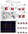
Treatment with felodipine daily for 9 days modulates LPS-induced short-term working memory impairment, hippocampal dendritic spine loss, and microgliosis in C57BL/6 mice. Three-month-old C57BL/6 mice were injected daily with vehicle (5% Tween-20 + 5% PEG300 in saline) or felodipine (5 mg/kg, i.p.), followed 30 min later by 250 μg/kg LPS or PBS for 9 days. (a) Y-maze tests were performed on day 7; spontaneous alternations and the number of total entries are shown (vehicle, n = 10 mice; LPS, n = 11 mice; felodipine+LPS, n = 10 mice). (b) Novel object recognition (NOR) training/tests were performed on days 8 and 9; object preference is shown (vehicle, n = 10 mice; LPS, n = 7 mice; felodipine+LPS, n = 9 mice). (c) Golgi staining was performed after the behavior experiments to measure dendritic spine number in the hippocampal AO and BS dendrites. (d) Quantification of data from (c) (vehicle, n = 19 slices from 5 mice; LPS, n = 15 slices from 4 mice; felodipine+LPS, n = 20 slices from 5 mice. (e) Microgliosis was assessed via immunofluorescence staining of Iba-1 in brain slices from the C57BL/6 mice used in the behavioral experiments. (f) Quantification of data from (e) (n = 4 mice/group, n = 16 brain slices). * p < 0.05, ** p < 0.01, *** p < 0.001. Golgi staining scale bar = 5 μM. IF scale bar = 100 μM. V: vehicle; L: LPS; F or Felodi: felodipine.
Figure 9
Figure 9
Schematic diagram of the effects of felodipine on LPS-induced neuroinflammatory responses and cognitive function. In microglial cells, felodipine downregulates LPS-induced proinflammatory cytokine COX-2 and IL-1β levels through L-type Ca2+ channel/TLR4/AKT signaling. Felodipine also inhibits LPS-stimulated nuclear STAT3 phosphorylation in microglia. In C57BL/6 (wild-type) mice, felodipine reduces LPS-induced micro/astrogliosis and proinflammatory cytokine COX-2 and IL-1β levels and reduces the short-term spatial memory impairment, decrease in hippocampal dendritic spinogenesis, and increase in microgliosis induced by chronic LPS administration. The capacity of felodipine to alter LPS-induced gliosis in vitro/in vivo and to modulate neuroinflammation-linked behavior suggests that felodipine may have potential for the treatment of neuroinflammation/cognitive function-linked diseases.

References

    1. DiSabato D.J., Quan N., Godbout J.P. Neuroinflammation: The devil is in the details. J. Neurochem. 2016;139((Suppl. 2)):136–153. doi: 10.1111/jnc.13607. - DOI - PMC - PubMed
    1. Corps K.N., Roth T.L., McGavern D.B. Inflammation and neuroprotection in traumatic brain injury. JAMA Neurol. 2015;72:355–362. doi: 10.1001/jamaneurol.2014.3558. - DOI - PMC - PubMed
    1. Del Rey A., Balschun D., Wetzel W., Randolf A., Besedovsky H.O. A cytokine network involving brain-borne IL-1beta, IL-1ra, IL-18, IL-6, and TNFalpha operates during long-term potentiation and learning. Brain Behav. Immun. 2013;33:15–23. doi: 10.1016/j.bbi.2013.05.011. - DOI - PubMed
    1. Morris G., Berk M., Walder K., Maes M. Central pathways causing fatigue in neuro-inflammatory and autoimmune illnesses. BMC Med. 2015;13:28. doi: 10.1186/s12916-014-0259-2. - DOI - PMC - PubMed
    1. Ransohoff R.M. How neuroinflammation contributes to neurodegeneration. Science. 2016;353:777–783. doi: 10.1126/science.aag2590. - DOI - PubMed