. 2022 Oct 31;58(11):1565.
doi: 10.3390/medicina58111565.

Development and Survival of Human Ovarian Cells in Chitosan Hydrogel Micro-Bioreactor

Affiliations

Development and Survival of Human Ovarian Cells in Chitosan Hydrogel Micro-Bioreactor

Elsa Labrune et al. Medicina (Kaunas). .

Abstract

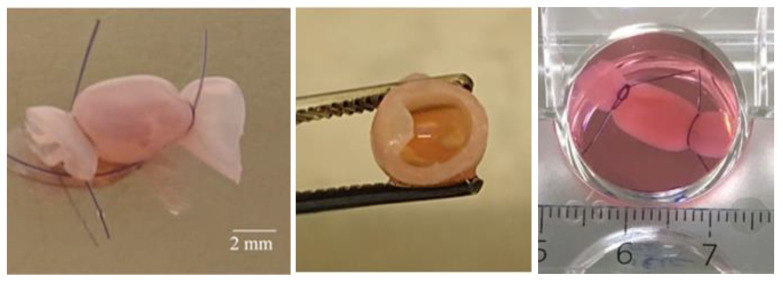
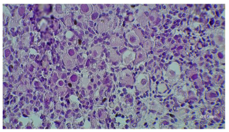
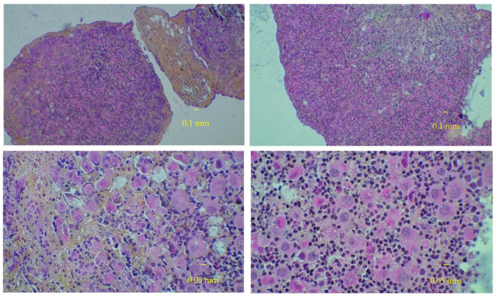
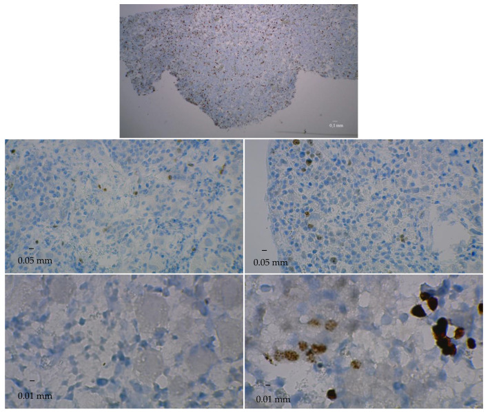
Background and Objectives: To test the long-term ability of human ovarian cortex cells to develop in unconventional culture conditions. Materials and Methods. Ovarian cortex cells from fetuses aged 23 to 39 weeks gestation were cultured for 90 days in hollow chitosan hydrogel micro-bioreactors and concurrently in traditional wells. Various cell-type counts were considered. Results: With intact follicles as a denominator, the percentage of growing intact follicles at Day 0 varied widely between ovaries (0 to 31.7%). This percentage tended to increase or stay relatively constant in bioreactor as in control cultures; it tended more toward an increase over time in bioreactor vs. control cultures. Modeled percentages showed differences (though not significant) in favor of bioreactor cultures (16.12% difference at D50 but only 0.12% difference at D90). With all follicles present as a denominator, the percentage of growing primary and secondary follicles at D0 varied widely between ovaries (0 to 29.3%). This percentage tended to increase over time in bioreactor cultures but to decrease in control cultures. Modeled percentages showed significant differences in favor of bioreactor cultures (8.9% difference at D50 and 11.1% difference at D90). At D50 and D90, there were only few and sparse apoptotic cells in bioreactor cultures vs. no apoptotic cells in control cultures. Conclusions: Over three months, bioreactor folliculogenesis outperformed slightly traditional culture. This is an interesting perspective for follicle preservation and long-term toxicological studies.

Keywords: bioreactor; chitosan; folliculogenesis; hydrogel; tissue culture technique.


Conflict of interest statement

The authors declare no conflict of interest.

Figures

Figure 1
Figure 1
Chitosan bioreactor. From left to right: bioreactor containing ovarian tissue and tied at each end with suture thread; cross-section of the bioreactor showing the inner cavity and wall; culture well with bioreactor in culture medium.
Figure 2
Figure 2
Fresh fetal ovarian tissue before culture. Slices colored before culture with hematoxylin-eosin-safran at low and high magnification.
Figure 2
Figure 2
Fresh fetal ovarian tissue before culture. Slices colored before culture with hematoxylin-eosin-safran at low and high magnification.
Figure 3
Figure 3
Fetal ovarian tissue in culture at day 50. Slices colored with hematoxylin-eosin-safran at low and high magnification. Left panels: control cultures. Right panels: bioreactor cultures.
Figure 4
Figure 4
Fetal ovarian tissue in culture at day 90. Slices colored with hematoxylin-eosin-safran at low and high magnification. Left panels: control cultures. Right panels: bioreactor cultures.
Figure 5
Figure 5
Fetal ovarian tissue in bioreactor at day 90. Slices colored with hematoxylin-eosin-safran. (A): primordial follicles. (B): primary follicles. (C): secondary follicle. (D): degenerated secondary follicle. Blue arrows indicate intact follicles. Orange arrows indicate degenerated follicles.
Figure 6
Figure 6
Ki67 immunostaining. Upper panel: Slice of fresh fetal ovarian tissue before culture. Middle row: Slices at day 50 of culture. Lower row: Slices at day 90 of culture. Left panels: control cultures. Right panels: bioreactor cultures. Positive (proliferating) cells have a brown color.
Figure 7
Figure 7
Cleaved caspase-3 immunostaining. Upper panel: Slice of fresh fetal ovarian tissue before culture. Middle row: Slices at day 50 of culture. Lower row: Slices at day 90 of culture. Left panels: control cultures. Right panels: bioreactor cultures. Apoptic cells—indicated by orange arrows—have brown cytoplasm.

References

    1. Takae S., Suzuki N. Current state and future possibilities of ovarian tissue transplantation. Reprod. Med. Biol. 2019;18:217–224. doi: 10.1002/rmb2.12268. - DOI - PMC - PubMed
    1. Donnez J., Dolmans M.-M. Fertility preservation in women. Nat. Rev. Endocrinol. 2013;9:735–749. doi: 10.1038/nrendo.2013.205. - DOI - PubMed
    1. Dolmans M.-M., Falcone T., Patrizio P. Importance of patient selection to analyze in vitro fertilization outcome with transplanted cryopreserved ovarian tissue. Fertil. Steril. 2020;114:279–280. doi: 10.1016/j.fertnstert.2020.04.050. - DOI - PubMed
    1. Donnez J., Dolmans M., Demylle D., Jadoul P., Pirard C., Squifflet J., Martinez-Madrid B., Van Langendonckt A. Livebirth after orthotopic transplantation of cryopreserved ovarian tissue. Lancet. 2004;364:1405–1410. doi: 10.1016/S0140-6736(04)17222-X. - DOI - PubMed
    1. Dolmans M.-M., Luyckx V., Donnez J., Andersen C.Y., Greve T. Risk of transferring malignant cells with transplanted frozen-thawed ovarian tissue. Fertil. Steril. 2013;99:1514–1522. doi: 10.1016/j.fertnstert.2013.03.027. - DOI - PubMed

LinkOut - more resources