Review
. 2022 Oct 25;27(21):7207.
doi: 10.3390/molecules27217207.

Potential of Therapeutic Small Molecules in Apoptosis Regulation in the Treatment of Neurodegenerative Diseases: An Updated Review

Affiliations
Review

Potential of Therapeutic Small Molecules in Apoptosis Regulation in the Treatment of Neurodegenerative Diseases: An Updated Review

Hamad Ghaleb Dailah. Molecules. .

Abstract

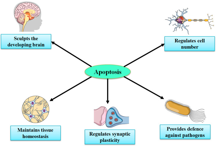
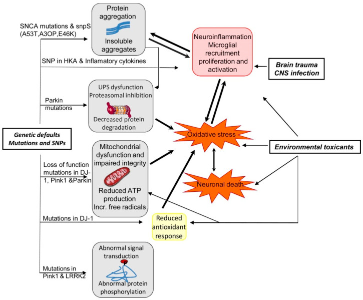
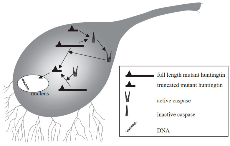
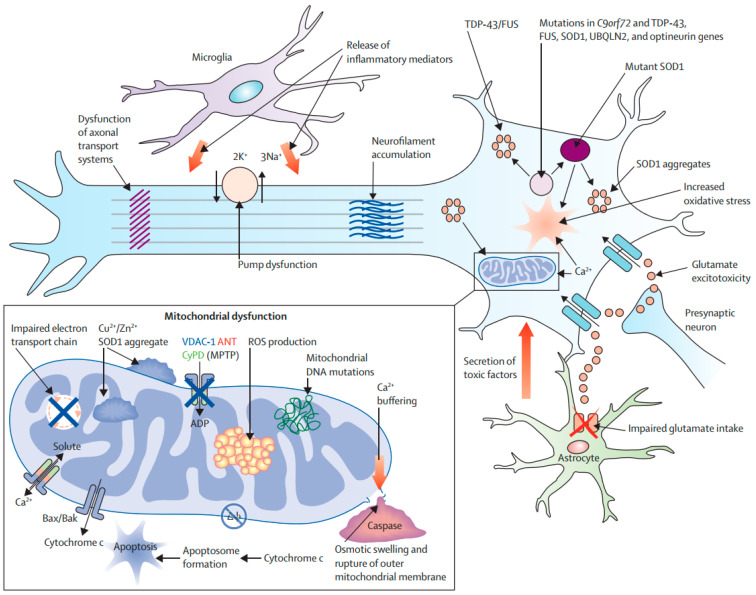
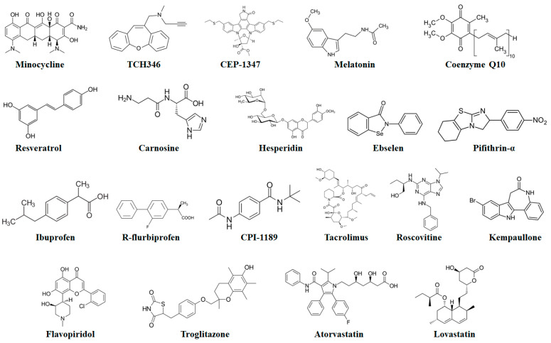
Neurodegenerative disorders (NDs) include Parkinson's disease (PD), Alzheimer's disease (AD), Huntington's disease (HD), and amyotrophic lateral sclerosis (ALS) and the common feature of NDs is the progressive death of specific neurons in the brain. Apoptosis is very important in developing the nervous system, nonetheless an elevated level of cell death has been observed in the case of NDs. NDs are different in terms of their neuronal vulnerability and clinical manifestations, however they have some overlapping neurodegenerative pathways. It has been demonstrated by several studies with cell lines and animal models that apoptosis has a significant contribution to make in advancing AD, ALS, HD, and PD. Numerous dying neurons were also identified in the brains of individuals with NDs and these conditions were found to be linked with substantial cell loss along with common characteristics of apoptosis including activation of caspases and cysteine-proteases, DNA fragmentation, and chromatin condensation. It has been demonstrated that several therapeutic agents including antioxidants, minocycline, GAPDH ligands, p53 inhibitors, JNK (c-Jun N-Terminal Kinase) inhibitors, glycogen synthase kinase-3 inhibitor, non-steroidal anti-inflammatory drugs, D2 dopamine receptor agonists, FK506, cell cycle inhibitors, statins, drugs targeting peroxisome proliferator-activated receptors, and gene therapy have the potential to provide protection to neurons against apoptosis. Therefore, the use of these potential therapeutic agents might be beneficial in the treatment of NDs. In this review, we have summarized the pathways that are linked with apoptotic neuronal death in the case of various NDs. We have particularly focused on the therapeutic agents that have neuroprotective properties and the potential to regulate apoptosis in NDs.

Keywords: DNA fragmentation; apoptosis; caspases; neurodegenerative disorders; neuronal death; neuroprotective drugs.


Conflict of interest statement

The author declares no conflict of interest.

Figures

Figure 1
Figure 1
The extrinsic and intrinsic pathways of apoptosis [92]. Abbreviations: AIF, apoptosis-inducing factor; APAF-1, apoptotic Protease Activating Factor-1; Apo2L/TRAIL, Apo2 ligand or tumor ne-crosis factor-related apoptosis-inducing ligand; BCL-2, B-cell lymphoma 2; BCL-XL, B-cell lym-phoma-extra-large; Bid, BH3-interacting domain death agonist; DISC, death-inducing signaling complex; DR4/5, death receptor 4/5; EndoG, endonuclease G; ER, endoplasmic reticulum; FADD, FAS-associated death domain protein; FasL, Fas Ligand; PUMA, p53 upregulated modulator of apoptosis; ROS, reactive oxygen species; tBid, truncated Bid; TNF-R1, tumor necrosis factor receptor 1. Figure adapted with permission from Ref [92]. Copyright 2014, Elsevier.
Figure 2
Figure 2
The signaling pathways involved in ferroptosis [101]. Abbreviations: ATF4, activating transcription factor 4; CoQ10, coenzyme Q10; Fe3+, ferric cation; Fe2+, ferrous cation; FSP1, ferroptosis suppressor protein 1; FTH1, ferritin heavy chain 1; FTL, ferritin light chain; GPx4, glutathione peroxidase 4; GSH, glutathione; HMG-CoA, 3-hydroxy-3-methylglutaryl coenzyme A; HO-1, heme oxygenase-1; HSPA5, heat shock protein 70 family protein 5; IPP, isopentenyl pyrophosphate; LOXs, lipoxy-genases; NADH, nicotinamide adenine dinucleotide; NCOA4, nuclear receptor coactivator 4; NOXs, NADPH oxidases; NRF2, nuclear factor erythroid 2-related factor 2; PL-PUFAs, phospho-lipids-containing PUFAs; PUFA-OOHs, hydroperoxides derivatives of PUFAs; PUFAs, polyun-saturated fatty acids; ROS, reactive oxygen species; SQS, squalene synthase; TF, transferrin; TFRC, transferrin receptor. Figure reproduced with permission from Ref [101]. Copyright 2021, Elsevier.
Figure 3
Figure 3
Important roles of apoptosis in the development of the central nervous system.
Figure 4
Figure 4
Amyloid beta-induced synapse loss in Alzheimer’s Disease [127]. Abbreviations: Akt, protein kinase B; AMPAR, 2-amino-3-(5-methyl-3-oxo-1,2-oxazol-4-yl)propanoic acid receptor; Aβ, amyloid beta; GSK3, glycogen synthase kinase-3; NFTs, neurofibrillary tangles. Figure reproduced with permis-sion from Ref [127]. Copyright 2020, Elsevier.
Figure 5
Figure 5
Summary of the effects of interaction between genetics and environmental factors in PD patho-genesis [92]. Abbreviations: ATP, adenosine triphosphate; CNS, central nervous system; LRRK2, leucine-rich repeat kinase 2; Pink1, PTEN (phosphatase and tensin homologue)-induced kinase1; SNCA, alpha synuclein; SNP, single-nucleotide polymorphism; UPS, ubiquitin proteasome system. Figure reproduced with permission from Ref [92]. Copyright 2014, Elsevier.
Figure 6
Figure 6
Mechanism of mutant huntingtin-mediated transcriptional dysregulation in Huntington’s disease [177]. Figure reproduced with permission from Ref [177]. Copyright 2003, Elsevier.
Figure 7
Figure 7
Molecular pathways of neurodegeneration in amyotrophic lateral sclerosis [43]. Abbreviations: BAK, Bcl-2 homologue antagonist/killer; Bax, Bcl-2-associated X; ROS, reactive oxygen species; SOD1, superoxide dismutase 1; MPTP, 1-methyl-4-phenyl-1,2,3,6-tetrahydropyridine; VDAC1, voltage-dependent anion channel 1. Figure reproduced with permission from Ref [43]. Copyright 2020, Springer.
Figure 8
Figure 8
Chemical structures of potential antiapoptotic drugs that might be useful in the treatment of neurodegenerative diseases.

References

    1. Muddapu V.R., Dharshini S.A.P., Chakravarthy V.S., Gromiha M.M. Neurodegenerative Diseases—Is Metabolic Deficiency the Root Cause? Front. Neurosci. 2020;14:213. doi: 10.3389/fnins.2020.00213. - DOI - PMC - PubMed
    1. Nichols E., Szoeke C.E.I., Vollset S.E., Abbasi N., Abd-Allah F., Abdela J., Aichour M.T.E., Akinyemi R.O., Alahdab F., Asgedom S.W., et al. Global, regional, and national burden of Alzheimer’s disease and other dementias, 1990–2016: A systematic analysis for the Global Burden of Disease Study 2016. Lancet Neurol. 2019;18:88–106. doi: 10.1016/S1474-4422(18)30403-4. - DOI - PMC - PubMed
    1. Livingston G., Huntley J., Sommerlad A., Ames D., Ballard C., Banerjee S., Brayne C., Burns A., Cohen-Mansfield J., Cooper C., et al. Dementia prevention, intervention, and care: 2020 report of the Lancet Commission. Lancet. 2020;396:413–446. doi: 10.1016/S0140-6736(20)30367-6. - DOI - PMC - PubMed
    1. Cannon J.R., Greenamyre J.T. The role of environmental exposures in neurodegeneration and neurodegenerative diseases. Toxicol. Sci. 2011;124:225–250. doi: 10.1093/toxsci/kfr239. - DOI - PMC - PubMed
    1. Castillo X., Castro-Obregón S., Gutiérrez-Becker B., Gutiérrez-Ospina G., Karalis N., Khalil A.A., Lopez-Noguerola J.S., Rodríguez L.L., Martínez-Martínez E., Perez-Cruz C., et al. Re-thinking the etiological framework of neurodegeneration. Front. Neurosci. 2019;13:728. doi: 10.3389/fnins.2019.00728. - DOI - PMC - PubMed