

Atomic-Resolution Experimental Structural Biology and Molecular Dynamics Simulations of Hyaluronan and Its Complexes
- PMID: 36364098
- PMCID: PMC9658939
- DOI: 10.3390/molecules27217276
Atomic-Resolution Experimental Structural Biology and Molecular Dynamics Simulations of Hyaluronan and Its Complexes
Abstract
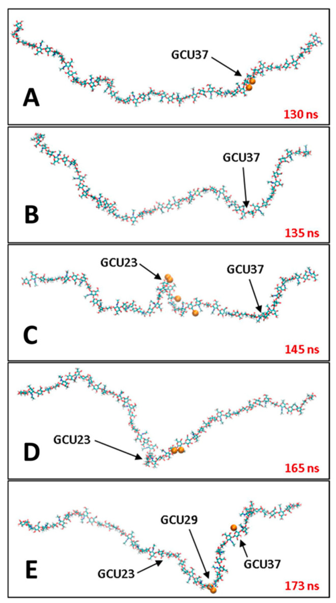
This review summarizes the atomic-resolution structural biology of hyaluronan and its complexes available in the Protein Data Bank, as well as published studies of atomic-resolution explicit-solvent molecular dynamics simulations on these and other hyaluronan and hyaluronan-containing systems. Advances in accurate molecular mechanics force fields, simulation methods and software, and computer hardware have supported a recent flourish in such simulations, such that the simulation publications now outnumber the structural biology publications by an order of magnitude. In addition to supplementing the experimental structural biology with computed dynamic and thermodynamic information, the molecular dynamics studies provide a wealth of atomic-resolution information on hyaluronan-containing systems for which there is no atomic-resolution structural biology either available or possible. Examples of these summarized in this review include hyaluronan pairing with other hyaluronan molecules and glycosaminoglycans, with ions, with proteins and peptides, with lipids, and with drugs and drug-like molecules. Despite limitations imposed by present-day computing resources on system size and simulation timescale, atomic-resolution explicit-solvent molecular dynamics simulations have been able to contribute significant insight into hyaluronan's flexibility and capacity for intra- and intermolecular non-covalent interactions.
Keywords: NMR; conformation; crystallography; flexibility; hyaluronan; hyaluronate; hyaluronic acid; interaction; molecular dynamics.
Conflict of interest statement
The author declares no conflict of interest.
Figures
Similar articles
-
Molecular dynamics and quantum mechanics of RNA: conformational and chemical change we can believe in.Acc Chem Res. 2010 Jan 19;43(1):40-7. doi: 10.1021/ar900093g. Acc Chem Res. 2010. PMID: 19754142 Free PMC article.
-
Hyaluronan-arginine enhanced and dynamic interaction emerges from distinctive molecular signature due to electrostatics and side-chain specificity.Carbohydr Polym. 2024 Feb 1;325:121568. doi: 10.1016/j.carbpol.2023.121568. Epub 2023 Nov 14. Carbohydr Polym. 2024. PMID: 38008475
-
Albumin-Hyaluronan Interactions: Influence of Ionic Composition Probed by Molecular Dynamics.Int J Mol Sci. 2021 Nov 16;22(22):12360. doi: 10.3390/ijms222212360. Int J Mol Sci. 2021. PMID: 34830249 Free PMC article.
-
Molecular Dynamics Simulation for All.Neuron. 2018 Sep 19;99(6):1129-1143. doi: 10.1016/j.neuron.2018.08.011. Neuron. 2018. PMID: 30236283 Free PMC article. Review.
-
Hyaluronan chain conformation and dynamics.Carbohydr Res. 2005 Apr 11;340(5):959-70. doi: 10.1016/j.carres.2005.01.030. Carbohydr Res. 2005. PMID: 15780260 Review.
Cited by
-
Extending the Martini 3 Coarse-Grained Force Field to Hyaluronic Acid.J Phys Chem B. 2025 Mar 6;129(9):2408-2425. doi: 10.1021/acs.jpcb.4c08043. Epub 2025 Feb 23. J Phys Chem B. 2025. PMID: 39988846 Free PMC article.
-
Molecular Dynamics Simulation of Structural Assembly and Hydration of Hyaluronic Acid in Salt Aqueous Buffer.Langmuir. 2025 Feb 18;41(6):3852-3864. doi: 10.1021/acs.langmuir.4c03966. Epub 2025 Feb 6. Langmuir. 2025. PMID: 39913243 Free PMC article.
References
-
- Simpson M., Schaefer L., Hascall V., Esko J.D. Hyaluronan. In: Varki A., Cummings R.D., Esko J.D., Stanley P., Hart G.W., Aebi M., Mohnen D., Kinoshita T., Packer N.H., Prestegard J.H., et al., editors. Essentials of Glycobiology. 4th ed. Cold Spring Harbor Laboratory Press; Cold Spring Harbor, NY, USA: 2022. pp. 205–216.
-
- Merry C.L.R., Lindahl U., Couchman J., Esko J.D. Proteoglycans and Sulfated Glycosaminoglycans. In: Varki A., Cummings R.D., Esko J.D., Stanley P., Hart G.W., Aebi M., Mohnen D., Kinoshita T., Packer N.H., Prestegard J.H., et al., editors. Essentials of Glycobiology. 4th ed. Cold Spring Harbor Laboratory Press; Cold Spring Harbor, NY, USA: 2022. pp. 217–232.
-
- Toole B.P. Hyaluronan: From extracellular glue to pericellular cue. Nat. Rev. Cancer. 2004;4:528–539. - PubMed
Publication types
MeSH terms
Substances
LinkOut - more resources
Full Text Sources
