Review
. 2022 Oct 28;27(21):7327.
doi: 10.3390/molecules27217327.

Recent Progress in Biosensors for Detection of Tumor Biomarkers

Affiliations
Review

Recent Progress in Biosensors for Detection of Tumor Biomarkers

Mantong Li et al. Molecules. .

Abstract

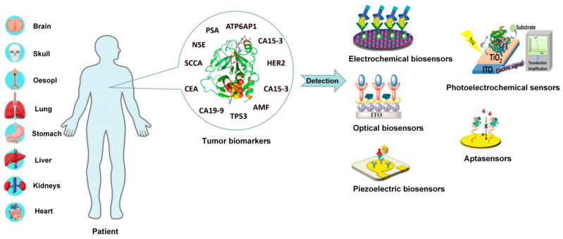
Cancer is a leading cause of death worldwide, with an increasing mortality rate over the past years. The early detection of cancer contributes to early diagnosis and subsequent treatment. How to detect early cancer has become one of the hot research directions of cancer. Tumor biomarkers, biochemical parameters for reflecting cancer occurrence and progression have caused much attention in cancer early detection. Due to high sensitivity, convenience and low cost, biosensors have been largely developed to detect tumor biomarkers. This review describes the application of various biosensors in detecting tumor markers. Firstly, several typical tumor makers, such as neuron-specific enolase (NSE), carcinoembryonic antigen (CEA), prostate-specific antigen (PSA), squamous cell carcinoma antigen (SCCA), carbohydrate, antigen19-9 (CA19-9) and tumor suppressor p53 (TP53), which may be helpful for early cancer detection in the clinic, are briefly described. Then, various biosensors, mainly focusing on electrochemical biosensors, optical biosensors, photoelectrochemical biosensors, piezoelectric biosensors and aptamer sensors, are discussed. Specifically, the operation principles of biosensors, nanomaterials used in biosensors and the application of biosensors in tumor marker detection have been comprehensively reviewed and provided. Lastly, the challenges and prospects for developing effective biosensors for early cancer diagnosis are discussed.

Keywords: biosensor; cancer; detection; nanomaterial; tumor biomarker.


Conflict of interest statement

The authors declare no conflict of interest.

Figures

Figure 2
Figure 2
Elements and selected components of a typical biosensor. Reprinted with permission from [19].
Figure 3
Figure 3
The working principle of optical biosensors. Reprinted with permission from [22].
Figure 4
Figure 4
(a) The working principle of the piezoelectric biosensor. (b) Influence of voltage on time. (c) The influence of amplitude on frequency. Reprinted with permission from [27].
Figure 5
Figure 5
(a) Schematic diagram of an electrochemical immunosensor for NSE detection. Reprinted with permission from [37]. (b) Schematic diagram of an electrochemical immunosensor structure. Reprinted with permission from [38]. (c) Schematic of a biofunctionalized black phosphorus-based fiber optic biosensor. Reprinted with permission from [32].
Figure 6
Figure 6
(a) The schematic illustration of the fabrication process of the sandwich-type electrochemical immunosensor. Reprinted with permission from [39]. (b) The detection procedure. Reprinted with permission from [40].
Figure 7
Figure 7
(a) Schematic diagram of detecting PSA by an electrochemical biosensor based on peptide cleavage. Reprinted with permission from [52]. (b) Schematic diagram of the PSA test. Reprinted with permission from [55]. (c) Schematic of the sandwich immunoassay format in the QCM sensor chip combine with gold-staining signal amplification. Reprinted with permission from [61].
Figure 8
Figure 8
(a) The overall fabrication process of the PEC immunosensor and the specific response to SCCA, and the mechanism for the photocurrent generation. Reprinted with permission from [65]. (b) Schematic diagram of the PEC immunosensor. Reprinted with permission from [67].
Figure 9
Figure 9
(a) Schematic diagram of the electrochemical immunosensor. Reprinted with permission from [69]. (b) Schematic diagram of a synthetic immunosensor made of PLL/HA/CNT hybrid materials. Reprinted with permission from [73].
Figure 10
Figure 10
(a) Schematic diagram of the ECL immunosensor based on CdS NCs and tGO-AuNPs. Reprinted with permission from [76]. (b) Schematic diagram of the Gr-Au tip platform for SERS detection, and a pattern diagram of cancer cells deposited on the Gr-Au tip platform. Reprinted with permission from [77]. (c) The relative calibration curves of different concentrations of fully complementary and mismatched oligonucleotides. Reprinted with permission from [81].
Figure 1
Figure 1
Schematic illustration of the application of electrochemical, optical, photoelectrochemical, piezoelectric sensors/biosensors and aptasensors for the detection of tumor biomarkers in a cancer patient.
Figure 11
Figure 11
Utilizing the salivary autoantibodies against ATP6AP1 to detect breast cancer through the biosensor. Reprinted with permission from [84].

References

    1. Yang G., Xiao Z., Tang C., Deng Y., Huang H., He Z. Recent advances in biosensor for detection of lung cancer biomarkers. Biosens. Bioelectron. 2019;141:111416. doi: 10.1016/j.bios.2019.111416. - DOI - PubMed
    1. Wu J., Fu Z., Yan F., Ju H. Biomedical and clinical applications of immunoassays and immunosensors for tumor markers. TrAC Trends Anal. Chem. 2007;26:679–688. doi: 10.1016/j.trac.2007.05.007. - DOI
    1. Schiffman J.D., Fisher P.G., Gibbs P. Early detection of cancer: Past, present, and future. Am. Soc. Clin. Oncol. Educ. Book. 2015;35:57–65. doi: 10.14694/EdBook_AM.2015.35.57. - DOI - PubMed
    1. Jayanthi V., Das A.B., Saxena U. Recent advances in biosensor development for the detection of cancer biomarkers. Biosens. Bioelectron. 2017;91:15–23. doi: 10.1016/j.bios.2016.12.014. - DOI - PubMed
    1. Xiao Z., Chen S., Feng S., Li Y., Zou J., Ling H., Zeng Y., Zeng X. Function and mechanisms of microRNA-20a in colorectal cancer. Exp. Ther. Med. 2020;19:1605–1616. doi: 10.3892/etm.2020.8432. - DOI - PMC - PubMed