. 2022 Oct 29;27(21):7362.
doi: 10.3390/molecules27217362.

Honokiol and Alpha-Mangostin Inhibit Mayaro Virus Replication through Different Mechanisms

Affiliations

Honokiol and Alpha-Mangostin Inhibit Mayaro Virus Replication through Different Mechanisms

Patricia Valdés-Torres et al. Molecules. .

Abstract

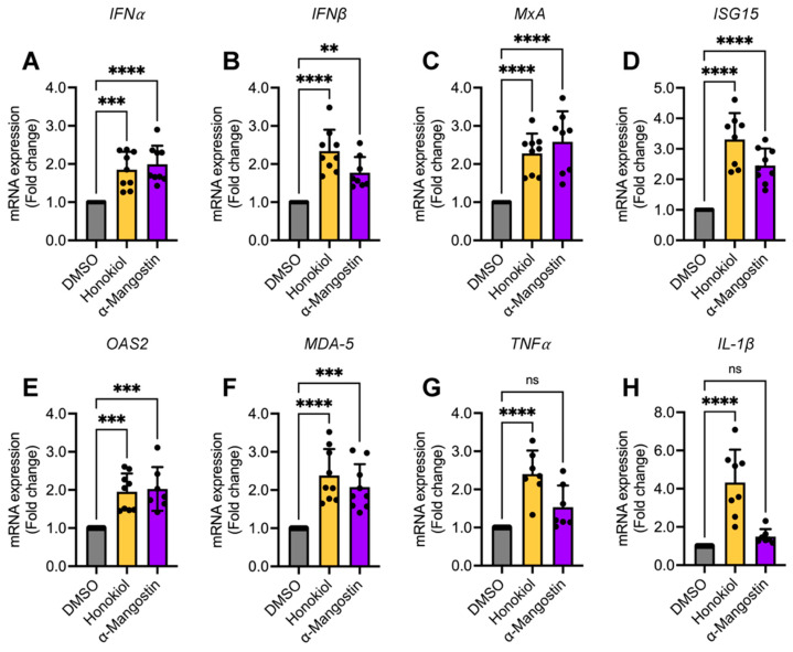
Mayaro virus (MAYV) is an emerging arbovirus with an increasing circulation across the Americas. In the present study, we evaluated the potential antiviral activity of the following natural compounds against MAYV and other arboviruses: Sanguinarine, (R)-Shikonin, Fisetin, Honokiol, Tanshinone IIA, and α-Mangostin. Sanguinarine and Shikonin showed significant cytotoxicity, whereas Fisetin, Honokiol, Tanshinone IIA, and α-Mangostin were well tolerated in all the cell lines tested. Honokiol and α-Mangostin treatment protected Vero-E6 cells against MAYV-induced damage and resulted in a dose-dependent reduction in viral progeny yields for each of the MAYV strains and human cell lines assessed. These compounds also reduced MAYV viral RNA replication in HeLa cells. In addition, Honokiol and α-Mangostin disrupted MAYV infection at different stages of the virus life cycle. Moreover, Honokiol and α-Mangostin decreased Una, Chikungunya, and Zika viral titers and downmodulated the expression of E1 and nsP1 viral proteins from MAYV, Una, and Chikungunya. Finally, in Honokiol- and α-Mangostin-treated HeLa cells, we observed an upregulation in the expression of type I interferon and specific interferon-stimulated genes, including IFNα, IFNβ, MxA, ISG15, OAS2, MDA-5, TNFα, and IL-1β, which may promote an antiviral cellular state. Our results indicate that Honokiol and α-Mangostin present potential broad-spectrum activity against different arboviruses through different mechanisms.

Keywords: Chikungunya; Honokiol; Mayaro; Una; Zika; antiviral activity; arboviruses; broad-spectrum activity; α-Mangostin.


Conflict of interest statement

The authors declare no conflict of interest. The funders had no role in the design of the study; in the collection, analyses, or interpretation of data; in the writing of the manuscript, or in the decision to publish the results.

Figures

Figure 1
Figure 1
Chemical structures of the natural compounds tested. (A) Sanguinarine chloride; (B) (R)-Shikonin; (C) Fisetin; (D) Honokiol; (E) Tanshinone IIA; and (F) α-Mangostin.
Figure 2
Figure 2
Cytotoxicity of the natural compounds evaluated in this study. Vero-E6 cells were treated with the indicated concentrations of Sanguinarine chloride (A), (R)-Shikonin (B), Fisetin (C), Honokiol (D), Tanshinone IIA, (E) or α-Mangostin (F). After 24 or 48 h of incubation, cell viability was determined using an MTT assay. Data represent the mean ± standard deviation of two independent experiments with five replicates. Data were analyzed with a one-way ANOVA test followed by Dunnett’s post hoc test. Statistically significant differences are denoted as follows: * p < 0.05; ** p < 0.01; **** p < 0.0001; and ns: non-significant.
Figure 3
Figure 3
Inhibition of MAYV-induced cytopathic effects by Honokiol and α-Mangostin in Vero-E6 cells is dose-dependent. Vero-E6 cells were infected with the MAYV strain AVR0565 at a multiplicity of infection (MOI) of 1. Further, after 1 h of virus adsorption, cells were treated with Fisetin, Honokiol, Tanshinone IIA (at doses of 5 or 10 μM), or α-Mangostin (at doses of 1 or 5 μM) for 48 h. DMSO (0.1%) served as a control. Cytopathic effects were evaluated using an inverted microscope. One representative microphotograph of at least 10 different fields is shown. Scale bar: 100 μm.
Figure 4
Figure 4
Honokiol and α-Mangostin promote a reduction in MAYV progeny production. Vero-E6 cells were infected with the MAYV strain AVR0565 using an MOI of 1. After 1 h of virus adsorption, cells were treated with the indicated doses of Fisetin (A), Honokiol (B), Tanshinone IIA (C), or α-Mangostin (D); further, DMSO (0.1%) was used as a control. After 24 h of incubation, viral progeny production in cell supernatants was quantified using a plaque-forming assay. Data represent the mean ± standard deviation of three independent experiments in triplicate. (E) Vero-E6 cells grown on glass coverslips were infected with MAYV and treated with Honokiol or α-Mangostin as indicated above. After 24 h of infection, cells were stained with an MAYV E1 antibody followed by a secondary antibody Alexa-Flour 568 and nuclei were stained with DAPI. Then, the cells were analyzed with an immunofluorescence confocal microscope, with a scale bar of: 30 μm. (F) The percentage of MAYV E1 protein-positive cells was determined in at least 10 different fields. Data were analyzed using a one-way ANOVA test followed by Dunnett post hoc test. Statistically significant differences are denoted as follows: ** p < 0.01; *** p < 0.001; **** p < 0.0001; and ns: non-significant.
Figure 5
Figure 5
Honokiol and α-Mangostin reduce MAYV progeny yields, regardless of the virus strain or human cell line tested. Vero-E6 cells were infected with the MAYV Guyane (A,B) or TRVL4675 (C,D) strains and then treated with Honokiol or α-Mangostin at the indicated concentrations for 24 h. After that, viral progeny production in cell supernatants was quantified using a plaque-forming assay. HDFs (E,F) or HeLa (G,H) cells were infected with the MAYV AVR0565 strain and treated as previously indicated. Following 24 h of incubation, viral titers in cell supernatants were analyzed as previously described. Cell viability in HDFs (I,J) or HeLa cells (K,L) treated with Honokiol (10 μM) or α-Mangostin (5 μM) for 24 h was evaluated using the MTT method. Data represent the mean ± standard deviation of three independent experiments in triplicate. Data were analyzed using one-way ANOVA test followed by Dunnett post hoc test or Mann–Whitney test. Statistically significant differences are denoted as follows: * p < 0.05; ** p < 0.01; *** p < 0.001; **** p < 0.0001; and ns: non-significant.
Figure 6
Figure 6
α-Mangostin pretreatment reduces MAYV progeny production. HDFs were pretreated with increasing doses of Honokiol (A) or α-Mangostin (B) for 2 h; after that, the compounds were removed, and the cells were infected with the MAYV AVR0565 strain. Following 1 h of virus adsorption, a fresh medium without the compounds was added to the cells, and they were incubated for 24 h. Next, viral titers were quantified as previously described. Following this, a 1 × 10⁵ UFP amount of the MAYV AVR0565 strain was incubated at 37 °C with the indicated concentration of Honokiol (C) or α-Mangostin (D) for 2 h. Then, the remaining virus in each experimental condition was directly calculated using plaque-forming assays. Data represent the mean ± standard deviation of three independent experiments in triplicate. Data were analyzed using one-way ANOVA test followed by Dunnett post hoc test. Statistically significant differences are denoted as follows: ** p < 0.01 and ns: non-significant.
Figure 7
Figure 7
Honokiol and α-Mangostin inhibit MAYV infection at different stages of the viral life cycle. HDFs infected with MAYV AVR0565 strain at an MOI 1 and the effect of Honokiol or α-Mangostin were assessed using binding (A,B), entry (C,D), and post-entry assays (E,F). Then, viral titers were quantified using a plaque-forming assay. Data represent the mean ± standard deviation of three independent experiments in triplicate. Data were analyzed using one-way ANOVA test followed by Dunnett post hoc test. Statistically significant differences are denoted as follows: * p < 0.5; ** p < 0.01; and ns: non-significant.
Figure 8
Figure 8
Honokiol and α-Mangostin reduce the expression of MAYV E1 and nsP1 proteins and promote a decrease in viral RNA. HeLa cells (A,B) were infected with the MAYV AVR0565 strain at an MOI of 1 and then treated with Honokiol or α-Mangostin at the indicated doses. After 24 h of incubation, protein extracts were obtained, and E1 and nsP1 viral protein levels were analyzed using Western blot. GAPDH protein was used as a loading control. Please note, kDa: kilodaltons and WB: Western blot. (C) HeLa cells were treated or untreated with Honokiol (10 μM) or α-Mangostin (5 μM) and viral RNA replication was assessed using RT-PCR. Data represent the mean ± standard deviation of two independent experiments in triplicate. Data were analyzed using one-way ANOVA test followed by Dunnett post hoc test. Statistically significant differences are denoted as follows: **** p < 0.0001.
Figure 9
Figure 9
Honokiol and α-Mangostin impair UNAV, CHIKV, and ZIKV replication. Vero-E6 cells were infected with UNAV (AD), CHIKV (EH), or ZIKV (I,J) at an MOI of 1. After viral adsorption, increasing doses of Honokiol or α-Mangostin were added to the cells, and they were incubated for 24 h. Next, viral titers and E1 and nsP1 protein expression were evaluated using a plaque-forming assay or Western blot, respectively. GAPDH protein was used as a loading control. Please note, kDa: Kilodaltons and WB: Western blot. Data represent the mean ± standard deviation of three independent experiments in triplicate. Data were analyzed using one-way ANOVA test followed by Dunnett post hoc test. Statistically significant differences are denoted as follows: * p < 0.5; ** p < 0.01; *** p < 0.001; **** p < 0.0001; and ns: non-significant.
Figure 10
Figure 10
Honokiol and α-Mangostin treatment stimulate the expression of type I IFN and specific IFN-stimulated genes. HeLa cells were treated with Honokiol (10 μM) or α-Mangostin (5 μM) for 24 h. Then, total RNA was extracted and the levels of the indicated immune response genes (AH) were assessed using quantitative RT-PCR. Relative mRNA expression in Honokiol- or α-Mangostin-treated cells was represented as fold changes as compared to DMSO-treated cells. Data represent the mean ± standard deviation of three independent experiments in triplicate. Data were analyzed using one-way ANOVA test followed by Dunnett post hoc test. Statistically significant differences are denoted as follows: ** p < 0.01; *** p < 0.001; **** p < 0.0001; and ns: non-significant.

Similar articles

Cited by

References

    1. Yactayo S., Staples J.E., Millot V., Cibrelus L., Ramon-Pardo P. Epidemiology of Chikungunya in the Americas. J. Infect. Dis. 2016;214:S441–S445. doi: 10.1093/infdis/jiw390. - DOI - PMC - PubMed
    1. Gubler D.J., Vasilakis N., Musso D. History and Emergence of Zika Virus. J. Infect. Dis. 2017;216:S860–S867. doi: 10.1093/infdis/jix451. - DOI - PMC - PubMed
    1. Ganjian N., Riviere-Cinnamond A. Mayaro virus in Latin America and the Caribbean. Rev. Panam. Salud Publica. 2020;44:e14. doi: 10.26633/RPSP.2020.14. - DOI - PMC - PubMed
    1. Aguilar-Luis M.A., Del Valle-Mendoza J., Sandoval I., Silva-Caso W., Mazulis F., Carrillo-Ng H., Tarazona-Castro Y., Martins-Luna J., Aquino-Ortega R., Pena-Tuesta I., et al. A silent public health threat: Emergence of Mayaro virus and co-infection with Dengue in Peru. BMC Res. Notes. 2021;14:1–7. doi: 10.1186/s13104-021-05444-8. - DOI - PMC - PubMed
    1. Gonzalez-Escobar G., Churaman C., Rampersad C., Singh R., Nathaniel S. Mayaro virus detection in patients from rural and urban areas in Trinidad and Tobago during the Chikungunya and Zika virus outbreaks. Pathog. Glob. Health. 2021;115:188–195. doi: 10.1080/20477724.2021.1878445. - DOI - PMC - PubMed