. 2022 Nov 5;27(21):7591.
doi: 10.3390/molecules27217591.

Targeting Proteolysis with Cyanogenic Glycoside Amygdalin Induces Apoptosis in Breast Cancer Cells

Affiliations

Targeting Proteolysis with Cyanogenic Glycoside Amygdalin Induces Apoptosis in Breast Cancer Cells

Valentina Cecarini et al. Molecules. .

Abstract

Background: Breast cancer is the most diagnosed cancer among women, and its incidence and mortality are rapidly growing worldwide. In this regard, plant-derived natural compounds have been shown to be effective as chemotherapeutic and preventative agents. Apricot kernels are a rich source of nutrients including proteins, lipids, fibers, and phenolic compounds and contain the aromatic cyanogenic glycoside amygdalin that has been shown to exert a cytotoxic effect on cancer cells by affecting the cell cycle, inducing apoptosis, and regulating the immune function.

Methods: Here, we describe a previously unexplored proapoptotic mechanism of action of amygdalin in breast cancer (MCF7) cells that involves the modulation of intracellular proteolysis. For comparative purposes, the same investigations were also conducted upon cell treatment with two apricot kernel aqueous extracts from Prunus armeniaca L.

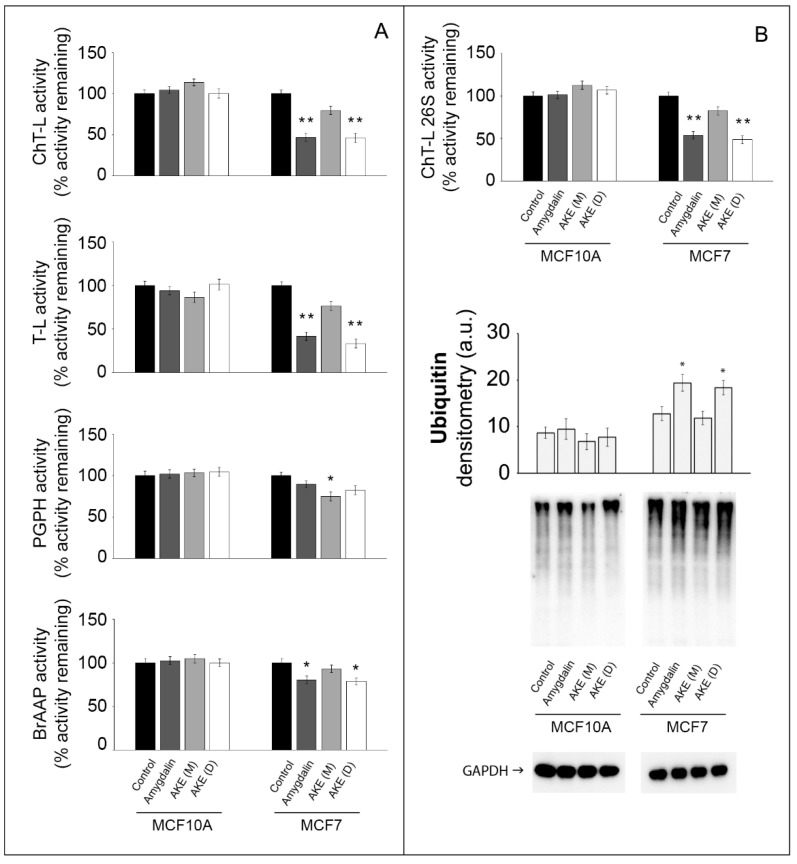
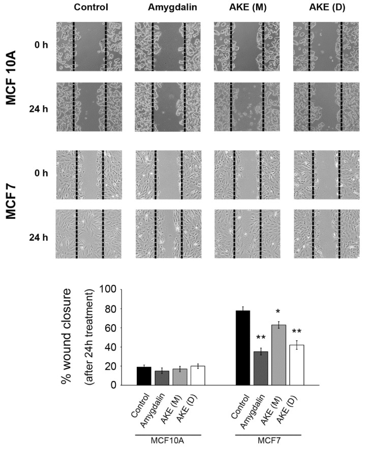
Results: We observed that both the 20S and 26S proteasome activities were downregulated in the MCF7 cells upon 24 h treatments. Simultaneously, the autophagy cascade resulted in being impaired due to cathepsin B and L inhibition that also contributed to a reduction in cancer cell migration. The inhibition of these proteolytic systems finally promoted the activation of apoptotic events in the MCF7 cells.

Conclusion: Collectively, our data unveil a novel mechanism of the anticancer activity of amygdalin, prompting further investigations for potential application in cancer preventative strategies.

Keywords: amygdalin; apoptosis; apricot kernel extract; autophagy; cancer; proteasome.


Conflict of interest statement

The authors declare no conflict of interest.

Figures

Figure 1
Figure 1
Phytochemical profile of the decoction (AKE (D), Panel (A)) and maceration (AKE (M), Panel (B)) extracts obtained upon chromatographic analysis. a, Shikimic acid; b, Gallic acid; c, Loganic acid; d, 5-caffeylquinic acid; e, Swertiamarin; f, Catechin hydrate; g, Delphinidin−3,5−diglucoside; h, Amygdalin; i, Sweroside; j, Chlorogenic acid; k, Vanillic acid; l, Caffeic acid; m, Epichatechin; n, Syringic acid; o, p-Coumaric acid; p, Ferulic acid; q, Naringin; r, Rutin hydrate; s, Hyperoside; t, Resveratrol; u, Amarogentin; v, Kaempferol−3−glucoside; z, Quercetin dihydrate.
Figure 2
Figure 2
Effect of amygdalin and apricot kernel extracts on cell viability and PCNA levels. The MTT assay was carried out to measure cell viability in MCF10A and MCF7 cells. Cells were treated with increasing concentrations of maceration extract and decoction extract (Panel (A)) and amygdalin (Panel (B)) for 24 h. Results are expressed as percent toward untreated cells. Data points marked with an asterisk are statistically significant compared with the respective untreated control cells (* p < 0.05). (C) Representative Western blots for PCNA and relative densitometry is reported. GAPDH was used as control for equal protein loading. Data points marked with an asterisk are statistically significant compared with the respective untreated control cells (* p < 0.05).
Figure 3
Figure 3
Effect of amygdalin and apricot kernel extracts on the 20S and 26S proteasome functionality. (A) 20S proteasome activities in normal and cancer cells following 24 h treatment with 50 µM amygdalin and 1 mg/mL apricot kernel extracts. Asterisks indicate significantly different values compared with respective untreated control cells (* p < 0.05, ** p < 0.01). Results are expressed as mean values and standard deviation and are obtained from five separate experiments. (B) 26S ChT-L proteasome activity in treated cells and a representative Western blot of ubiquitin-conjugates with the relative densitometry. GAPDH was used as equal loading control. * p < 0.05, ** p < 0.01 indicates significantly different values compared with respective untreated control cells. Results are expressed as mean values and standard deviation and are obtained from five separate experiments. Statistical analysis was performed with one-way ANOVA, followed by the Bonferroni test using SigmaStat 3.1 software.
Figure 4
Figure 4
Effect of amygdalin and apricot kernel extracts on apoptosis markers. Representative Western blot of p53, p27, and Bax (Panel (A)) and relative densitometry (Panel (B)). GAPDH was used as equal loading control. Results are expressed as mean values and standard deviation and are obtained from five separate experiments. * p < 0.05 indicates significantly different values compared with respective untreated control cells. (C) DEVDase activity was measured in normal and cancer cells following 24 h treatment with 50 µM amygdalin and 1 mg/mL apricot kernel extracts. Asterisks indicate significantly different values compared with respective untreated control cells (* p < 0.05). Results are expressed as mean values and standard deviation and are obtained from five separate experiments. Statistical analysis was performed with one-way ANOVA, followed by the Bonferroni test using SigmaStat 3.1 software.
Figure 5
Figure 5
Effect of amygdalin and apricot kernel extracts on autophagy. (A) A representative Western blot of p62 protein and relative densitometry. GAPDH was used as equal loading control. Results are expressed as mean values and standard deviation and are obtained from five separate experiments. Statistical analysis was performed with one-way ANOVA, followed by the Bonferroni test using SigmaStat 3.1 software. * p < 0.05 indicates significantly different values compared with respective untreated control cells. (B) MDC staining of MCF10A and MCF7 treated cells. Cells were treated with amygdalin and kernel extracts and then exposed to the autofluorescent dye MDC to detect autophagic vacuoles. 10× and 60× magnifications are shown for MC7 cells. * p < 0.05 indicates significantly different values compared with respective untreated control cells. (C) Cathepsin B and cathepsin L activity measured in control and treated cells. Activity was measured using fluorogenic peptides as substrates as described in the Materials and Methods section. Data are indicated as percentage vs. untreated control cells (* p < 0.05, ** p < 0.01).
Figure 6
Figure 6
Computed binding modes of amygdalin to β1, β2, and β5 subunits of human constitutive 20S proteasome (pdb ID: 6rgq). Panels (AC) report the comparative visualization of 3D models of amygdalin and the residues close to the active sites of β1, β2, and β5 subunits, respectively, that are directly involved in the formation of the enzyme-inhibitor complexes (displayed as light blue and pink sticks, respectively). H-bonds are indicated as yellow dashed solid lines. Panels (A’C’): comparative 2D visualization of amygdalin and the residues close to the active site of β1, β2, and β5 subunits, respectively, that are directly involved in the formation of H-bonds (purple arrows), polar (light blue ribbon), and VdW interactions (green ribbon). Atoms/groups exposed to solvent are indicated with grey circles.
Figure 7
Figure 7
Computed binding modes of amygdalin to cathepsin B (PDB ID: 1csb) and L (PDB ID: 3hha). Panels (A,B) report the comparative visualization of 3D models of amygdalin and the residues close to the active site of cathepsin B and L, respectively, that are directly involved in the formation of the enzyme-inhibitor complexes (displayed as light blue and pink sticks, respectively). H-bonds are indicated as yellow dashed solid lines. Panels (A’,B’) report the comparative 2D visualization of amygdalin and the residues close to the active site of cathepsin B and L, respectively, that are directly involved in the formation of H-bonds (purple arrows), polar (light blue ribbon), and VdW interactions (green ribbon). Atoms/groups exposed to solvent are indicated with grey circles.
Figure 8
Figure 8
Effect of treatments on cell migration. Confluent monolayers were scratched, and wound closure was monitored using a microscope equipped with a camera after 24 h exposure to amygdalin or extracts. Each experiment was repeated at least five times. Results were expressed as percentage wound closure (* p < 0.05 and ** p < 0.01 compared to control).

Similar articles

Cited by

References

    1. Hazafa A., Iqbal M.O., Javaid U., Tareen M.B.K., Amna D., Ramzan A., Piracha S., Naeem M. Inhibitory effect of polyphenols (phenolic acids, lignans, and stilbenes) on cancer by regulating signal transduction pathways: A review. Clin. Transl. Oncol. 2022;24:432–445. doi: 10.1007/s12094-021-02709-3. - DOI - PubMed
    1. Bonfili L., Amici M., Cecarini V., Cuccioloni M., Tacconi R., Angeletti M., Fioretti E., Keller J.N., Eleuteri A.M. Wheat sprout extract-induced apoptosis in human cancer cells by proteasomes modulation. Biochimie. 2009;91:1131–1144. doi: 10.1016/j.biochi.2009.06.001. - DOI - PubMed
    1. Cuccioloni M., Bonfili L., Mozzicafreddo M., Cecarini V., Scuri S., Cocchioni M., Nabissi M., Santoni G., Eleuteri A.M., Angeletti M. Mangiferin blocks proliferation and induces apoptosis of breast cancer cells via suppression of the mevalonate pathway and by proteasome inhibition. Food Funct. 2016;7:4299–4309. doi: 10.1039/C6FO01037G. - DOI - PubMed
    1. Acquaviva R., Tomasello B., Di Giacomo C., Santangelo R., La Mantia A., Naletova I., Sarpietro M.G., Castelli F., Malfa G.A. Protocatechuic Acid, a Simple Plant Secondary Metabolite, Induced Apoptosis by Promoting Oxidative Stress through HO-1 Downregulation and p21 Upregulation in Colon Cancer Cells. Biomolecules. 2021;11:1485. doi: 10.3390/biom11101485. - DOI - PMC - PubMed
    1. Aamazadeh F., Ostadrahimi A., Rahbar Saadat Y., Barar J. Bitter apricot ethanolic extract induces apoptosis through increasing expression of Bax/Bcl-2 ratio and caspase-3 in PANC-1 pancreatic cancer cells. Mol. Biol. Rep. 2020;47:1895–1904. doi: 10.1007/s11033-020-05286-w. - DOI - PubMed