. 2022 Nov 3;14(21):4659.
doi: 10.3390/nu14214659.

Antioxidant Capacity and Protective Effect of Cow Placenta Extract on D-Galactose-Induced Skin Aging in Mice

Affiliations

Antioxidant Capacity and Protective Effect of Cow Placenta Extract on D-Galactose-Induced Skin Aging in Mice

Liu-Hong Shen et al. Nutrients. .

Abstract

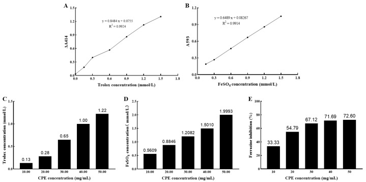
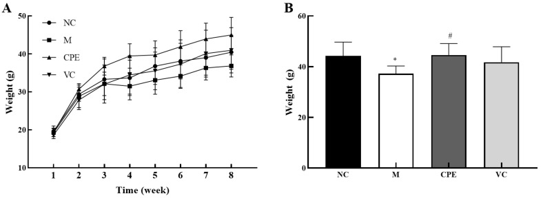
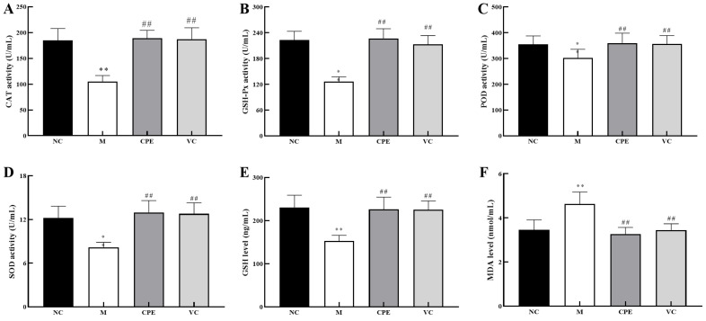
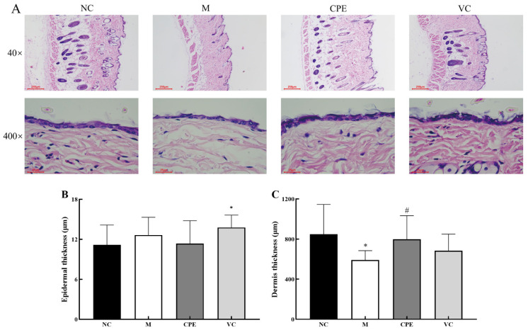
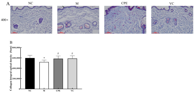
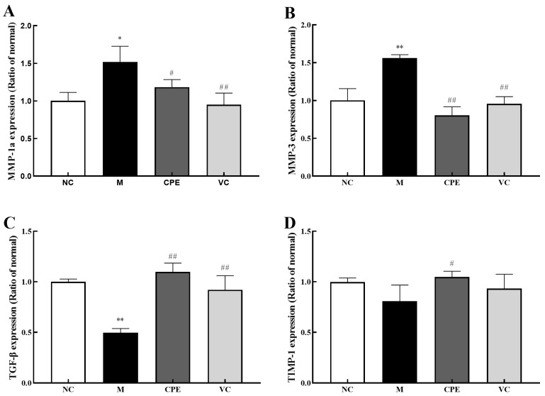
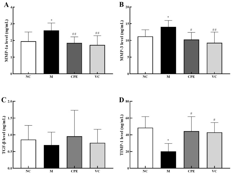
Placental extract has been used for skin care and delaying skin aging. Cow placenta is an abundant resource with a large mass, which has not been harnessed effectively. Cow placenta extract (CPE) has the functions of antioxidation, anti-inflammatory, promoting growth and development, and promoting hair growth. However, little is known about the effect of oral administration of cow placenta extract on skin conditions. Therefore, the present study aimed to investigate the antioxidant capacity of CPE in vitro and in vivo and its protective effect on d-galactose (D-gal) induced skin aging in mice. The results showed that CPE had strong free radical scavenging, reducing and metal chelating activities. CPE can increase the activity of catalase (CAT), glutathione peroxidase (GSH-Px), peroxidase (POD), superoxide dismutase (SOD), and the content of glutathione (GSH), decrease the content of malondialdehyde (MDA). Moreover, CPE can decrease the gene and protein expression of matrix metalloproteinase 1a (MMP-1a) and matrix metalloproteinase 3 (MMP-3) and increase the expression of transforming growth factor-β (TGF-β) and tissue inhibitor of metalloproteinase 1 (TIMP-1) of mouse skin. Histopathological analysis showed CPE reduced the collagen damage caused by D-gal, increased collagen synthesis and reduced its degradation to delay skin aging.

Keywords: D-galactose; antioxidant; cow; placenta; skin aging.


Conflict of interest statement

The authors declare no conflict of interest.

Figures

Figure 1
Figure 1
Antioxidant capacity of CPE in vitro. Note: (A): Trolox standard curve; (B): FeSO4 standard curve; (C): TEAC of 10–50 mg/mL CPE; (D): FeSO4 concentration corresponding to 10–50 mg/mL CPE; (E): Metal chelating capacity of 10–50 mg/mL CPE. CPE, cow placenta extract; A, absorbance; ∆, change; FeSO4, ferrous sulfate; TEAC, Trolox-Equivalent antioxidant capacity.
Figure 2
Figure 2
Body weight changes of mice during the experiment. Note: (A): the weight growth of mice during the experiment; (B): the weight of mice at the eighth week. “*” indicated that the date had statistically significant differences (p < 0.05) compared with group NC. “#” indicated that the date had statistically significant differences (p < 0.05) compared with group M. NC, negative control group; M, model group; CPE, treatment group; VC, positive control group.
Figure 3
Figure 3
Antioxidant capacity of CPE in the serum. Note: (A): Serum CAT activity in each group; (B): Serum GSH-Px level in each group; (C): Serum POD activity in each group; (D): Serum SOD level in each group; (E): Serum GSH activity in each group; (F): Serum MDA activity in each group. “*” and “**” indicated that the date had statistically significant differences (p < 0.05) and extremely significant (p < 0.01) compared with group NC, respectively. “##” indicated that the date had extremely significant (p < 0.01) compared with group M. CAT, catalase; GSH-Px, glutathione peroxidase; POD, peroxidase; SOD, superoxide dismutase; GSH, glutathione; MDA, malondialdehyde.
Figure 4
Figure 4
Results of epidermal and dermal thickness in mice skin with HE staining. Note: (A): HE-stained sections of skin tissue in each group; (B): Epidermal thickness of skin tissue in each group; (C): Dermal thickness of skin tissue in each group. “*” indicated that the date had statistically significant differences (p < 0.05) compared with group NC. “#” indicated that the date had statistically significant differences (p < 0.05) compared with group M. HE, hematoxylin-eosin.
Figure 5
Figure 5
Results of collagen in mice skin with Masson staining. Note: (A): Masson -stained sections of skin tissue in each group; (B): Collagen IOD of skin tissue in each group. “*” indicated that the date had statistically significant differences (p < 0.05) compared with group NC. “#” indicated that the date had statistically significant differences (p < 0.05) compared with group M. IOD, integral optical density.
Figure 6
Figure 6
MMP-1a, MMP-3, TIMP-1 and TGF-β levels in mice skin. Note: (A): MMP-1a content in mouse skin tissue; (B): MMP-3 content in mouse skin tissue; (C): TGF-β content in mouse skin tissue; (D): TIMP-1 content in mouse skin tissue. “*” and “**” indicated that the date had statistically significant differences (p < 0.05) and extremely significant (p < 0.01) compared with group NC, respectively. “#” and “##” indicated that the date had statistically significant differences (p < 0.05) and extremely significant (p < 0.01) compared with group M, respectively. MMP-1a, matrix metalloproteinase 1a; MMP-3, matrix metalloproteinase 3; TGF-β, transforming growth factor-β; TIMP-1, metalloproteinase 1.
Figure 7
Figure 7
MMP-1a, MMP-3, TIMP-1 and TGF-β levels in mice skin. Note: (A): MMP-1a content in mouse skin tissue; (B): MMP-3 content in mouse skin tissue; (C): TGF-β content in mouse skin tissue; (D): TIMP-1 content in mouse skin tissue. “*” indicated that the date had statistically significant differences (p < 0.05) compared with group NC. “#” and “##” indicated that the date had statistically significant differences (p < 0.05) and extremely significant (p < 0.01) compared with group M, respectively.

References

    1. Tobin D.J. Introduction to skin aging. J. Tissue Viability. 2017;26:37–46. doi: 10.1016/j.jtv.2016.03.002. - DOI - PubMed
    1. Gracia-Cazana T., Gonzalez S., Parrado C., Juarranz A., Gilaberte Y. Influence of the Exposome on Skin Cancer. Actas Dermosifiliogr. 2020;111:460–470. doi: 10.1016/j.adengl.2020.04.011. - DOI - PubMed
    1. Nagae M., Nagata M., Teramoto M., Yamakawa M., Matsuki T., Ohnuki K., Shimizu K. Effect of Porcine Placenta Extract Supplement on Skin Condition in Healthy Adult Women: A Randomized, Double-Blind Placebo-Controlled Study. Nutrients. 2020;12:1671. doi: 10.3390/nu12061671. - DOI - PMC - PubMed
    1. Chang P.Y., Chin L.C., Kimura K., Nakahata Y. Human placental extract activates a wide array of gene expressions related to skin functions. Sci. Rep. 2022;12:11031–11038. doi: 10.1038/s41598-022-15270-y. - DOI - PMC - PubMed
    1. Zhang Y., You L.C. LC-MS/MS based analysis of dairy cow placenta hydrolysis products enzymolysis by different proteases. J. China Agric. Univ. 2021;26:133–141.