Randomized Controlled Trial
. 2022 Nov 4;14(21):4674.
doi: 10.3390/nu14214674.

Small-Scale Randomized Controlled Trial to Explore the Impact of β-Hydroxy-β-Methylbutyrate Plus Vitamin D3 on Skeletal Muscle Health in Middle Aged Women

Affiliations
Randomized Controlled Trial

Small-Scale Randomized Controlled Trial to Explore the Impact of β-Hydroxy-β-Methylbutyrate Plus Vitamin D3 on Skeletal Muscle Health in Middle Aged Women

William D Fairfield et al. Nutrients. .

Abstract

β-Hydroxy-β-methylbutyrate (HMB), a leucine metabolite, can increase skeletal muscle size and function. However, HMB may be less effective at improving muscle function in people with insufficient Vitamin D3 (25-OH-D < 30 ng/mL) which is common in middle-aged and older adults. Therefore, we tested the hypothesis that combining HMB plus Vitamin D3 (HMB + D) supplementation would improve skeletal muscle size, composition, and function in middle-aged women. In a double-blinded fashion, women (53 ± 1 yrs, 26 ± 1 kg/m2, n = 43) were randomized to take placebo or HMB + D (3 g Calcium HMB + 2000 IU D per day) during 12 weeks of sedentary behavior (SED) or resistance exercise training (RET). On average, participants entered the study Vitamin D3 insufficient while HMB + D increased 25-OH-D to sufficient levels after 8 and 12 weeks. In SED, HMB + D prevented the loss of arm lean mass observed with placebo. HMB + D increased muscle volume and decreased intermuscular adipose tissue (IMAT) volume in the thigh compared to placebo but did not change muscle function. In RET, 12-weeks of HMB + D decreased IMAT compared to placebo but did not influence the increase in skeletal muscle volume or function. In summary, HMB + D decreased IMAT independent of exercise status and may prevent the loss or increase muscle size in a small cohort of sedentary middle-aged women. These results lend support to conduct a longer duration study with greater sample size to determine the validity of the observed positive effects of HMB + D on IMAT and skeletal muscle in a small cohort of middle-aged women.

Keywords: hypertrophy; intermuscular adipose tissue (IMAT); resistance exercise; sarcopenia.


Conflict of interest statement

J.A.R. is a current employee and L.M.P. is a former employee of MTI BioTech which has a partnership with TSI USA, LLC. TSI markets HMB. J.A.R. and L.M.P. are co-inventors on several HMB-related patents. J.A.R. and L.M.P. were not involved in data collection, randomization, or final analysis and remained blinded to study groups like the rest of the investigative team.

Figures

Figure 1
Figure 1
Workflow for segmentation of skeletal muscle and adipose tissue depots. A representative image is shown where the original images are segmented into adipose and skeletal muscle tissue in each cross-section using pixel density and thresholding. Each cross-sectional image is then used to reconstruct a 3D rendering of each tissue across the thigh. The 3D images allow visualization of spatial organization of each tissue and how IMAT infiltrates into skeletal muscle throughout the length of the thigh.
Figure 2
Figure 2
HMB + D increased 25OHD and HMB. Serum 25-OH-D (A,C) and urine HMB (B,C) concentration at week 0, 4, 8 and 12 during sedentary control or resistance exercise training. Data analyzed using a two-way ANOVA to detect treatment by time interaction. * p < 0.05, ** p < 0.01, *** p < 0.001 vs. placebo. $ Time x Group interaction; # main effect for time, and ^ main effect for group. Data presented as mean ± SEM.
Figure 3
Figure 3
HMB + D decreased thigh IMAT volume during 12 weeks of sedentary behavior or resistance exercise training. Influence of HMB + D on thigh skeletal muscle volume and IMAT during sedentary control (Placebo, n = 10; HMB, n = 8) (A,B) and resistance exercise training (Placebo, n = 10; HMB + D, n = 10) (C,D). On the left side of each panel, data presented as mean ± SEM. Data analyzed using a two-way ANOVA to detect treatment by time interaction. $ p < 0.05 for time × treatment interaction, # p < 0.05 main effect for time. On the right side of each panel, data presented as individual changes (Δ). p values are provided.
Figure 4
Figure 4
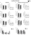
No impact of HMB + D on skeletal muscle function after 12 weeks of sedentary behavior or resistance exercise training. HMB + D did not influence 1-RM leg extension flexion, press nor leg extension isokinetic torque production during sedentary control (Placebo, n = 10; HMB, n = 9) (AD). HMB + D did not augment the increase in 1RM for leg extension flexion, press, or leg extension isokinetic torque production after resistance exercise training (Placebo, n = 10; HMB + D, n = 10) (EH). On the left side of each panel, data presented as mean ± SEM. Data analyzed using a two-way ANOVA to detect treatment by time interaction. # p < 0.05 main effect for time. On the right side of each panel, data presented as individual changes (Δ).
Figure 5
Figure 5
Leg muscle quality after 12 weeks of HMB + D or placebo during sedentary control or resistance exercise training. Leg muscle quality is expressed as specific force (kg per unit of skeletal muscle volume (cm3)). Leg extension specific force decreased in HMB + D compared to placebo (A). HMB + D did not influence specific force for leg press or leg flexion during sedentary control (Placebo, n = 10; HMB, n = 8) (B,C). HMB + D did not influence the increase in specific force for leg extension, leg press, or leg flexion after resistance exercise training (Placebo, n = 10; HMB + D, n = 10) (DF). On the left side of each panel, data presented as mean ± SEM. Data analyzed using a two-way ANOVA to detect treatment by time interaction. # p < 0.05 main effect for time. On the right side of each panel, data presented as individual changes (Δ).
Figure 6
Figure 6
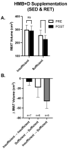
Contribution of Vitamin D3 on IMAT after HMB + D supplementation in middle-aged women. Data from women in both the sedentary control and resistance exercise training groups were pooled and then stratified by those who completed the study Vitamin D3 insufficient versus sufficient. IMAT was decreased after HMB + D in women who were Vitamin D3 sufficient (n = 11; SED, n = 6; RET, n = 5) but not insufficient (n = 7, SED, n = 3; RET, n = 4) (A). Women were further stratified into those who remained Vitamin D3 insufficient (Insufficient ➔ Insufficient, SED, n = 4; RET, n = 3), were Vitamin D3 sufficient at the start and end of study (Sufficient ➔ Sufficient, SED, n = 3, RET, n = 4)) and those who transitioned from Vitamin D3 insufficient to sufficient (Insufficient ➔ sufficient, SED, n = 2, RET, n = 3) (B). Data presented as mean ± SEM.

References

    1. Delmonico M.J., Harris T.B., Visser M., Park S.W., Conroy M.B., Velasquez-Mieyer P., Boudreau R., Manini T.M., Nevitt M., Newman A.B., et al. Longitudinal Study of Muscle Strength, Quality, and Adipose Tissue Infiltration. Am. J. Clin. Nutr. 2009;90:1579–1585. doi: 10.3945/ajcn.2009.28047. - DOI - PMC - PubMed
    1. Buford T.W., Lott D.J., Marzetti E., Wohlgemuth S.E., Vandenborne K., Pahor M., Leeuwenburgh C., Manini T.M. Age-Related Differences in Lower Extremity Tissue Compartments and Associations with Physical Function in Older Adults. Exp. Gerontol. 2012;47:38–44. doi: 10.1016/j.exger.2011.10.001. - DOI - PMC - PubMed
    1. Goodpaster B.H., Chomentowski P., Ward B.K., Rossi A., Glynn N.W., Delmonico M.J., Kritchevsky S.B., Pahor M., Newman A.B. Effects of Physical Activity on Strength and Skeletal Muscle Fat Infiltration in Older Adults: A Randomized Controlled Trial. J. Appl. Physiol. 2008;105:1498–1503. doi: 10.1152/japplphysiol.90425.2008. - DOI - PMC - PubMed
    1. Goodpaster B.H., Carlson C.L., Visser M., Kelley D.E., Scherzinger A., Harris T.B., Stamm E., Newman A.B. Attenuation of Skeletal Muscle and Strength in the Elderly: The Health ABC Study. J. Appl. Physiol. 2001;90:2157–2165. doi: 10.1152/jappl.2001.90.6.2157. - DOI - PubMed
    1. Marcus R.L., Addison O., LaStayo P.C. Intramuscular Adipose Tissue Attenuates Gains in Muscle Quality in Older Adults at High Risk for Falling. A Brief Report. J. Nutr. Health Aging. 2013;17:215–218. doi: 10.1007/s12603-012-0377-5. - DOI - PubMed

Publication types

Substances

LinkOut - more resources