

An "Instantaneous" Response of a Human Visual System to Hue: An EEG-Based Study
- PMID: 36366181
- PMCID: PMC9657469
- DOI: 10.3390/s22218484
An "Instantaneous" Response of a Human Visual System to Hue: An EEG-Based Study
Abstract
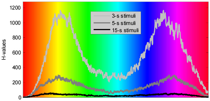
(1) The article presents a new technique to interpret biomedical data (EEG) to assess cortical responses to continuous color/hue variations. We propose an alternative approach to analyze EEG activity evoked by visual stimulation. This approach may augment the traditional VEP analysis. (2) Considering ensembles of EEG epochs as multidimensional spatial vectors evolving over time (rather than collections of time-domain signals) and evaluating the similarity between such vectors across different EEG epochs may result in a more accurate detection of colors that evoke greater responses of the visual system. To demonstrate its suitability, the developed analysis technique was applied to the EEG data that we previously collected from 19 participants with normal color vision, while exposing them to stimuli of continuously varying hue. (3) Orange/yellow and dark blue/violet colors generally aroused better-pronounced cortical responses. The selection of EEG channels allowed for assessing the activity that predominantly originates from specific cortical regions. With such channel selection, the strongest response to the hue was observed from Parieto-Temporal region of the right hemisphere. The statistical test-Kruskal-Wallis one-way analysis of variance-indicates that the distance evaluated for spatial EEG vectors at different post-stimulus latencies generally originate from different statistical distributions with a probability exceeding 99.9% (α = 0.001).
Keywords: EEG analysis; EEG spatial vectors; Kruskal–Wallis test; response to solid color changing over time; similarity between multidimensional vectors.
Conflict of interest statement
The authors declare no conflict of interest.
Figures


References
-
- Yeh Y.-Y., Lee D.-S., Ko Y.-H. Color combination and exposure time on legibility and EEG response of icon presented on visual display terminal. Displays. 2013;34:33–38. doi: 10.1016/j.displa.2012.11.007. - DOI
-
- Wang H., Zhang N. The analysis on vehicle color evoked EEG based on ERP method; Proceedings of the 4th International Conference on Bioinformatics and Biomedical Engineering (iCBBE) 2010; Chengdu, China. 18–20 June 2010.
-
- Tripathy J., Fuss F.K., Kulish V.V., Yang S. Influence of colour hue on fractal EEG dimensions; Proceedings of the International Conference on Biomedical and Pharmaceutical Engineering, ICBPE2006; Singapore. 11–14 December 2006; pp. 186–187.
MeSH terms
LinkOut - more resources
Full Text Sources
