. 2022 Nov 3;10(11):1862.
doi: 10.3390/vaccines10111862.

Evolutionary Trajectories of Avian Avulaviruses and Vaccines Compatibilities in Poultry

Affiliations

Evolutionary Trajectories of Avian Avulaviruses and Vaccines Compatibilities in Poultry

Mohammed A Rohaim et al. Vaccines (Basel). .

Abstract

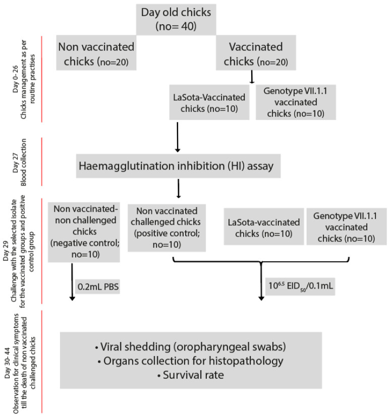
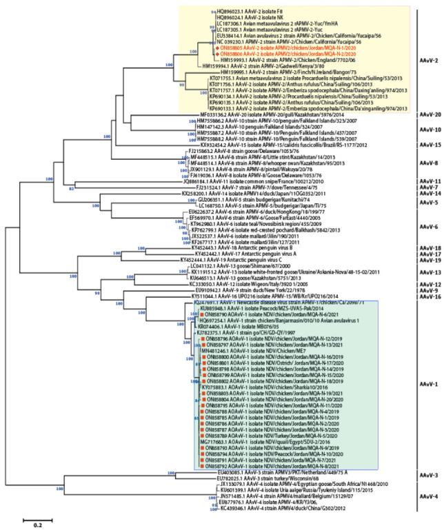
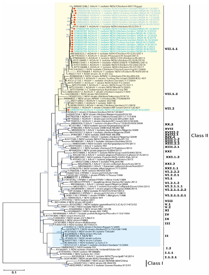
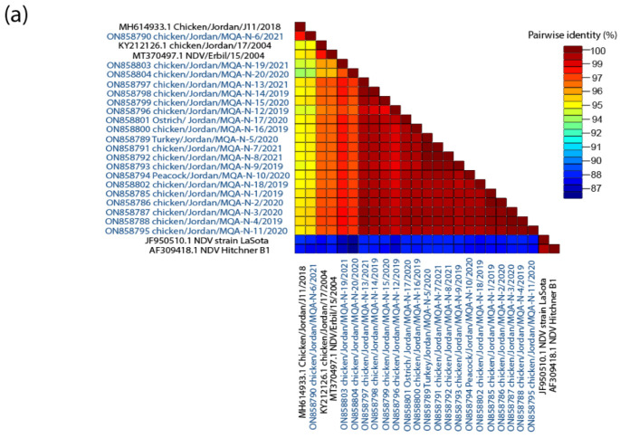
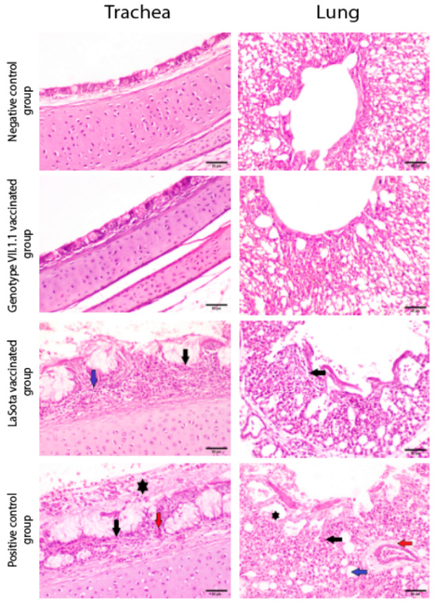
Newcastle disease virus (NDV) causes one of the highly infectious avian diseases in poultry leading to genuine financial misfortunes around the world. Recently, there has been an increasing trend in the number of ND-associated outbreaks in commercial Jordanian poultry flocks indicating a possible complex evolutionary dynamic of NDV infections in the country. To underpin the dynamics of circulating NDV strains and to assess the vaccine-escape potential, a total of 130 samples were collected from different poultry flocks in six Jordanian Governorates during 2019-2021. Twenty positive isolates, based on real-time reverse transcriptase PCR, were used for further genetic characterization and evolutionary analysis. Our results showed that there is a high evolutionary distance between the newly identified NDV strains (genotype VII.1.1) in this study and the commercially used vaccines (genotypes I and II), suggesting that circulating NDV field strains are under constant evolutionary pressure. These mutations may significantly affect flocks that have received vaccinations as well as flocks with insufficient immunity in terms of viral immunity and disease dynamics. To assess this further, we investigated the efficacy of the heterologous inactivated LaSota or homologous genotype VII.1.1 vaccine for their protection against virulent NDV in chicken. Vaccine-induced immunity was evaluated based on the serology, and protection efficacy was assessed based on clinical signs, survival rates, histopathology, and viral shedding. Chickens vaccinated with the inactivated genotype VII.1.1 based vaccine showed 100% protection with a significant reduction in virus shedding, and ameliorated histopathology lesions compared to LaSota vaccinated chicks that showed 60% protection. These results revealed that the usage of NDV inactivated vaccine from the circulating field strains can successfully ameliorate the clinical outcome and virus pathobiology in vaccinated chicks and will serve as an effective vaccine against the threat posed by commonly circulating NDV strains in the poultry industry.

Keywords: Jordan; avian orthoavulaviruses 1; efficacy; evolutionary pressure; vaccine.


Conflict of interest statement

The authors declare no conflict of interest. The funders had no role in the design of the study; in the collection, analyses, or interpretation of data; in the writing of the manuscript; or in the decision to publish the results.

Figures

Figure 1
Figure 1
Experimental layout for the assessment of vaccines effectiveness in chicks.
Figure 2
Figure 2
Phylogenomic revealed the clustering of 20 isolates within the AOAVv-1 while two isolates were allocated within AAvV-2/APMV-2. Unrooted phylogenetic trees were generated using the distance-based using maximum likelihood method and MEGA 6 software. Statistical support for tree branches was assessed by bootstrap analysis using 1000 replications of bootstrap re-sampling; numbers above branches indicate neighbor-joining bootstrap values that were ≥80%; the tree is drawn to scale, with branch lengths measured in the number of substitutions per site. The reported AAvV-1 isolates in this study are marked with red square within light green box; however, AAvV-2 isolates are marked with red hexagon labelled within light yellow box.
Figure 3
Figure 3
Phylogenetic analysis of the studied AOAvV-1 isolates and their clustering patterns with representative AOAvV-1 isolates. Full-length F-gene (1662 nt)-based phylogenetic analysis of our AOAvV-1 isolates with representative strains of each genotype. Reported isolates clustered in the genotype VII.1.1 of class II. Unrooted phylogenetic trees were generated using the distance-based using maximum likelihood method and MEGA 6 software. Statistical support for tree branches was assessed by bootstrap analysis using 1000 replications of bootstrap re-sampling; numbers above branches indicate neighbor-joining bootstrap values that were ≥80%; the tree is drawn to scale, with branch lengths measured in the number of substitutions per site. The reported AOAvV-1 isolates in this study are marked with red square within yellow box, however, NDV genotype II including LaSota (commonly used vaccine) was labelled within light blue box.
Figure 4
Figure 4
(a) The pairwise identities plot of fusion protein sequences aligned by MAFFT and displayed by Sequence Demarcation Tool (SDT) software. (b) WebLogo graphs illustrating the amino acid divergence between AOAVv-1 isolates reported in this study compared to LaSota vaccine and previously reported isolates in Jordan.
Figure 4
Figure 4
(a) The pairwise identities plot of fusion protein sequences aligned by MAFFT and displayed by Sequence Demarcation Tool (SDT) software. (b) WebLogo graphs illustrating the amino acid divergence between AOAVv-1 isolates reported in this study compared to LaSota vaccine and previously reported isolates in Jordan.
Figure 5
Figure 5
(a) Structure-based Antibody Prediction. X-axis contains the position of residues while the y-axis shows the propensity line indicates the threshold value. Regions above the threshold value, shown in green, are representing the residues under positive prediction. (b) The cumulative dN/dS of the average synonymous and non-synonymous substitutions moving codon by codon across F protein of AOAVv-1 isolates reported in Jordan including the reported isolates in this study with highlighting the most affected domains (high selective pressure) within the F protein.
Figure 5
Figure 5
(a) Structure-based Antibody Prediction. X-axis contains the position of residues while the y-axis shows the propensity line indicates the threshold value. Regions above the threshold value, shown in green, are representing the residues under positive prediction. (b) The cumulative dN/dS of the average synonymous and non-synonymous substitutions moving codon by codon across F protein of AOAVv-1 isolates reported in Jordan including the reported isolates in this study with highlighting the most affected domains (high selective pressure) within the F protein.
Figure 6
Figure 6
Survival rates and Evaluation of viral shedding. (a) Percentage survival rates and (b) Viral shedding from oropharyngeal swabs of genotype VII.1.1 and LaSota vaccinated challenged chicks with virulent NDV compared to negative and positive control groups. Bars represent the standard deviation means. **** indicates the level of significance at p value < 0.05.
Figure 7
Figure 7
Photomicrographs representing H&E stained sections of tracheas and lungs collected from genotype VII.1.1 and LaSota vaccinated- challenged chicks with virulent NDV compared to mock chicks (negative control) and positive control groups (non-vaccinated challenged chicks). Non-vaccinated- non-challenged chicks and genotype VII.1.1 vaccinated- challenged chicks showing normal histological architecture for Tracheas and normal histological architecture of parabronchus and air capillaries in lungs. On the other hand, LaSota vaccinated challenged chicks showed necrosis of lamina epithelialis and mucosal glands (black arrow) associated with mononuclear cells infiltration in lamina propria (blue arrow) in Trachea and inflammatory cells infiltration (black arrow) in lungs. In addition, non-vaccinated-challenged chicks (positive control group) showing multifocal necrosis of lamina epithelialis (black arrow), congestion (red arrow) and accumulation of mucous exudate in the tracheal lumen (asterisk) in Trachea and showing inflammatory cells infiltration in the air capillaries (black arrow), perivascular edema (red arrow), dilatation of atria (asterisk) and dilatation of air capillaries (blue arrow) in lungs (scale bar 50 µm).

Similar articles

Cited by

References

    1. Subbiah M., Xiao S., Khattar S.K., Dias F.M., Collins P.L., Samal S.K. Pathogenesis of two strains of avian paramyxovirus serotype 2, Yucaipa and Bangor, in chickens and turkeys. Avian Dis. 2010;54:1050–1057. doi: 10.1637/9380-041910-Reg.1. - DOI - PMC - PubMed
    1. Lamb R.A., Parks G.D. Paramyxoviridae: The viruses and their replication. In: Knipe D.M., Howley P.M., editors. Fields Virology. 5th ed. Lippincott Williams & Wilkins; Philadelphia, PA, USA: 2006. pp. 1449–1496.
    1. Mayo M.A. A summary of taxonomic changes recently approved by ICTV. Arch. Virol. 2002;147:1655–1663. doi: 10.1007/s007050200039. - DOI - PubMed
    1. Kolakofsky D., Roux L., Garcin D., Ruigrok R.W. Paramyxovirus mRNA editing, the ‘rule of six’ and error catastrophe: A hypothesis. J. Gen. Virol. 2005;86:1869–1877. doi: 10.1099/vir.0.80986-0. - DOI - PubMed
    1. Hisanaga T., Soos C., Lewis N., Lung O., Suderman M., Berhane Y. Genetic and Antigenic Characterization of Avian Avulavirus Type 6 (AAvV-6) Circulating in Canadian Wild Birds (2005–2017) Viruses. 2021;13:543. doi: 10.3390/v13040543. - DOI - PMC - PubMed

LinkOut - more resources