. 2022 Oct 31;14(11):2411.
doi: 10.3390/v14112411.

H1N1 Influenza A Virus Protein NS2 Inhibits Innate Immune Response by Targeting IRF7

Affiliations

H1N1 Influenza A Virus Protein NS2 Inhibits Innate Immune Response by Targeting IRF7

Bo Zhang et al. Viruses. .

Abstract

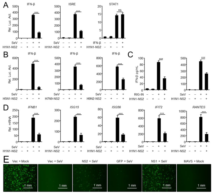
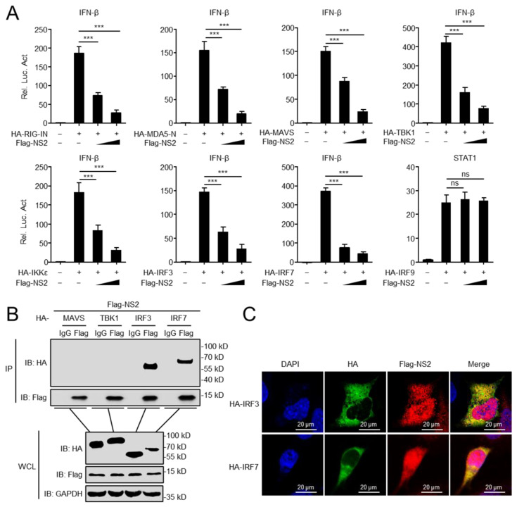
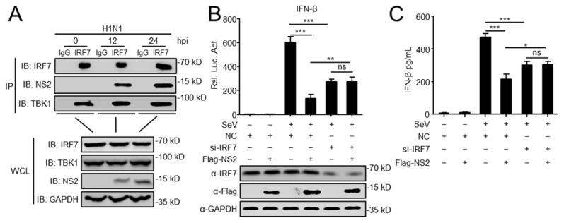
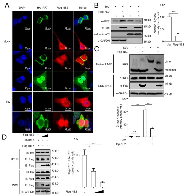
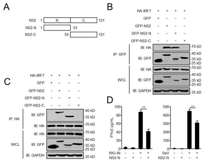
Influenza A virus (IAV) is a globally distributed zoonotic pathogen and causes a highly infectious respiratory disease with high morbidity and mortality in humans and animals. IAV has evolved various strategies to counteract the innate immune response, using different viral proteins. However, the mechanisms are not fully elucidated. In this study, we demonstrated that the nonstructural protein 2 (NS2) of H1N1 IAV negatively regulate the induction of type-I interferon. Co-immunoprecipitation experiments revealed that NS2 specifically interacts with interferon regulatory factor 7 (IRF7). NS2 blocks the nuclear translocation of IRF7 by inhibiting the formation of IRF7 dimers, thereby prevents the activation of IRF7 and inhibits the production of interferon-beta. Taken together, these findings revealed a novel mechanism by which the NS2 of H1N1 IAV inhibits IRF7-mediated type-I interferon production.

Keywords: influenza A virus; interferon; interferon regulatory factor 7; nonstructural protein 2.


Conflict of interest statement

The authors declare no conflict of interest. The funders had no role in the design of the study, in the collection, analyses, or interpretation of data; in the writing of the manuscript, or in the decision to publish the results.

Figures

Figure 1
Figure 1
NS2 inhibits IFN-I induction. (A) NS2 specifically inhibits SeV-triggered activation of IFN-β and ISRE promoter, but not IFN-β-triggered activation of the STAT1 reporter. HEK293 cells were transfected with H1N1-NS2 (200 ng) or empty vector (Vec) and the IFN-β, ISRE or STAT1 luciferase reporter, and indicated plasmids. At twenty hours after transfection, the cells were left untreated or treated with SeV for 12 h or IFN-β for 4 h before reporter assays. (B) NS2 proteins from different subtypes of IAVs inhibit SeV-triggered activation of IFN-β promoter. HEK293 cells were transfected with H5N1/H7N9/H9N2-NS2 (200 ng) or Vec and IFN-β luciferase reporter and indicated plasmids. At twenty hours after transfection, cells were left uninfected or infected with SeV for 12 h before reporter assays. (C) The effects of overexpressed NS2 on RIG-IN and SeV-induced IFN-β secretion. HEK293 cells were transfected with H1N1-NS2 (200 ng) or Vec, and subsequently transfected with RIG-IN or infected with SeV. The concentration of IFN-β in supernatants was determined using human IFN-β DuoSet ELISA kit. (D) NS2 inhibits SeV-triggered transcription of IFN-β and downstream genes. A549 cells were transfected with Flag-NS2 or Vec, and subsequently left uninfected or infected with SeV (MOI = 1) for 12 h before qPCR analysis. (E) NS2 facilitates VSV-GFP replication. HEK293 cells were transfected with indicated plasmids for 24 h, and cells were then infected with SeV (MOI = 1) or not (Mock) for 24 h. Cell supernatants were inactivated by ultraviolet radiation for 20 min and collected to treat fresh HEK293 cells for another 24 h. Cells then were infected with VSV-GFP (MOI = 0.01) for 12 h, and then observed microscopically. Scale bar, 1 mm. The data shown represent three independent experiments (p < 0.05 (*), p < 0.01 (**), p < 0.001 (***); ‘ns’ indicates no significant difference).
Figure 2
Figure 2
NS2 interacts with IRF3 and IRF7. (A) NS2 inhibits activation of the IFN-β promoter induced by RIG-IN, MDA5-Card, MAVS, TBK1, IKKε, IRF3, and IRF7. HEK293 cells were transfected with indicated plasmids along with increasing amounts (0, 200 ng, and 400 ng/mL) of Flag-NS2 expression plasmids. The activation of STAT1-luciferase triggered by IRF9 was used as a negative control. Reporter assays were performed 24 h after transfection. (B) Overexpressed NS2 interacts with IRF3 and IRF7. HEK293 cells were transfected with indicated plasmids for 24 h. Then, Co-IP and immunoblotting analyses were performed using indicated antibodies. (C) NS2 co-localizes with IRF3 and IRF7 in the cytoplasm. U2OS cells were transfected with Flag-NS2 and HA-IRF3/IRF7 plasmid for 24 h, and then stained with Flag antibody or HA antibody and secondary antibodies. Nuclei were counter-stained with DAPI. Scale bar, 20 μm. The data shown represent three independent experiments (p < 0.05 (*), p < 0.01 (**), p < 0.001 (***), ‘ns’ indicates no significant difference).
Figure 3
Figure 3
NS2 targets IRF7 and inhibits IFN-I production. (A) Endogenous NS2 is associated with IRF7. A549 cells were infected with H1N1 virus (MOI = 0.1) for the indicated times before co-immunoprecipitation and immunoblot analysis. (B) Luciferase reporter plasmids (IFNβ-Luc) and the pRL-TK plasmid were co-transfected into HEK293 cells, along with Flag-NS2 and IRF7 siRNA or scrambled siRNA (NC). At twenty hours after transfection, cells were left untreated or treated with SeV for 12 h before reporter assays. (C) HEK293 cells were transfected with H1N1-NS2 or Vec, along with Flag-NS2 and IRF7 siRNA or NC for 24 h. Then cells were infected with SeV for 12 h. The concentrations of IFN-β in the supernatants were then detected by using human IFN-β DuoSet ELISA kit. The data shown represent three independent experiments (p < 0.05 (*), p < 0.01 (**), p < 0.001 (***), ‘ns’ indicates no significant difference).
Figure 4
Figure 4
NS2 blocks the nuclear translocation of IRF7. (A) U2OS cells were transfected with HA-IRF7 expression plasmid, along with Flag-NS2 or Vec. At twenty hours later, cells were either infected with SeV (4 h) or left untreated. Cells were then stained with rabbit anti-HA and mouse anti-Flag antibodies. Alexa Fluor 488-anti-rabbit immunoglobulin (green) and Alexa Fluor Cy3-anti-mouse immunoglobulin (red) were used as secondary antibodies. Cell nuclei (blue) were stained with DAPI. Scale bar, 20 μm. (B) HEK293 cells were transfected with Flag-NS2 or Vec and infected with SeV (4 h) or not. Cell lysates were separated into cytoplasmic and nuclear fractions, and the expression of IRF7 and Flag-NS2 were analyzed by Western blot (fractions: C, purified cytosol; N, purified nucleus). The intensities of the indicated protein bands were determined by using ImageJ (National Institutes of Health, Bethesda, MD, USA), and the ratio of nuclear IRF7 to cytosolic IRF7 was shown in the right panel. (C) NS2 suppressed IRF7 dimerization. HEK293 cells were transfected with Flag-NS2 or Vec, and treated with SeV for another 4 h. Native PAGE assays were performed to detect IRF7 dimerization. SDS PAGE assay were performed to detect IRF7, Flag-NS2, and GAPDH. The intensities of the dimer and monomer of IRF7 were determined by using ImageJ (National Institutes of Health, Bethesda, MD, USA), and the ratio of IRF7 dimer to IRF7 monomer was shown in the bottom panel. (D) HEK293 cells were transfected with plasmids expressing HA-IRF7 and Flag-IRF7, together with Vec or Flag-NS2 (100 ng and 500 ng) for 24 h. Cell lysates were then immunoprecipitated with anti-HA and immunoblotted with the indicated antibodies. The intensities of the Flag-IRF7 and HA-IRF7 in IP were determined by using ImageJ (National Institutes of Health, Bethesda, MD, USA), and the ratio of Flag-IRF7 to HA-IRF7 was shown in the right panel. The data shown represent three independent experiments (p < 0.05 (*), p < 0.01 (**), p < 0.001 (***)).
Figure 5
Figure 5
The N-terminal domain of NS2 is essential for the interaction with IRF7. (A) GFP-tagged NS2, NS2-N (amino acids (aa) 1 to 53), NS2-C (aa 54 to 121) were constructed and cloned into pEGFP-C1 by using standard molecular biology techniques. (B,C) The N-terminal domain of NS2 influences the interaction between NS2 and IRF7. HEK293 cells were transfected with HA-IRF7 along with GFP-NS2, GFP-NS2-N, and GFP-NS2-C expression plasmid, followed by IP with anti-GFP or anti-HA. (D) The effects of overexpression of the N-terminal domain of NS2 on RIG-IN and SeV-induced IFN-β secretion. HEK293 cells were transfected with GFP-NS2-N or GFP, and subsequently treated with RIG-IN or infected with SeV. The concentration of IFN-β in the supernatants was assessed by using human IFN-β DuoSet ELISA kit. The data shown represent three independent experiments (p < 0.05 (*), p < 0.01 (**)).

References

    1. Lipsitch M. Avian influenza: Ferret H7N9 flu model questioned. Nature. 2013;501:33. doi: 10.1038/501033e. - DOI - PubMed
    1. Medina R.A., Garcia-Sastre A. Influenza A viruses: New research developments. Nat. Rev. Microbiol. 2011;9:590–603. doi: 10.1038/nrmicro2613. - DOI - PMC - PubMed
    1. Zhang Q., Shi J., Deng G., Guo J., Zeng X., He X., Kong H., Gu C., Li X., Liu J., et al. H7N9 influenza viruses are transmissible in ferrets by respiratory droplet. Science. 2013;341:410–414. doi: 10.1126/science.1240532. - DOI - PubMed
    1. Cui P., Zeng X., Li X., Li Y., Shi J., Zhao C., Qu Z., Wang Y., Guo J., Gu W., et al. Genetic and biological characteristics of the globally circulating H5N8 avian influenza viruses and the protective efficacy offered by the poultry vaccine currently used in China. Sci. China Life Sci. 2022;65:795–808. doi: 10.1007/s11427-021-2025-y. - DOI - PubMed
    1. Yin X., Deng G., Zeng X., Cui P., Hou Y., Liu Y., Fang J., Pan S., Wang D., Chen X., et al. Genetic and biological properties of H7N9 avian influenza viruses detected after application of the H7N9 poultry vaccine in China. PLoS Pathog. 2021;17:e1009561. doi: 10.1371/journal.ppat.1009561. - DOI - PMC - PubMed

Publication types

MeSH terms

Substances

LinkOut - more resources