. 2022 Nov 5;14(11):2454.
doi: 10.3390/v14112454.

Pathogenicity and Transmissibility of Goose-Origin H5N6 Avian Influenza Virus Clade 2.3.4.4h in Mammals

Affiliations

Pathogenicity and Transmissibility of Goose-Origin H5N6 Avian Influenza Virus Clade 2.3.4.4h in Mammals

Cheng Zhang et al. Viruses. .

Abstract

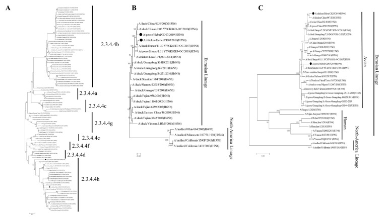
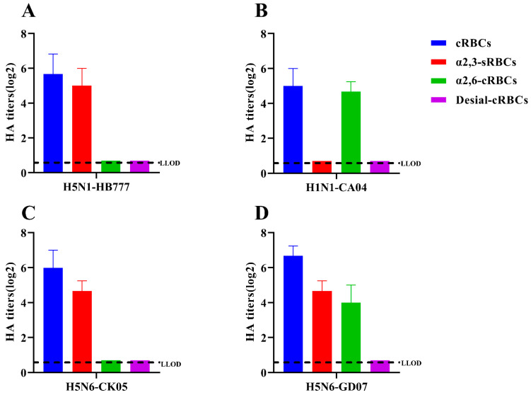
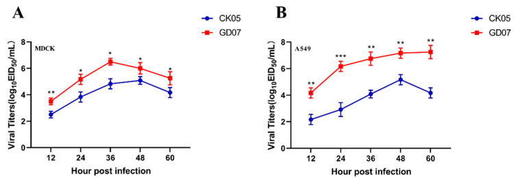
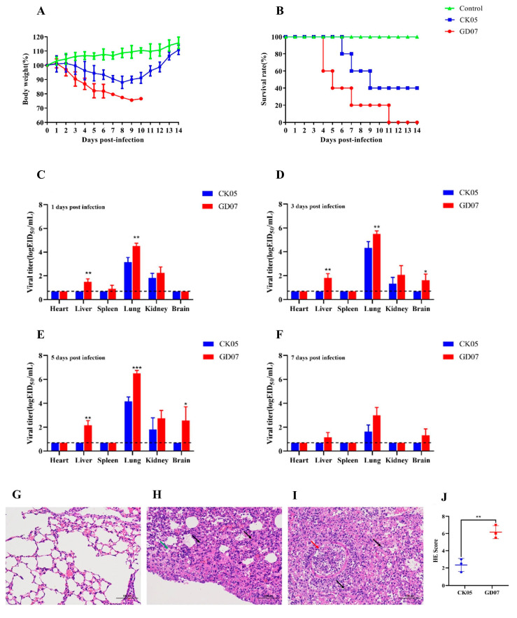
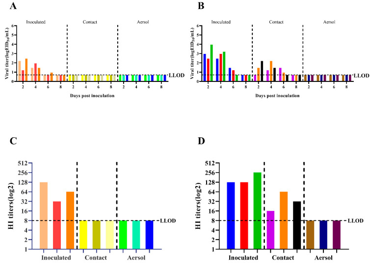
Throughout the last decade, H5N6 avian influenza viruses (AIVs) circulating in poultry and infecting humans have caused increasing global concerns that they might become a pandemic threat to global health. Since AIVs could occasionally cause asymptomatic infections in geese, virus monitoring in such a host should be critical to the control of cross-species infection. In addition, previous studies showed that clade 2.3.4.4h H5N6 AIVs could infect mammals without adaptation. However, the pathogenicity and transmissibility of goose-origin clade 2.3.4.4h H5N6 AIVs in mammals remain unknown. In this study, two H5N6 AIVs were isolated from a domestic chicken (A/chicken/Hebei CK05/2019 (H5N6)) and a goose (A/goose/Hebei/GD07/2019(H5N6)). This study is the first to evaluate the pathogenicity and transmissibility of goose-origin clade 2.3.4.4h H5N6 AIVs in mammals by comparison with chicken-origin 2.3.4.4h H5N6 AIVs. The CK05 virus had an affinity for α-2,3-receptors, while the GD07 virus had an affinity for both α-2,3-and α-2,6-receptors. The GD07 virus had a higher replication capacity in vitro and more severe pathogenicity in mice than the CK05 virus. The CK05 virus could not be transmitted effectively among guinea pigs, whereas the GD07 virus could be transmitted through direct contact among guinea pigs. The results of this study indicated the potential health threat of clade 2.3.4.4h H5N6 AIVs to mammals and emphasized the importance of continuous monitoring of H5N6 AIVs, especially in waterfowl.

Keywords: H5N6; chicken; goose; pathogenicity; transmissibility.


Conflict of interest statement

The authors declare no conflict of interest.

Figures

Figure 1
Figure 1
Phylogenetic trees of the HA, NA, and PB2 genes of the H5N6 (CK05 and GD07) viruses. (A) Phylogenetic tree of HA. (B) Phylogenetic trees of NA. (C) Phylogenetic trees of PB2. Sequences in this study are marked with black chicken or goose.
Figure 2
Figure 2
Receptor-binding specificity of H5N6 (CK05 and GD07) viruses. The receptor-binding specificity of viruses is determined by RBCs containing different SA receptors. Only avian-like (α-2,3) receptors were found in the control group, HB777(H5N1) (A), while only human-like (α-2,6) receptors were found in CA04(H1N1) (B). Avian-like (-2,3) receptors were found in both CK05 (C) and GD07(D). Human-like (-2,6) receptors are present only in the GD07 (D) strain. In each group, three separate experiments were carried out. The lower limit of detection (LLOD) is indicated with a black dotted line.
Figure 3
Figure 3
Viral titers at different time points in MDCK (A) or A549 (B) cells. The two strains (CK05 and GD07) infected cells with an MOI of 0.01 (105 cells). At 12, 24, 36, 48, and 60 hpi, cell supernatants were collected and inoculated into SPF chicken embryos. The titer of the virus at different time points was determined by EID50. Three independent experiments were performed in each group. * p < 0.05, ** p < 0.01, *** p < 0.001.
Figure 4
Figure 4
Pathogenicity of the isolated viruses in mice. All mice were intranasally inoculated with the H5N6 viruses at 106 EID50. (A) The daily weight of each group was monitored for 14 days. (B) The survival rate of each group was recorded for 14 days. (C) The tissue distribution of the CK05 and GD07 virus in BALB/c mice at 1 dpi. (D) The tissue distribution of the CK05 and GD07 virus in BALB/c mice at 3 dpi. (E) The tissue distribution of the CK05 and GD07 virus in BALB/c mice at 5 dpi. (F) The tissue distribution of the CK05 and GD07 virus in BALB/c mice at 7 dpi. (G) Lung pathological sections of the BALB/c mice in the control group at 5 dpi. (H) Lung pathological sections of the BALB/c mice in the CK05 group at 5 dpi. (I) Lung pathological sections of the BALB/c mice in the GD07 group at 5 dpi. (J) Pathological scores in the lungs of infected BALB/c mice. Images were acquired using a ×20 magnification objective. Alveolar wall thickening, lymphocyte infiltration (arrow black); acidophilic protein-like exudation (arrow green); epithelial cell necrosis (arrow red). * p < 0.05, ** p < 0.01, *** p < 0.001. LLOD for viral titers is indicated with a black dotted line.
Figure 5
Figure 5
Transmission of H5N6 (CK05 and GD07) viruses in guinea pigs. (A) X-axis: guinea pigs in the groups infected with CK05, exposed to direct contact, and transmitted by aerosols. Y-axis: influenza virus titers in guinea pig nasal washes. (B) X-axis: guinea pigs in the groups infected with GD07, exposed to direct contact, and transmitted by aerosols. Y-axis: influenza virus titers in guinea pig nasal washes. (C) X-axis: guinea pigs in the CK05 infection group, direct contact group, and aerosol transmission group. Y-axis: HI antibody titers of different guinea pigs. (D) X-axis: guinea pigs infected with GD07 group, directly exposed group and aerosol transmitted group. Y-axis: HI antibody titers of different guinea pigs. Each color bar represents an individual guinea pig. LLOD is indicated with a black dotted line.

Similar articles

Cited by

References

    1. Henning J., Wibawa H., Morton J., Usman T.B., Junaidi A., Meers J. Scavenging ducks and transmission of highly pathogenic avian influenza, Java, Indonesia. Emerg. Infect. Dis. 2010;16:1244–1250. doi: 10.3201/eid1608.091540. - DOI - PMC - PubMed
    1. Lv X., Li X., Sun H., Li Y., Peng P., Qin S., Wang W., Li Y., An Q., Fu T., et al. Highly Pathogenic Avian Influenza A(H5N8) Clade 2.3.4.4b Viruses in Satellite-Tracked Wild Ducks, Ningxia, China, 2020. Emerg. Infect. Dis. 2022;28:1039–1042. doi: 10.3201/eid2805.211580. - DOI - PMC - PubMed
    1. Li X., Sun J., Lv X., Wang Y., Li Y., Li M., Liu W., Zhi M., Yang X., Fu T., et al. Novel Reassortant Avian Influenza A(H9N2) Virus Isolate in Migratory Waterfowl in Hubei Province, China. Front. Microbiol. 2020;11:220. doi: 10.3389/fmicb.2020.00220. - DOI - PMC - PubMed
    1. Pantin-Jackwood M.J., Costa-Hurtado M., Bertran K., DeJesus E., Smith D., Swayne D.E. Infectivity, transmission and pathogenicity of H5 highly pathogenic avian influenza clade 2.3.4.4 (H5N8 and H5N2) United States index viruses in Pekin ducks and Chinese geese. Vet. Res. 2017;48:33. doi: 10.1186/s13567-017-0435-4. - DOI - PMC - PubMed
    1. Kang Y., Liu L., Feng M., Yuan R., Huang C., Tan Y., Gao P., Xiang D., Zhao X., Li Y., et al. Highly pathogenic H5N6 influenza A viruses recovered from wild birds in Guangdong, southern China, 2014–2015. Sci. Rep. 2017;7:44410. doi: 10.1038/srep44410. - DOI - PMC - PubMed

Publication types