

Alveolar epithelial cells and microenvironmental stiffness synergistically drive fibroblast activation in three-dimensional hydrogel lung models
- PMID: 36366982
- PMCID: PMC9729409
- DOI: 10.1039/d2bm00827k
Alveolar epithelial cells and microenvironmental stiffness synergistically drive fibroblast activation in three-dimensional hydrogel lung models
Abstract
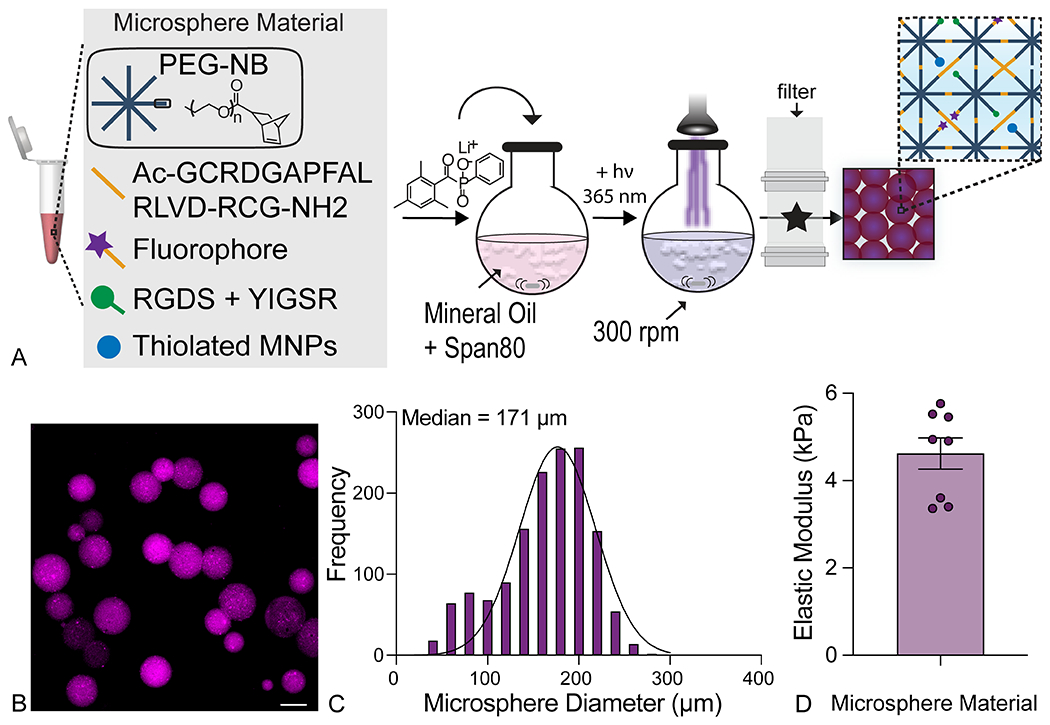
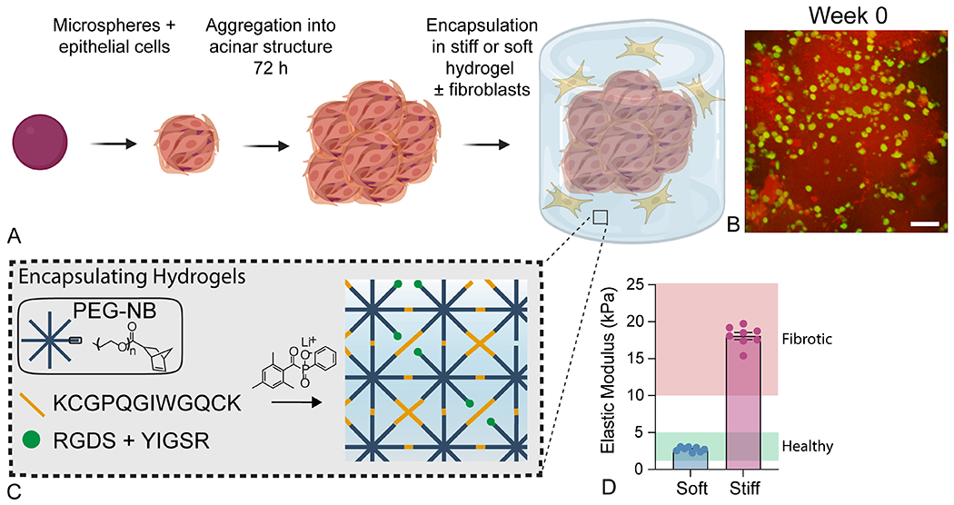
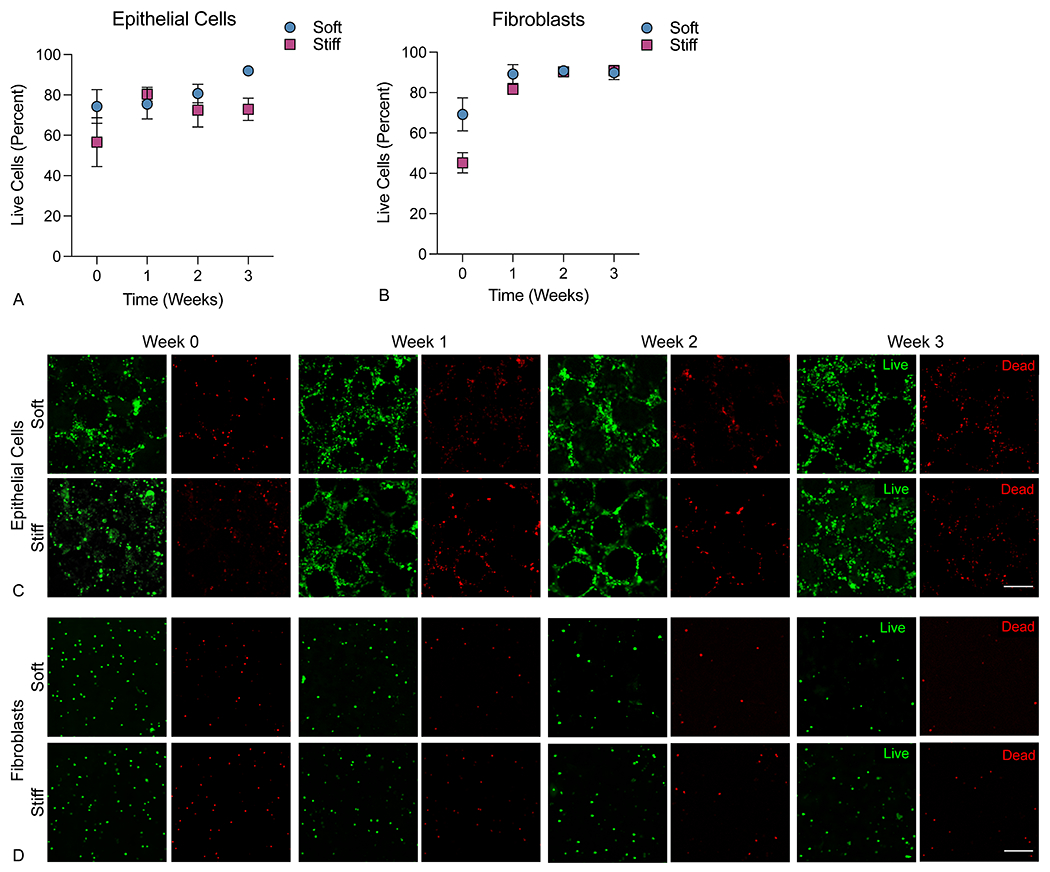
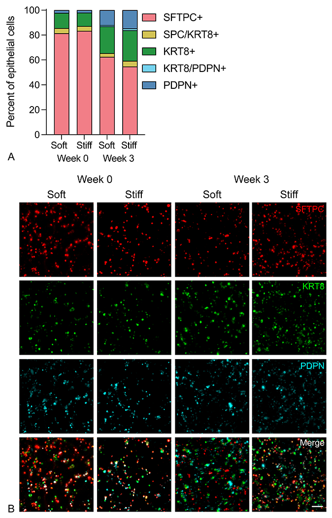
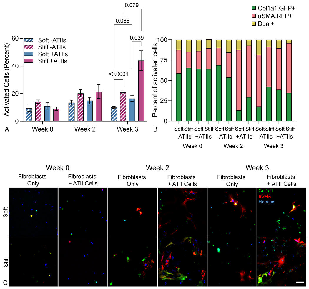
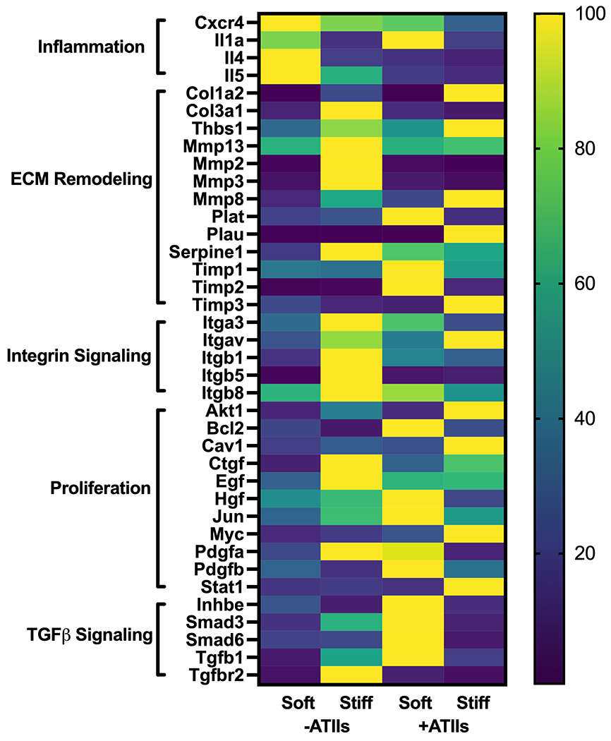
Idiopathic pulmonary fibrosis (IPF) is a devastating lung disease that progressively and irreversibly alters the lung parenchyma, eventually leading to respiratory failure. The study of this disease has been historically challenging due to the myriad of complex processes that contribute to fibrogenesis and the inherent difficulty in accurately recreating the human pulmonary environment in vitro. Here, we describe a poly(ethylene glycol) PEG hydrogel-based three-dimensional model for the co-culture of primary murine pulmonary fibroblasts and alveolar epithelial cells that reproduces the micro-architecture, cell placement, and mechanical properties of healthy and fibrotic lung tissue. Co-cultured cells retained normal levels of viability up to at least three weeks and displayed differentiation patterns observed in vivo during IPF progression. Interrogation of protein and gene expression within this model showed that myofibroblast activation required both extracellular mechanical cues and the presence of alveolar epithelial cells. Differences in gene expression indicated that cellular co-culture induced TGF-β signaling and proliferative gene expression, while microenvironmental stiffness upregulated the expression of genes related to cell-ECM interactions. This biomaterial-based cell culture system serves as a significant step forward in the accurate recapitulation of human lung tissue in vitro and highlights the need to incorporate multiple factors that work together synergistically in vivo into models of lung biology of health and disease.
Conflict of interest statement
Conflicts of Interest
Chelsea Magin reports a relationship with Vanderbilt University that includes: speaking and lecture fees. Chelsea Magin has patent #PCT/US2019/012722 pending to University of Colorado.
Figures
Similar articles
-
Collagen 3D matrices as a model for the study of cell behavior in pulmonary fibrosis.Exp Lung Res. 2022 Apr;48(3):126-136. doi: 10.1080/01902148.2022.2067265. Epub 2022 May 20. Exp Lung Res. 2022. PMID: 35594338
-
Three dimensional fibrotic extracellular matrix directs microenvironment fiber remodeling by fibroblasts.Acta Biomater. 2024 Mar 15;177:118-131. doi: 10.1016/j.actbio.2024.02.008. Epub 2024 Feb 11. Acta Biomater. 2024. PMID: 38350556
-
Epithelial-mesenchymal crosstalk influences cellular behavior in a 3D alveolus-fibroblast model system.Biomaterials. 2018 Feb;155:124-134. doi: 10.1016/j.biomaterials.2017.11.008. Epub 2017 Nov 15. Biomaterials. 2018. PMID: 29175081 Free PMC article.
-
Resident interstitial lung fibroblasts and their role in alveolar stem cell niche development, homeostasis, injury, and regeneration.Stem Cells Transl Med. 2021 Jul;10(7):1021-1032. doi: 10.1002/sctm.20-0526. Epub 2021 Feb 24. Stem Cells Transl Med. 2021. PMID: 33624948 Free PMC article. Review.
-
Angiotensin-TGF-beta 1 crosstalk in human idiopathic pulmonary fibrosis: autocrine mechanisms in myofibroblasts and macrophages.Curr Pharm Des. 2007;13(12):1247-56. doi: 10.2174/138161207780618885. Curr Pharm Des. 2007. PMID: 17504233 Review.
Cited by
-
Mechanomemory of pulmonary fibroblasts demonstrates reversibility of transcriptomics and contraction phenotypes.Biomaterials. 2025 Mar;314:122830. doi: 10.1016/j.biomaterials.2024.122830. Epub 2024 Sep 10. Biomaterials. 2025. PMID: 39276408
-
Integrating mechanical cues with engineered platforms to explore cardiopulmonary development and disease.iScience. 2023 Nov 15;26(12):108472. doi: 10.1016/j.isci.2023.108472. eCollection 2023 Dec 15. iScience. 2023. PMID: 38077130 Free PMC article. Review.
-
Hydrogel-embedded precision-cut lung slices support ex vivo culture of in vivo-induced premalignant lung lesions.Physiol Rep. 2025 Jul;13(13):e70459. doi: 10.14814/phy2.70459. Physiol Rep. 2025. PMID: 40650429 Free PMC article.
-
Biomaterial-based 3D human lung models replicate pathological characteristics of early pulmonary fibrosis.bioRxiv [Preprint]. 2025 Feb 17:2025.02.12.637970. doi: 10.1101/2025.02.12.637970. bioRxiv. 2025. Update in: Acta Biomater. 2025 Aug 7:S1742-7061(25)00591-4. doi: 10.1016/j.actbio.2025.08.010. PMID: 40027659 Free PMC article. Updated. Preprint.
-
Hydrogel-Embedded Precision-Cut Lung Slices Model Lung Cancer Premalignancy Ex Vivo.Adv Healthc Mater. 2024 Feb;13(4):e2302246. doi: 10.1002/adhm.202302246. Epub 2023 Nov 27. Adv Healthc Mater. 2024. PMID: 37953708 Free PMC article.
References
-
- L R, HR C and MG J, Lancet (London, England), 2017, 389. - PubMed
-
- Taniguchi H, Ebina M, Kondoh Y, Ogura T, Azuma A, Suga M, Taguchi Y, Takahashi H, Nakata K, Sato A, Takeuchi M, Raghu G, Kudoh S, Nukiwa T and the J Pirfenidone Clinical Study Group in, European Respiratory Journal, 2010, 35, 821. - PubMed
-
- Richeldi L, du Bois RM, Raghu G, Azuma A, Brown KK, Costabel U, Cottin V, Flaherty KR, Hansell DM, Inoue Y, Kim DS, Kolb M, Nicholson AG, Noble PW, Selman M, Taniguchi H, Brun M, Le Maulf F, Girard M, Stowasser S, Schlenker-Herceg R, Disse B and Collard HR, New England Journal of Medicine, 2014, 370, 2071–2082. - PubMed
-
- Strunz M, Simon LM, Ansari M, Kathiriya JJ, Angelidis I, Mayr CH, Tsidiridis G, Lange M, Mattner LF, Yee M, Ogar P, Sengupta A, Kukhtevich I, Schneider R, Zhao Z, Voss C, Stoeger T, Neumann JHL, Hilgendorff A, Behr J, O’Reilly M, Lehmann M, Burgstaller G, Königshoff M, Chapman HA, Theis FJ and Schiller HB, Nature Communications, 2020, 11, 1–20. - PMC - PubMed
MeSH terms
Substances
Grants and funding
LinkOut - more resources
Full Text Sources
Other Literature Sources