

Mammillary Body Atrophy in Temporal Lobe Epilepsy With Hippocampal Sclerosis
- PMID: 36367061
- PMCID: PMC9669561
- DOI: 10.3988/jcn.2022.18.6.635
Mammillary Body Atrophy in Temporal Lobe Epilepsy With Hippocampal Sclerosis
Abstract
Background and purpose: We aimed to determine 1) the frequency of mammillary body (MB) atrophy in patients with temporal lobe epilepsy (TLE) and hippocampal sclerosis (HS), 2) the clinical significance of MB atrophy, and 3) the association between MB atrophy and volume changes in other subcortical limbic structures.
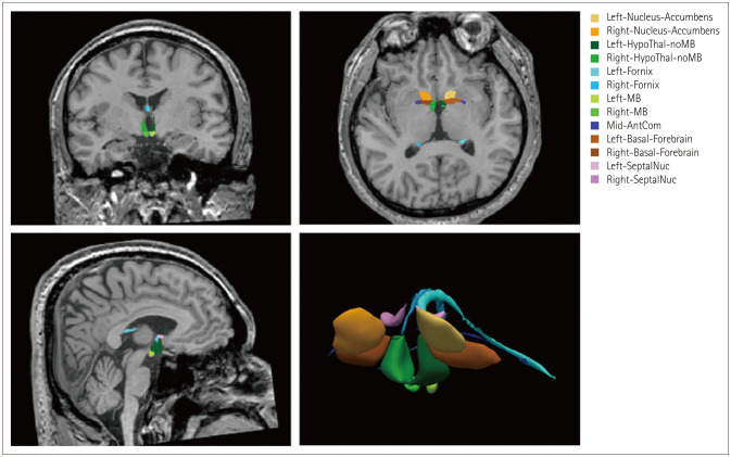
Methods: We enrolled 69 patients with pathologically confirmed TLE with HS, who underwent a standard anterior temporal lobectomy, as well as 40 healthy controls. We used the FreeSurfer deep-learning tool of U-Net to obtain the volumes of the subcortical limbic structures, including the MB, hypothalamus, basal forebrain, septal nuclei, fornix, and nucleus accumbens. MB atrophy was considered to be present when the MB volume was decreased relative to the healthy controls.
Results: MB atrophy was present in 18 (26.1%) of the 69 patients with TLE and HS. Among the clinical characteristics, the mean age at seizure onset was higher (25.5 vs. 15.9 years, p=0.027) and the median duration of epilepsy was shorter (149 vs. 295 months, p=0.003) in patients with than without MB atrophy. The basal forebrain (0.0185% vs. 0.0221%, p=0.004) and septal nuclei (0.0062% vs. 0.0075%, p=0.003) in the ipsilateral hemisphere of HS were smaller in the patients with MB atrophy.
Conclusions: We observed ipsilateral MB atrophy in about one-quarter of patients with TLE and HS. The severity of subcortical limbic structure abnormalities was greater in patients without MB atrophy. These findings suggest that MB atrophy in TLE with HS is not rare, but it has little clinical significance.
Keywords: epilepsy; magnetic resonance imaging; mammillary body.
Copyright © 2022 Korean Neurological Association.
Conflict of interest statement
The authors have no potential conflicts of interest to disclose.
Figures
Similar articles
-
Temporal lobe volumes in patients with hippocampal sclerosis with or without cortical dysplasia.Neurology. 2004 May 25;62(10):1729-35. doi: 10.1212/01.wnl.0000127301.33384.36. Neurology. 2004. PMID: 15159469
-
Lateralizing Characteristics of Morphometric Changes to Hippocampus and Amygdala in Unilateral Temporal Lobe Epilepsy with Hippocampal Sclerosis.Medicina (Kaunas). 2022 Mar 26;58(4):480. doi: 10.3390/medicina58040480. Medicina (Kaunas). 2022. PMID: 35454319 Free PMC article.
-
Frequent seizures are associated with a network of gray matter atrophy in temporal lobe epilepsy with or without hippocampal sclerosis.PLoS One. 2014 Jan 27;9(1):e85843. doi: 10.1371/journal.pone.0085843. eCollection 2014. PLoS One. 2014. PMID: 24475055 Free PMC article.
-
Progression of gray matter atrophy in seizure-free patients with temporal lobe epilepsy.Epilepsia. 2016 Apr;57(4):621-9. doi: 10.1111/epi.13334. Epub 2016 Feb 11. Epilepsia. 2016. PMID: 26865066
-
Quantification of thalamic nuclei in patients diagnosed with temporal lobe epilepsy and hippocampal sclerosis.Neuroradiology. 2020 Feb;62(2):185-195. doi: 10.1007/s00234-019-02299-6. Epub 2019 Oct 31. Neuroradiology. 2020. PMID: 31673749
References
-
- Blümcke I. Neuropathology of focal epilepsies: a critical review. Epilepsy Behav. 2009;15:34–39. - PubMed
-
- Labate A, Cerasa A, Aguglia U, Mumoli L, Quattrone A, Gambardella A. Voxel-based morphometry of sporadic epileptic patients with mesiotemporal sclerosis. Epilepsia. 2010;51:506–510. - PubMed
-
- Pail M, Brázdil M, Marecek R, Mikl M. An optimized voxel-based morphometric study of gray matter changes in patients with left-sided and right-sided mesial temporal lobe epilepsy and hippocampal sclerosis (MTLE/HS) Epilepsia. 2010;51:511–518. - PubMed
-
- Lee HJ, Seo SA, Park KM. Quantification of thalamic nuclei in patients diagnosed with temporal lobe epilepsy and hippocampal sclerosis. Neuroradiology. 2020;62:185–195. - PubMed
LinkOut - more resources
Full Text Sources
Miscellaneous
