. 2023 Jan;27(1):6.
doi: 10.3892/mmr.2022.12893. Epub 2022 Nov 11.

Ginsenoside Rg1 alleviates learning and memory impairments and Aβ disposition through inhibiting NLRP1 inflammasome and autophagy dysfunction in APP/PS1 mice

Affiliations

Ginsenoside Rg1 alleviates learning and memory impairments and Aβ disposition through inhibiting NLRP1 inflammasome and autophagy dysfunction in APP/PS1 mice

Xuewang Li et al. Mol Med Rep. 2023 Jan.

Abstract

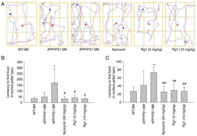
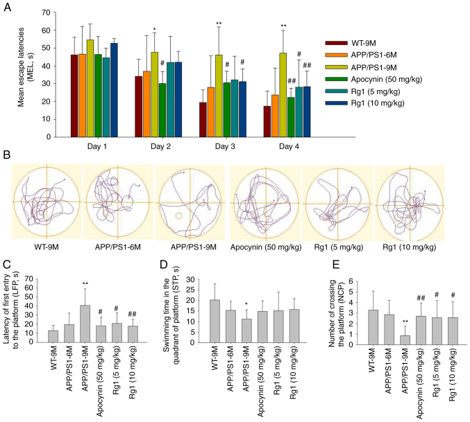
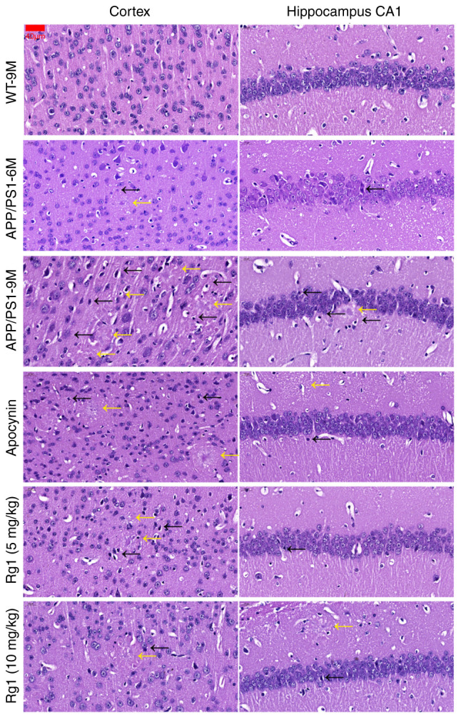
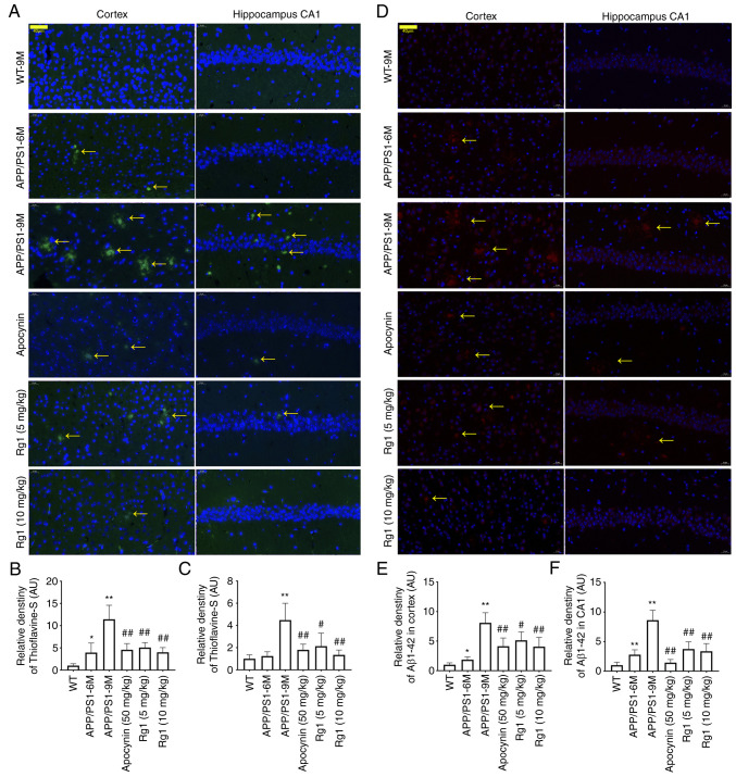
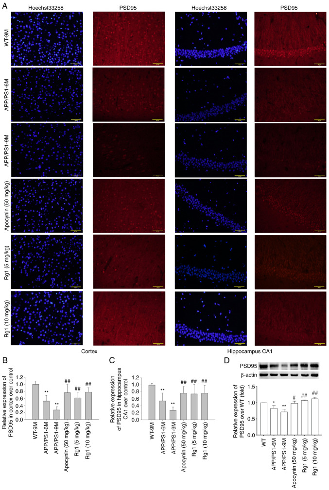
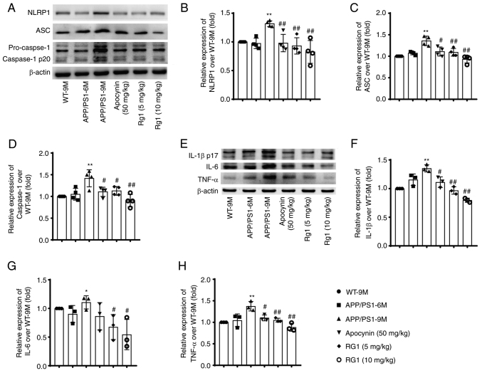
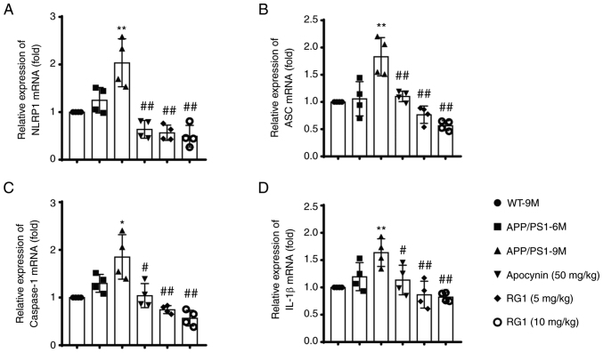
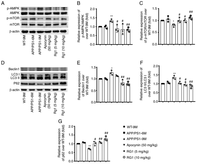
Alzheimer's disease (AD) is a common neurodegenerative disorder. Amyloid β (Aβ) deposition is considered an important pathological feature of AD. Growing evidence has linked neuroinflammation and autophagy to Aβ deposition in the progression of AD. However, there are few drug options for inhibiting neuroinflammation and autophagy to prevent AD. Ginsenoside Rg1 (Rg1), a steroidal saponin extracted from ginseng, has been reported to possess multiple neuroprotective effects. The present study aimed to evaluate whether Rg1 treatment could attenuate cognitive disorders and neuronal injuries by inhibiting NLRP1 inflammasome and autophagy dysfunction in an AD model of APP/PS1 mice. The results of behavioral tests indicated that Rg1 treatment for 12 weeks could significantly improve olfactory dysfunction as well as learning and memory impairments. The results of histopathological tests indicated that Rg1 treatment could reduce Aβ deposition and neuronal damages in APP/PS1‑9M mice. Additionally, the results of immunoblot, reverse transcription‑quantitative PCR or immunohistochemistry demonstrated that Rg1 treatment significantly downregulated the expression levels of inflammation‑related proteins of NLRP1, caspase1, IL‑1β and TNF‑α, as well as autophagy‑related proteins of p‑AMPK/AMPK, Beclin1 and LC3 II/LC3 I, and increased the expression levels of p‑mTOR/mTOR and P62 in APP/PS1‑9M mice. In addition, the molecular docking analysis showed that there was favorable binding result between Rg1 and NLRP1. The present study suggested that Rg1 may alleviate learning and memory impairments and Aβ disposition by inhibiting NLRP1 inflammasome and improving autophagy dysfunction, suggesting that Rg1 may be a potential therapeutic agent for delaying AD.

Keywords: AMPK/mTOR; APP/PS1 mice; Alzheimer's disease; Ginsenoside Rg1; NLRP1 inflammasome; autophagy.


Conflict of interest statement

The authors declare that they have no competing interests.

Figures

Figure 1.
Figure 1.
Rg1 treatment improves olfactory dysfunction in APP/PS1 mice. (A) The track of BFT. (B) Latency to find food in buried pellet (sec). (C) Latency to find food in surface pellet (sec). Results are expressed as the mean ± SD (n=10). *P<0.05 and **P<0.01 vs. the WT-9M group; #P<0.05 and ##P<0.01 vs. the APP/PS1-9M group. Rg1, Ginsenoside Rg1; BFT, buried food test.
Figure 2.
Figure 2.
Rg1 treatment improves learning and memory ability in APP/PS1 mice. (A) The mean escape latencies. (B) The track of Morris water maze in the fifth day. (C) The latency of first entry platform. (D) Swimming time in platform quadrant. (E) Number of crossing the platform. Results are expressed as the mean ± SD (n=10). *P<0.05 and **P<0.01 vs. the WT-9M group; #P<0.05 and ##P<0.01 vs. APP/PS1-9M group. Rg1, Ginsenoside Rg1.
Figure 3.
Figure 3.
Rg1 treatment alleviates neuronal damage in APP/PS1 mice. The results of H&E staining in the cortex and hippocampus CA1 (magnification, ×400; scale bar, 40 µm; n=4). Rg1, Ginsenoside Rg1.
Figure 4.
Figure 4.
Rg1 treatment alleviates Aβ deposition in APP/PS1 mice. (A) The results of Thioflavin-S staining in the cortex and hippocampus CA1 (magnification, ×400; scale bar, 40 µm). (B) Relative density of Thioflavin-S staining in cortex over WT. (C) Relative density of Thioflavin-S staining in hippocampus CA1 over WT. (D) The results of Aβ1-42 in the cortex and hippocampus CA1 (immunofluorescence, ×400; scale bar, 40 µm). (E) Relative density of Aβ1-42 in cortex over WT. (F) Relative density of Aβ1-42 in hippocampus CA1 over WT. Results are expressed as the mean ± SD (n=4). *P<0.05 and **P<0.01 vs. the WT-9M group; #P<0.05 and ##P<0.01 vs. the APP/PS1-9M group. Rg1, Ginsenoside Rg1.
Figure 5.
Figure 5.
Rg1 treatment increases the expression of PSD95 in APP/PS1 mice. (A) The expression of PSD95 in the cortex and hippocampus CA1 (magnification, ×400; scale bar, 50 µm; n=4). (B) Relative expression of PSD95 in cortex over WT. (C) Relative expression of PSD95 in hippocampus CA1 over WT. (D) Relative expression of PSD95 (western blot analysis, n=4). Results are expressed as the mean ± SD. *P<0.05 and **P<0.01 vs. the WT-9M group; #P<0.05 and ##P<0.01 vs. the APP/PS1-9M group. PSD95, post-synaptic density scaffolding protein 95; Rg1, Ginsenoside Rg1.
Figure 6.
Figure 6.
Rg1 treatment decreases the expression of NLRP1 inflammasome in APP/PS1 mice (western blot analysis). (A) The bands of NLRP1, ASC, Caspase-1 and β-actin. (B-D) Relative expression of (B) NLRP1, (C) ASC and (D) Caspase-1 over WT (n=4). (E) The bands of IL-1β, IL-6, TNF-α and β-actin (n=3). (F-H) Relative expression of (F) IL-1β, (G) IL-6 and (H) TNF-α over WT. Results are expressed as the mean ± SD. *P<0.05 and **P<0.01 vs. the WT-9M group; #P<0.05 and ##P<0.01 vs. the APP/PS1-9M group. Rg1, Ginsenoside Rg1.
Figure 7.
Figure 7.
Rg1 treatment decreases the mRNA expression levels of NLRP1 inflammasome in APP/PS1 mice (reverse transcription-quantitative PCR). (A-D) Relative expression of (A) NLRP1, (B) ASC, (C) Caspase-1 and (D) IL-1β mRNA over WT. Results are expressed as the mean ± SD (n=4). *P<0.05 and **P<0.01 vs. the WT-9M group; #P<0.05 and ##P<0.01 vs. the APP/PS1-9M group. Rg1, Ginsenoside Rg1.
Figure 8.
Figure 8.
Rg1 treatment improves autophagy dysfunction in APP/PS1 mice (Western blot analysis). (A) The bands of p-AMPK, AMPK, p-mTOR, mTOR and β-actin. (B and C) Relative expression of (B) p-AMPK/AMPK and (C) p-mTOR/mTOR over WT. (D) The bands of Beclin1, LC3, P62 and β-actin. (E-G) Relative expression of (E) Beclin1, (F) LC3 II/LC3 I and (G) P62 over WT. Results are expressed as the mean ± SD (n=4). *P<0.05 and **P<0.01 vs. the WT-9M group; #P<0.05 and ##P<0.01 vs. the APP/PS1-9M group. Rg1, Ginsenoside Rg1.

Similar articles

Cited by

References

    1. Gouras GK, Olsson TT, Hansson O. β-Amyloid peptides and amyloid plaques in Alzheimer's disease. Neurotherapeutics. 2015;12:3–11. doi: 10.1007/s13311-014-0313-y. - DOI - PMC - PubMed
    1. Zolezzi JM, Bastias-Candia S, Santos MJ, Inestrosa NC. Alzheimer's disease: Relevant molecular and physiopathological events affecting amyloid-β brain balance and the putative role of PPARs. Front Aging Neurosci. 2014;6:176. doi: 10.3389/fnagi.2014.00176. - DOI - PMC - PubMed
    1. Hardy J, Selkoe DJ. The amyloid hypothesis of Alzheimer's disease: Progress and problems on the road to therapeutics. Science. 2002;297:353–356. doi: 10.1126/science.1072994. - DOI - PubMed
    1. Kuang H, Tan CY, Tian HZ, Liu LH, Yang MW, Hong FF, Yang SL. Exploring the bi-directional relationship between autophagy and Alzheimer's disease. CNS Neurosci Ther. 2020;26:155–166. doi: 10.1111/cns.13216. - DOI - PMC - PubMed
    1. Newcombe EA, Camats-Perna J, Silva ML, Valmas N, Huat TJ, Medeiros R. Inflammation: The link between comorbidities, genetics, and Alzheimer's disease. J Neuroinflammation. 2018;15:276. doi: 10.1186/s12974-018-1313-3. - DOI - PMC - PubMed