. 2023 Jan;49(1):4.
doi: 10.3892/or.2022.8441. Epub 2022 Nov 11.

Loss of CD24 promotes radiation‑ and chemo‑resistance by inducing stemness properties associated with a hybrid E/M state in breast cancer cells

Affiliations

Loss of CD24 promotes radiation‑ and chemo‑resistance by inducing stemness properties associated with a hybrid E/M state in breast cancer cells

Isaline Bontemps et al. Oncol Rep. 2023 Jan.

Abstract

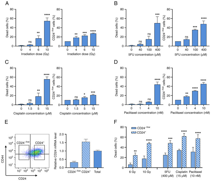
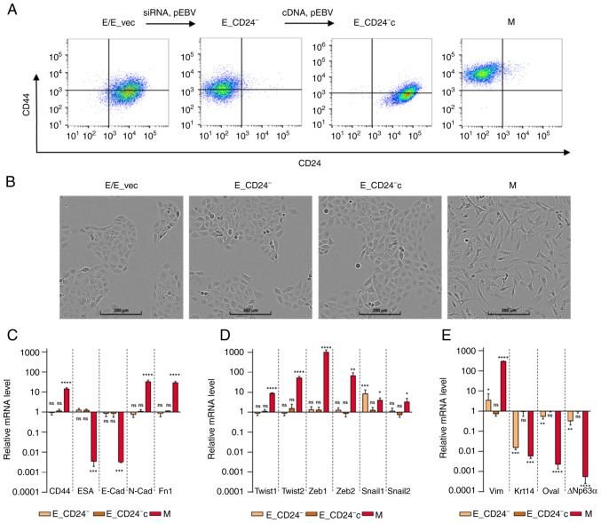
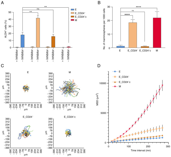
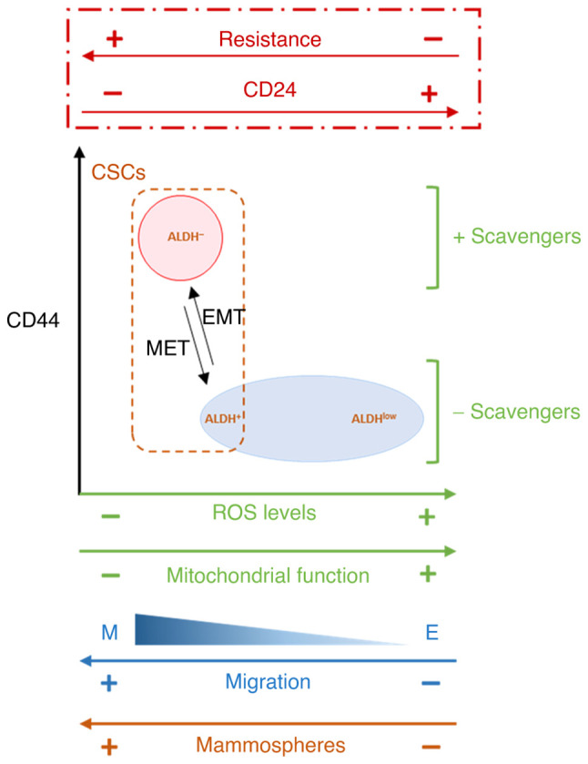
Cancer stem cells (CSCs) serve an essential role in failure of conventional antitumor therapy. In breast cancer, CD24‑/low/CD44+ phenotype and high aldehyde dehydrogenase activity are associated with CSC subtypes. Furthermore, CD24‑/low/CD44+ pattern is also characteristic of mesenchymal cells generated by epithelial‑mesenchymal transition (EMT). CD24 is a surface marker expressed in numerous types of tumor, however, its biological functions and role in cancer progression and treatment resistance remain poorly documented. Loss of CD24 expression in breast cancer cells is associated with radiation resistance and control of oxidative stress. Reactive oxygen species (ROS) mediate the effects of anticancer drugs as well as ionizing radiation; therefore, the present study investigated if CD24 mediates radiation‑ and chemo‑resistance of breast cancer cells. Using a HMLE breast cancer cell model, CD24 expression has been artificially modulated and it was observed that loss of CD24 expression induced stemness properties associated with acquisition of a hybrid E/M phenotype. CD24‑/low cells were more radiation‑ and chemo‑resistant than CD24+ cells. The resistance was associated with lower levels of ROS; CD24 controlled ROS levels via regulation of mitochondrial function independently of antioxidant activity. Together, these results suggested a key role of CD24 in de‑differentiation of breast cancer cells and promoting acquisition of therapeutic resistance properties.

Keywords: CD24; breast cancer; cancer stem cells; epithelial-mesenchymal transition; resistance.


Conflict of interest statement

The authors declare that they have no competing interests.

Figures

Figure 1.
Figure 1.
Low CD24 expression defines a radio- and chemo-resistant subpopulation of HMLE cells. CD24−/low subpopulation was defined as the 10% of cells expressing the lowest fluorescence in control cells. (A) Percentage of dead and CD24−/low HMLE cells at five days following high dose irradiation (4–10 Gy). Percentage of dead and CD24−/low HMLE cells following three day exposure to high concentrations of (B) 5-fluorouracil (5FU), (C) cisplatin and (D) paclitaxel. Data are presented as the mean ± SD of 3 independent experiments. Significant differences (compared with untreated cells) were analyzed by one-way ANOVA followed by Dunnett's multiple comparisons correction test. *P<0.05, **P<0.01, ***P<0.001 and ****P<0.0001. ns, not significant. (E) CD24−/low and CD24+ HMLE cell subpopulations were analyzed by flow cytometry following CD24/CD44 staining. CD24 expression was analyzed by reverse transcription-quantitative PCR. (F) Percentage of CD24−/low and CD24+ dead cells five days after 4 and 6 Gy irradiation and following three day exposure to 400 µM 5FU, 15 µM cisplatin and 10 nM paclitaxel. Data are presented as the mean ± SD of 3 independent experiments. Significant differences (compared with untreated cells) were analyzed by 2 way ANOVA followed by Sidak's multiple comparisons correction test. *P<0.05, **P<0.01, ***P<0.001 and ****P<0.0001. ns, not significant.
Figure 2.
Figure 2.
Characterization of E, M and E-transfected HMLE cells. (A) Flow cytometry characterization following CD24/CD44 labelling of parental E cells, E cells transfected with control p-EBV vector (E_vec) and p-EBV-plasmid expressing a CD24 small interfering RNA (E_CD24), E_CD24 cells transfected with p-EBV-plasmid expressing CD24 mRNA (E_CD24c) and M cells obtained following FACS analysis of CD24−/low/CD44+ cells induced by prolonged exposure to TGFβ1. (B) Phase-contrast images of E, E_vec, E_CD24, E_CD24c and M cells. Analysis by reverse transcription-quantitative PCR of relative expression of mRNAs encoding (C) EMT-associated factors (CD44, ESA, E-cad, N-cad and Fn1), (D) primary transcription factors of EMT (Twist-1, Twist-2, Snail-1, Snail-2, Zeb-1 and Zeb-2) and (E) other factors involved in EMT (Vim, Krt14, DTP63a and OVOL2) in E_CD24-, E_CD24-c and M compared with E cells. Results were normalized to expression levels in E cells. Data are presented as the mean ± SD of 3 independent experiments. Significant differences (compared with E cells) were analyzed by one-way ANOVA followed by Dunnett's multiple comparisons correction test. *P<0.05, **P<0.01, ***P<0.001 and ****P<0.0001. ns, not significant; E, epithelial; M, mesenchymal; EMT, E-M transition; ESA, epithelial cell adhesion molecule; Cad, cadherin; Fn, fibronectin; Zeb, zinc finger E-box binding homeobox; Vim, vimentin; Krt, keratin; OVOL2, ovo-like zinc finger 2.
Figure 3.
Figure 3.
Stemness properties and migration potential of E_CD24 HMLE cells. (A) Percentage of Aldefluor-positive subpopulation defined by Aldefluor assay. (B) Mammosphere formation efficiency. Data are presented as the mean ± SD of 3 independent experiments. Significant differences were analyzed by ANOVA, followed by Sidak's for (A) and Dunnett's multiple comparisons correction test for (B). **P<0.01, ****P<0.0001. Cell migration potential analysis using DiPer program. (C) Visualization of cell trajectory. (D) Measurement of cell surface area using MSD analysis. Data are presented as the mean ± SD of 3 independent experiments. ns, not significant; E, epithelial; M, mesenchymal; MSD, mean square displacement.
Figure 4.
Figure 4.
Decreased CD24 expression enhances radio- and chemo-resistance in HMLE.E cells. (A) Time course of death of 10 Gy-irradiated cells. Data are presented as the mean ± SD of 3 independent experiments. (B) Percentage of dead cells following three day exposure to 400 µM 5FU, 15 µM cisplatin and 10 nM paclitaxel. Data are presented as the mean ± SD of 4–12 independent experiments. Significant differences (compared with E cells) were analyzed by one-way ANOVA with Kruskal-Wallis followed by Dunn's multiple comparisons correction test. *P<0.05, **P<0.01, ***P<0.001, ****P<0.0001. (C) Clonogenic cell survival curves following 2–6 Gy irradiation, 4–16 µM 5FU, 2–6 µM cisplatin and 1–3 nM paclitaxel treatment. ns, non-significant; 5FU, 5-fluorouracil; E, epithelial; M, mesenchymal.
Figure 5.
Figure 5.
CD24 downregulation decreases total and mitochondrial ROS levels. (A) Cellular ROS levels were assessed by DHE and (B) mitochondrial ROS levels were assessed by Mitosox-Red probe. ROS levels were studied in untreated control cells and three days following 10 Gy irradiation and exposure to 400 µM 5FU. Data are presented as the mean ± SD of 4–10 independent experiments. Significant differences were analyzed by one-way ANOVA with Kruskal-Wallis followed by Dunn's multiple comparisons correction test. *P<0.05, ***P<0.001. ns, non-significant; ROS, reactive oxygen species; 5FU, 5-fluorouracil; E, epithelial; M, mesenchymal.
Figure 6.
Figure 6.
CD24 downregulation has no impact on ROS scavengers but decreases mitochondrial mass and membrane potential. (A) Analysis by reverse transcription-quantitative PCR of the relative expression of mRNAs encoding stress-associated factors involved in ROS metabolism in E_CD24-, E_CD24-c and M compared with E cells. For each gene, expression in E cells was normalized to 1 and ratio of relative mRNA level of E to E_CD24-, E_CD24-c and M cells is presented. Data are presented as the mean ± SD of 3 independent experiments. Significant differences (compared with E cells) were analyzed by one-way ANOVA followed by Dunnett's multiple comparisons correction test. *P<0.05, **P<0.01, ***P<0.001 and ****P<0.0001. ns: not significant. Mitochondrial (B) mass was assessed by Mitotracker Green probe and (C) membrane potential was assessed using TMRE staining. Mitochondrial mass and membrane potential were studied in untreated cells and at three days following 10 Gy irradiation and exposure to 400 µM 5FU. Data are presented as the mean ± SD of 4–10 independent experiments. Significant differences were analyzed by one-way ANOVA with Kruskal-Wallis followed by Dunn's multiple comparisons correction test. *P<0.05, **P<0.01, ***P<0.001. ns, non-significant. SOD, superoxide dismutase 2; HMOX1, heme oxygenase 1; GSR, glutathione-sulfide reductase; TXNDR1, thioredoxin reductase 1; TMRE, tetramethylrhodamine, ethyl ester; E, epithelial; M, mesenchymal; 5FU, 5-fluorouracil; ROS, reactive oxygen species.
Figure 7.
Figure 7.
Proposed model of the association between CD24 expression and EMT, stemness properties, ROS levels and scavengers and mitochondrial function. In HMLE cells cultures, CD24 expression is heterogeneous and CD24−/low subpopulation is selected following radiation and drug treatment. Artificial modulation of CD24 expression shows that radio- and chemo-sensitivity are directly controlled by CD24, and that loss of CD24 expression in E HMLE cells increase the presence of ALDH+ E-like CSCs, in a hybrid E/M state, in association with decreased mitochondrial mass and membrane potential. EMT, epithelial-mesenchymal transition; ROS, reactive oxygen species; CSC, cancer stem cell; ALDH, aldehyde dehydrogenase; MET, mesenchymal-to-epithelial transition.

Similar articles

Cited by

References

    1. Marusyk A, Almendro V, Polyak K. Intra-tumour heterogeneity: A looking glass for cancer? Nat Rev Cancer. 2012;12:323–334. doi: 10.1038/nrc3261. - DOI - PubMed
    1. Pece S, Tosoni D, Confalonieri S, Mazzarol G, Vecchi M, Ronzoni S, Bernard L, Viale G, Pelicci PG, Di Fiore PP. Biological and molecular heterogeneity of breast cancers correlates with their cancer stem cell content. Cell. 2010;140:62–73. doi: 10.1016/j.cell.2009.12.007. - DOI - PubMed
    1. Batlle E, Clevers H. Cancer stem cells revisited. Nat Med. 2017;23:1124–1134. doi: 10.1038/nm.4409. - DOI - PubMed
    1. Al-Hajj M, Wicha MS, Benito-Hernandez A, Morrison SJ, Clarke MF. Prospective identification of tumorigenic breast cancer cells. Proc Natl Acad Sci USA. 2003;100:3983–3988. doi: 10.1073/pnas.0530291100. - DOI - PMC - PubMed
    1. Ginestier C, Hur MH, Charafe-Jauffret E, Monville F, Dutcher J, Brown M, Jacquemier J, Viens P, Kleer CG, Liu S, et al. ALDH1 is a marker of normal and malignant human mammary stem cells and a predictor of poor clinical outcome. Cell Stem Cell. 2007;1:555–567. doi: 10.1016/j.stem.2007.08.014. - DOI - PMC - PubMed

MeSH terms