. 2022 Dec 21;33(12):2348-2360.
doi: 10.1021/acs.bioconjchem.2c00418. Epub 2022 Nov 11.

Engineering and Validation of a Peptide-Stabilized Poly(lactic- co-glycolic) Acid Nanoparticle for Targeted Delivery of a Vascular Disruptive Agent in Cancer Therapy

Affiliations

Engineering and Validation of a Peptide-Stabilized Poly(lactic- co-glycolic) Acid Nanoparticle for Targeted Delivery of a Vascular Disruptive Agent in Cancer Therapy

Sylwia A Dragulska et al. Bioconjug Chem. .

Abstract

Developing a biocompatible and biodegradable nanoparticle (NP) carrier that integrates drug-loading capability, active targeting, and imaging modality is extremely challenging. Herein, we report an NP with a core of poly(lactic-co-glycolic) acid (PLGA) chemically modified with the drug combretastatin A4 (CA4), a vascular disrupting agent (VDA) in clinical development for ovarian cancer (OvCA) therapy. The NP is stabilized with a short arginine-glycine-aspartic acid-phenylalanine x3 (RGDFFF) peptide via self-assembly of the peptide on the PLGA surface. Importantly, the use of our RGDFFF coating replaces the commonly used polyethylene glycol (PEG) polymer that itself often induces an unwanted immunogenic response. In addition, the RGD motif of the peptide is well-known to preferentially bind to αvβ3 integrin that is implicated in tumor angiogenesis and is exploited as the NP's targeting component. The NP is enhanced with an optical imaging fluorophore label via chemical modification of the PLGA. The RGDFFF-CA4 NPs are synthesized using a nanoprecipitation method and are ∼75 ± 3.7 nm in diameter, where a peptide coating comprises a 2-3 nm outer layer. The NPs are serum stable for 72 h. In vitro studies using human umbilical cord vascular endothelial cells (HUVEC) confirmed the high uptake and biological activity of the RGDFFF-CA4 NP. NP uptake and viability reduction were demonstrated in OvCA cells grown in culture, and the NPs efficiently accumulated in tumors in a preclinical OvCA mouse model. The RGDFFF NP did not induce an inflammatory response when cultured with immune cells. Finally, the NP was efficiently taken up by patient-derived OvCA cells, suggesting a potential for future clinical applications.


Figures

Figure 1.
Figure 1.
Schematic representation of PLGA-CA4 conjugate and RGDFFF peptide (top), and RGDFFF-CA4 NP (bottom right). The TEM image (bottom left) shows a large area with RGDFFF-CA4 NPs.
Figure 2.
Figure 2.
TEM images of the NPs: (A) RGDFFF NP, (B) RGDFFF-CA4 NP, (C) plain PLGA NP (no coating). The XPS spectrum of RGDFFF-PLGA NPs shows the peak of N 1s (D).
Figure 3.
Figure 3.
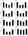
The stability of RGDFFF-CA4 NPs in 10% serum (A). Intracellular production of ROS in the presence of RGDFFF NPs. Error bars represent the mean ± standard deviation of three independent trials (B). NO production of RAW264.7 cell lines in the presence of RGDFFF NPs (C) and plain (uncoated) PLGA NPs (D). In ROS and NO experiments, the concentrations of the NPs are as follows: x 2 – 0.034 mg/ml, x 1 – 0.017 mg/ml, x 0.2 – 0.0034 mg/ml, and x 0.04 – 0.00068 mg/ml. PC – positive control (100 ng/ml LPS), NC – negative control: untreated RAW264.7 cells (PBS).
Figure 4.
Figure 4.
Western blots of HUVEC and various OvCA-relevant cell line lysates demonstrating expression of αv and β3 subunits. 80 μg of protein was loaded per lane. The panels shown are representative of N=3 independent trials.
Figure 5.
Figure 5.
Flow cytometry analysis of RGDFFF-Cy5.5 NP uptake. All cell lines were incubated with the NPs and harvested after 15 min. Control cells receiving no NPs, are highlighted in red, and cells treated with NPs are shown in blue. The table characterizes the percent of NP-positive cells.
Figure 6.
Figure 6.
Top: Cellular internalization of RGDFFF-Cy5.5 NPs (red) in OvCA cell lines after 48 h incubation. The membrane (green) was stained with Alexa fluor 488-WGA, and the nucleus (blue) was labeled with DAPI. The scale bars are 20 μm. Bottom: Time-lapse images of RGDFFF NPs (red) uptake by CP70 cells during the first 30 min of incubation. Arrows point to the NPs. The cellular membrane (green) was stained as above. The scale bar is 10 μm.
Figure 7.
Figure 7.
In vitro viability with RGDFFF-CA4 NP. Each column represents the mean and standard deviation of N=3 and p<0.005 (HUVEC, OVCAR-3), p<0.04 (TOV-21G, OV-90), p<0.03 (CP70), p<0.02 (A2780), p<0.01 (ES-2), p< 0.12 (SKOV-3). The concentrations correspond to IC50 values of CA4 for each cell line and are as follows: 2 nM (HUVEC), 5 nM (A2780), 4 nM (CP70), 5 nM (ES-2), 100 nM (OV-90), 2.5 nM (OVCAR-3), 10 nM (SKOV-3), 25 nM (TOV-21G).
Figure 8.
Figure 8.
The images of NPs biodistribution in vivo (top) in the OvCA mouse model and excised tumors (bottom) from a NP-injected mouse (left) and a control PBS-injected mouse (right). The images were acquired with the help of the Small Animal Imaging Center in the Translational and Molecular Imaging Institute.
Figure 9.
Figure 9.
Confocal images of frozen sections of OVCAR-3 tumors excised after 4 days from the mouse injected with RGDFFF-Cy7 NPs. The CD31 staining of vascular endothelial cells appears in red, and RGDFFF-Cy7 NPs are in green; the nuclei are stained with DAPI (blue). Scale bars represent 10 μm. The sections clearly show the association of the NPs with the blood vessels and tumor cells.
Figure 10.
Figure 10.
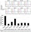
Flow Cytometry analysis of RGDFFF-Cy5.5 NP uptake by PDCLs. Red shows blank (non-treated cell), and blue shows cells incubated with RGDFFF-Cy5.5 NPs. The X-axis is the Cy5.5 intensity, and Y-axis is the number of cells. The table characterizes the percent of NP-positive cells from three trials.

Similar articles

Cited by

References

    1. Peer D; Karp JM; Hong S; Farokhzad OC; Margalit R; Langer R, Nanocarriers as an emerging platform for cancer therapy. Nat Nanotechnol 2007, 2 (12), 751–60. - PubMed
    1. Jafari S; Derakhshankhah H; Alaei L; Fattahi A; Varnamkhasti BS; Saboury AA, Mesoporous silica nanoparticles for therapeutic/diagnostic applications. Biomed Pharmacother 2019, 109, 1100–1111. - PubMed
    1. Singh P; Pandit S; Mokkapati V; Garg A; Ravikumar V; Mijakovic I, Gold Nanoparticles in Diagnostics and Therapeutics for Human Cancer. Int J Mol Sci 2018, 19 (7). - PMC - PubMed
    1. Zhu L; Zhou Z; Mao H; Yang L, Magnetic nanoparticles for precision oncology: theranostic magnetic iron oxide nanoparticles for image-guided and targeted cancer therapy. Nanomedicine (Lond) 2017, 12 (1), 73–87. - PMC - PubMed
    1. Ahlawat J; Henriquez G; Narayan M, Enhancing the Delivery of Chemotherapeutics: Role of Biodegradable Polymeric Nanoparticles. Molecules 2018, 23 (9). - PMC - PubMed

Publication types