. 2023 Dec;14(6):970-986.
doi: 10.1007/s12975-022-01102-8. Epub 2022 Nov 11.

Intracranial Pressure Dysfunction Following Severe Intracerebral Hemorrhage in Middle-Aged Rats

Affiliations

Intracranial Pressure Dysfunction Following Severe Intracerebral Hemorrhage in Middle-Aged Rats

Anna C J Kalisvaart et al. Transl Stroke Res. 2023 Dec.

Abstract

Rising intracranial pressure (ICP) aggravates secondary injury and heightens risk of death following intracerebral hemorrhage (ICH). Long-recognized compensatory mechanisms that lower ICP include reduced cerebrospinal fluid and venous blood volumes. Recently, we identified another compensatory mechanism in severe stroke, a decrease in cerebral parenchymal volume via widespread reductions in cell volume and extracellular space (tissue compliance). Here, we examined how age affects tissue compliance and ICP dynamics after severe ICH in rats (collagenase model). A planned comparison to historical young animal data revealed that aged SHAMs (no stroke) had significant cerebral atrophy (9% reduction, p ≤ 0.05), ventricular enlargement (9% increase, p ≤ 0.05), and smaller CA1 neuron volumes (21%, p ≤ 0.05). After ICH in aged animals, contralateral striatal neuron density and CA1 astrocyte density significantly increased (12% for neurons, 7% for astrocytes, p ≤ 0.05 vs. aged SHAMs). Unlike young animals, other regions in aged animals did not display significantly reduced cell soma volume despite a few trends. Nonetheless, overall contralateral hemisphere volume was 10% smaller in aged ICH animals compared to aged SHAMs (p ≤ 0.05). This age-dependent pattern of tissue compliance is not due to absent ICH-associated mass effect (83.2 mm3 avg. bleed volume) as aged ICH animals had significantly elevated mean and peak ICP (p ≤ 0.01), occurrence of ICP spiking events, as well as bilateral evidence of edema (e.g., 3% in injured brain, p ≤ 0.05 vs. aged SHAMs). Therefore, intracranial compliance reserve changes with age; after ICH, these and other age-related changes may cause greater fluctuation from baseline, increasing the chance of adverse outcomes like mortality.

Keywords: Aging; Cell volume; Edema; Intracerebral hemorrhage; Intracranial pressure; Stroke.


Conflict of interest statement

The authors declare no competing interests.

The authors declare no competing interests.

Figures
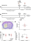
Fig. 1
Fig. 1
Experimental design and timelines (a, b); a representative cresyl violet section (c) demonstrates aged ICH hematoma volume at 24 h post-ICH in experiment 1 (d); note that portions of the hematoma often wash away during tissue processing, which is evident in fresh tissue. There were no significant differences in cortical thickness across hemispheres in aged ICH animals vs. aged SHAMs (e); however, aged ICH animals had significantly smaller right (contralateral) hemisphere parenchymal volume (total volume − ventricle volume), assessing from 4.5 to − 3.55 mm anterior to bregma (f). *p < 0.05 versus aged SHAMs
Fig. 2
Fig. 2
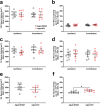
In experiment 1, neuron soma volume (a, c, e) and density (b, d, f) were assessed in aged rodents after either ICH or sham procedure. There were no significant differences in cell volume or density when assessing bilaterally in the hippocampal CA1 layer (a, b) and cortical area S1 (c, d). Assessment of the striatum was limited to the contralateral side of the brain, as the ipsilateral striatum was largely destroyed by the hematoma in ICH animals; there were no significant differences in neuron cell volume here (e), but there was a significant increase in aged ICH striatal cell density vs. aged SHAMs (f). *p < 0.05 versus aged SHAMs
Fig. 3
Fig. 3
In experiment 1, astrocyte soma volume (a, c, e) and density (b, d, f) were assessed in aged rodents after a striatal ICH or sham procedure. Regions analyzed bilaterally included the CA1 zone (a, b) and area S1 (c, d), while striatum was analyzed only in the right hemisphere (e, f). Astrocyte volume in CA1 was significantly different across hemispheres, but not across experimental groups, a baseline effect observed prior in other aging experiments (a). Astrocyte density in CA1 was significantly higher in aged ICH animals vs. aged SHAMs (b). There were no significant differences across experimental groups or hemispheres in area S1 and striatum; however, striatal astrocyte density trended on being significantly higher in aged ICH animals, similar to what was observed in neuronal morphological data
Fig. 4
Fig. 4
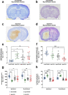
Representative brain sections from adult SHAM and aged SHAM animals (a, b), as well as adult ICH and aged ICH animals (c, d) demonstrating an increased ventricular volume (e) that is accompanied by a reduction in total right hemispheric parenchymal volume (d) as this rodent strain ages. Additionally, CA1 neuron volume (f) and density (g) in aged SHAM and aged ICH animals were significantly lower compared to adult SHAMs, nearing values observed in adult ICH animals. Therefore, following an ICH in an aged animal, it is possible that a lesser degree of tissue compliance is needed to accommodate the mass of a hematoma compared to their younger counterparts, as there is likely less resistance to CSF outflow through the ventricular system (c, d). *p < 0.05, ** p < 0.01, ***p < 0.001, ****p < 0.0001 versus adult SHAMs
Fig. 5
Fig. 5
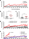
In experiment 2, ICP recordings were taken for 24 h in aged ICH animals and aged SHAMs (n = 10 per group); average 60-min ICP increased significantly in the aged ICH group at 12–24 h post-ICH vs. aged SHAMs (a). Aged ICH animals had significantly higher mean and peak ICP over 60-min epochs compared to aged SHAMs across the entire 24-h period (b, c), indicating acute ICH mass effect. Aged ICH and aged SHAM groups were further divided into those that spontaneously experienced ICP events (5 aged ICH animals, 2 aged SHAMs), and those that did not (5 aged ICH animals, 8 aged SHAMs; d). These ICP events are associated with adverse outcomes both in rodents and humans, displaying uncharacteristic deviations away from baseline, such as DIICP and RICP events (defined in main body of text). The aged ICH animals that experienced both RICP and DIICP events (n = 2), RICP events only (n = 2), or DIICP events only (n = 1; spontaneous mortality) are shown in panel e; such events are indicative of poor intracranial compliance. **p < 0.01 versus aged SHAMs
Fig. 6
Fig. 6
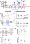
Representative ICP trace in an aged ICH animal displaying all ICP events flagged over the 24 h recording period in experiment 2 (a); examples of the baseline reference period are shown (average ICP over the preceding 60 min prior to the DIICP/RICP event), in panels b and c. In aged ICH animals, the 60-min baseline reference period prior to each event was split into 15-min epochs, and change in mean ICP slope was calculated for each epoch, comparing to equivalent periods averaged across aged SHAMs (d); a significant increase in pre-event slope occurred over the 15–0 min epoch immediately prior to DIICP events across aged ICH events vs. time-matched aged SHAMs, indicating worse control of cerebral compliance reserves (e). Following ICP recordings, animals were euthanized, and the striatum and hippocampi were dissected out bilaterally for BWC, along with the cerebellum (f). In the striatum of aged ICH animals, BWC was significantly elevated across both hemispheres vs. aged SHAMs, with significantly higher BWC in the ipsilateral (left) vs. contralateral (right) hemisphere (g). Aged ICH animals had significantly higher brain water content bilaterally in the hippocampus vs. aged SHAMs, also indicating widespread edema (main effect of group; p ≤ 0.05; h). As expected, brain water content in the cerebellum was not elevated in aged ICH animals compared to controls (i). *p < 0.05, ** p < 0.01, ***p < 0.001 vs. aged SHAMs
Fig. 7
Fig. 7
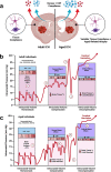
A summary of theorized tissue compliance dynamics that take place with age (a); increased ventricular size and decreased parenchymal volume that occur in the brain with age play a role in ICP compliance, changing the dynamics of accommodating ICH-associated mass effect (b, c). This theorized difference in ICP dynamics in young adults vs. aged individuals in response to cranial mass effect is illustrated by (1) brain at intracranial volume homeostasis. Typical “P1,” “P2,” and P3,” idealized ICP waveforms are shown (red line), reflecting intracranial compliance and respiratory rhythms; (2) brain at intracranial volume compensation. Added mass results in redirection of blood and CSF from and within the cranium, and a reduction in brain tissue volume as compensatory measures, deviating from typical ICP waveform patterns; (3–4) balancing the fluctuation in CSF and venous/capillary blood volume can cause dramatic spikes (3) and undershoots (4) in ICP away from baseline as mass effect grows more severe, reflecting periods of hyper and hypo cerebrovascular perfusion. Lastly, (5) as mass effect reaches a critical point and compliance mechanisms are exhausted, ICP begins to increase and loses its distinctive waveform as it climbs (e.g., intracranial volume decompensation). This is associated with a high risk of cerebellar herniation and mortality. With age, cerebrovascular reactivity, CSF redirection, and potentially tissue, compliance does not occur as rapidly or as effectively, causing refractory dynamic instability in ICP away from baseline and increasing risk of poor outcome or mortality. Figure created using BioRender.com (Toronto, Ontario)

Similar articles

Cited by

References

    1. Wilkinson CM, Kung TFC, Jickling GC, Colbourne F. A translational perspective on intracranial pressure responses following intracerebral hemorrhage in animal models. Brain Hemorrhages. 2021;2:34–48. doi: 10.1016/J.HEST.2020.10.002. - DOI
    1. Wilson MH. Monro-Kellie 2.0: the dynamic vascular and venous pathophysiological components of intracranial pressure. J Cereb Blood Flow Metab. 2016;36:1338–50. doi: 10.1177/0271678X16648711. - DOI - PMC - PubMed
    1. Nadeau CA, Dietrich K, Wilkinson CM, Crawford AM, George GN, Nichol HK, et al. Prolonged blood-brain barrier injury occurs after experimental intracerebral hemorrhage and is not acutely associated with additional bleeding. Transl Stroke Res. 2019;10:287–297. doi: 10.1007/s12975-018-0636-9. - DOI - PMC - PubMed
    1. McDowell MM, Ducruet AF, Friedlander RM. Management of cerebral edema/intracranial pressure in ischemic stroke. Primer on cerebrovascular diseases: Academic Press; 2017. pp. 738–742.
    1. Kim D-J, Czosnyka Z, Kasprowicz M, Smieleweski P, Baledent O, Guerguerian A-M, et al. Continuous monitoring of the Monro-Kellie Doctrine: is it possible? J Neurotrauma. 2012;29:1354–1363. doi: 10.1089/neu.2011.2018. - DOI - PMC - PubMed