. 2022 Dec 1;323(6):H1331-H1342.
doi: 10.1152/ajpheart.00286.2022. Epub 2022 Nov 11.

Renal TLR-7/TNF-α pathway as a potential female-specific mechanism in the pathogenesis of autoimmune-induced hypertension

Affiliations

Renal TLR-7/TNF-α pathway as a potential female-specific mechanism in the pathogenesis of autoimmune-induced hypertension

Sarika Chaudhari et al. Am J Physiol Heart Circ Physiol. .

Abstract

Hypertension is prevalent in patients with systemic lupus erythematosus (SLE). The goal of the current study is to track the pathogenesis of hypertension and renal injury in SLE, identify contributory mechanisms, and highlight differences in disease development among sexes. Mean arterial pressure was measured in conscious male and female SLE (NZBWF1) and control (NZW) mice at 34-35 wk of age using indwelling arterial catheters. Measures of renal injury, renal inflammation, and renal hemodynamics were used to monitor the potential contributors to latent sex differences. Both male and female SLE mice were hypertensive at 35 wk of age, and the hypertension was linked to renal injury in females, but not in males. A known contributor of renal pathology in SLE, Toll-like receptor (TLR)-7, and its downstream effector, the proinflammatory cytokine tumor necrosis factor (TNF)-α, were lower in male SLE mice than in females. Male SLE mice also had higher glomerular filtration rate (GFR) and lower renal vascular resistance (RVR) than females. Our data suggest that although hypertension in female SLE mice is associated with renal mechanisms, hypertension in male SLE mice may develop independent of renal changes. Future studies will continue to dissect sex-specific factors that should be considered when treating patients with hypertension with underlying chronic inflammation and/or autoimmunity.NEW & NOTEWORTHY There is a high prevalence of hypertension in male and female SLE; however, male SLE mice are hypertensive without renal involvement. The development of hypertension in female SLE mice is renocentric and strongly associated with injurious renal mechanisms like the TLR-7→TNF-α pathway. This clear difference in the pathogenesis among the sexes could have a significant impact on how we treat patients with hypertension with underlying chronic autoimmune/inflammatory diseases.

Keywords: Toll-like receptor 7; blood pressure; renal hemodynamics; renal injury; systemic lupus erythematosus.


Conflict of interest statement

No conflicts of interest, financial or otherwise, are declared by the authors.

Figures

Figure 1.
Figure 1.
Both male and female mice develop characteristic autoantibodies of SLE. Plasma levels of double-stranded DNA (dsDNA) autoantibodies (activity units) in male and female control and SLE mice at 30 wk and 34/35 wk of age (n = 14–29/group). The results of the two-way ANOVA with repeated measures are indicated on the graph. P values were calculated using the Holm–Sidak post hoc analysis. P < 0.05 was considered statistically significant (n = 5 or 6 mice/group); ****P vs. SLE/male and control mice. SLE, systemic lupus erythematosus.
Figure 2.
Figure 2.
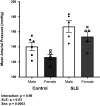
Both male and female SLE mice are hypertensive. Mean arterial pressure in male and female control and SLE mice at 35 wk of age analyzed by two-way ANOVA (n = 5 or 6 mice/group). The results of the two-way ANOVA are indicated on the graph. P < 0.05 was considered statistically significant. Mean arterial pressure is higher in SLE mice, but since there is no significant interaction following a two-way ANOVA, no group comparisons are included. Mean arterial pressure in male and female SLE mice when compared with our historical control only (control female mice) analyzed by one-way ANOVA indicate both male and female SLE mice had significantly higher blood pressure (mmHg) than female controls (data not shown). Also, if compared with normal C57BL6/J mice with mean arterial pressure of 100 mmHg, the male and female SLE mice are hypertensive (data not shown). SLE, systemic lupus erythematosus.
Figure 3.
Figure 3.
Male SLE mice do not develop renal injury. The percentage of mice with positive albuminuria (A) and albumin-to-creatinine ratio (µg/mg; B) in male and female control and SLE mice (n = 15 mice/group). The results of the two-way ANOVA are indicated. P < 0.05 was considered statistically significant. **P vs. SLE/male and control mice. SLE, systemic lupus erythematosus.
Figure 4.
Figure 4.
Female SLE mice demonstrate higher glomerular and tubulointerstitial injury. Paraffin-embedded kidney sections were stained with PAS stain (A) and Masson trichrome stain (C) to assess the glomerular injury and to assess the tubule-interstitial fibrosis respectively. P < 0.05 was considered statistically significant. The glomerulosclerosis index (B) was higher in female SLE mice (n = 6–10 mice/group; ****P vs. SLE/male and control mice). The percentage of area of blue staining indicating renal fibrosis (D) was also higher in female SLE mice as compared with the other groups (n = 6–10 mice/group; ****P vs. SLE/male and control mice). Similarly, female SLE mice had higher concentration of the markers of tubular injury, urinary KIM-1 normalized to creatinine (E), as compared with the male SLE mice (n = 8–12 mice/group; **P vs. SLE/male) and higher but not significant concentration of urinary NGAL normalized to creatinine (F), as compared with the control and male SLE mice (n = 7–12/group). Statistical comparisons were made using a two-way ANOVA followed by the Holm–Sidak post hoc analysis. NGAL, neutrophil gelatinase-associated lipocalin; SLE, systemic lupus erythematosus.
Figure 5.
Figure 5.
Female SLE mice had higher renal vascular resistance and lower GFR than male SLE mice. Conscious, anesthetized mice were implanted with a catheter in the carotid artery to obtain anesthetized blood pressure (mmHg; B). Renal blood flow (RBF, mL/min) was obtained using a Transonics probe (C). Renal vascular resistance (RVR) was calculated dividing anesthetized blood pressure by RBF. Data were normalized by dividing by body weight (g). P < 0.05 was considered statistically significant. Female SLE mice had significantly higher RVR adjusted for body weight (mmHg·min·kg·mL−1) than male SLE mice (A) (n = 6–9 mice/group; *P vs. SLE/male). Glomerular filtration rate (µL/min/100 g body wt) was measured in conscious, freely moving mice and was higher for male SLE mice (D). Statistical comparisons for all were made using unpaired t test. (n = 4–9 mice/group; *P vs. SLE/male). GFR, glomerular filtration rate; SLE, systemic lupus erythematosus.
Figure 6.
Figure 6.
Female SLE mice express higher levels of renal TLR-7 expression. Protein expression of TLR-7 was assessed via Western blot in the renal cortices (A) and medullas (B) of male and female SLE and control mice. Female SLE mice displayed a significantly higher expression of TLR-7 (at 66 kDa, the cleaved version) than both male SLE and female control mice. Statistical comparisons were made using a two-way ANOVA. The results of the two-way ANOVA are indicated on both graphs. P values were calculated using the Holm–Sidak post hoc analysis. P < 0.05 was considered statistically significant (n = 5 or 6 mice/group; ****P and **P indicates significant difference vs. SLE/male and control mice for cortical and medullary renal TLR-7, respectively). SLE, systemic lupus erythematosus; TLR-7, Toll-like receptor-7.
Figure 7.
Figure 7.
Female SLE mice express higher levels of renal TNF-α expression. Protein expression of was assessed via Western blot in the renal cortices (A) and medullas (B) of male and female SLE and control mice. Female SLE mice displayed a significantly higher expression of TNF-α (at 26 kDa, the transmembrane form) than male SLE and female control mice in both the renal cortex and medulla. Statistical comparisons were made using a two-way ANOVA. The results of the two-way ANOVA are indicated on both graphs. The P values were calculated using the Holm–Sidak post hoc analysis. P < 0.05 was considered statistically significant (n = 5 or 6 mice/group; *P and **P indicate significant difference vs. SLE/male and control mice for cortical and medullary renal TNF-α, respectively). SLE, systemic lupus erythematosus.

Comment in

References

    1. Flint AC, Conell C, Ren X, Banki NM, Chan SL, Rao VA, Melles RB, Bhatt DL. Effect of systolic and diastolic blood pressure on cardiovascular outcomes. N Engl J Med 381: 243–251, 2019. doi: 10.1056/NEJMoa1803180. - DOI - PubMed
    1. Rapsomaniki E, Timmis A, George J, Pujades-Rodriguez M, Shah AD, Denaxas S, White IR, Caulfield MJ, Deanfield JE, Smeeth L, Williams B, Hingorani A, Hemingway H. Blood pressure and incidence of twelve cardiovascular diseases: lifetime risks, healthy life-years lost, and age-specific associations in 1·25 million people. Lancet 383: 1899–1911, 2014. doi: 10.1016/s0140-6736(14)60685-1. - DOI - PMC - PubMed
    1. Doroszko A, Janus A, Szahidewicz-Krupska E, Mazur G, Derkacz A. Resistant hypertension. Adv Clin Exp Med 25: 173–183, 2016. doi: 10.17219/acem/58998. - DOI - PubMed
    1. Yaxley JP, Thambar SV. Resistant hypertension: an approach to management in primary care. J Family Med Prim Care 4: 193–199, 2015. doi: 10.4103/2249-4863.154630. - DOI - PMC - PubMed
    1. Sarwar MS, Islam MS, Al Baker SME, Hasnat A. Resistant hypertension: underlying causes and treatment. Drug Res (Stuttg) 63: 217–223, 2013. doi: 10.1055/s-0033-1337930. - DOI - PubMed

Substances

LinkOut - more resources