. 2022 Dec 1;323(6):H1262-H1269.
doi: 10.1152/ajpheart.00578.2022. Epub 2022 Nov 11.

SARS-CoV-2 infection downregulates myocardial ACE2 and potentiates cardiac inflammation in humans and hamsters

Affiliations

SARS-CoV-2 infection downregulates myocardial ACE2 and potentiates cardiac inflammation in humans and hamsters

Anissa Viveiros et al. Am J Physiol Heart Circ Physiol. .

Abstract

Myocardial pathologies resulting from SARS-CoV-2 infections are consistently rising with mounting case rates and reinfections; however, the precise global burden is largely unknown and will have an unprecedented impact. Understanding the mechanisms of COVID-19-mediated cardiac injury is essential toward the development of cardioprotective agents that are urgently needed. Assessing novel therapeutic strategies to tackle COVID-19 necessitates an animal model that recapitulates human disease. Here, we sought to compare SARS-CoV-2-infected animals with patients with COVID-19 to identify common mechanisms of cardiac injury. Two-month-old hamsters were infected with either the ancestral (D614) or Delta variant (B.1.617.2) of SARS-CoV-2 for 2 days, 7 days, and/or 14 days. We measured viral RNA and cytokine expression at the earlier time points to capture the initial stages of infection in the lung and heart. We assessed myocardial angiotensin-converting enzyme 2 (ACE2), the entry receptor for the SARS-CoV-2 virus, and cardioprotective enzyme, as well as markers for inflammatory cell infiltration in the hamster hearts at days 7 and 14. In parallel, human hearts were stained for ACE2, viral nucleocapsid, and inflammatory cells. Indeed, we identify myocardial ACE2 downregulation and myeloid cell burden as common events in both hamsters and humans infected with SARS-CoV-2, and we propose targeting downstream ACE2 downregulation as a therapeutic avenue that warrants clinical investigation.NEW & NOTEWORTHY Cardiac manifestations of COVID-19 in humans are mirrored in the SARS-CoV-2 hamster model, recapitulating myocardial damage, ACE2 downregulation, and a consistent pattern of immune cell infiltration independent of viral dose and variant. Therefore, the hamster model is a valid approach to study therapeutic strategies for COVID-19-related heart disease.

Keywords: ACE2; COVID-19; heart; inflammation.


Conflict of interest statement

Z. Kassiri is an editor of American Journal of Physiology-Heart and Circulatory Physiology and was not involved and did not have access to information regarding the peer-review process or final disposition of this article. An alternate editor oversaw the peer-review and decision-making process for this article. None of the other authors had any conflicts of interest, financial or otherwise, to disclose.

Figures

Figure 1.
Figure 1.
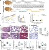
SARS-CoV-2 infection induces lung injury and upregulates cytokine expression in hamsters. A: experimental timeline for Delta (collected at days 2 and 7 for RT-PCR) and ancestral (collected at days 7 and 14 for histopathology) SARS-CoV-2-infected hamsters. Vehicle-inoculated control hamsters were analyzed in parallel. B: anthropometric data of body weight and lung weight of ancestral SARS-CoV-2-infected hamsters. Body weight was measured and recorded daily and reflected as percent of weight change relative to day 0. Time points were compared using two-way repeated-measures ANOVA and Sidak’s multiple comparisons test. Lungs were harvested and weighed at the end of the 14-day study period. C: SARS-CoV-2 nasal swabs and plaque measurement D: gross histopathological assessment of hamster lungs using hematoxylin and eosin staining (H&E) and Masson’s trichrome staining at day 7 after ancestral SARS-CoV-2 infection E: staining for SARS-CoV-2 nucleocapsid protein in the lungs at day 7 after viral challenge F: SARS-CoV-2 viral RNA copies in the lung following SARS-CoV-2 Delta challenge G: expression of Il-1β, Tnf-α, Il-6, and Il-10 in hamster lungs at day 2 and day 7 following Delta SARS-CoV-2 infection. Cytokines are visualized as a relative expression compared with controls. Data are represented as means ± SE, and each point represents biological replicates (n = 4 hamsters/group). Unpaired Student’s t test was performed for comparisons of controls to SARS-CoV-2-treated animals. One-way ANOVA with Dunnett’s multiple comparisons test or Kruskal Wallis test with Dunn’s multiple comparisons were used to compare parametric or nonparametric data, respectively; *P < 0.05, **P < 0.01.
Figure 2.
Figure 2.
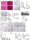
SARS-CoV-2 infection downregulates myocardial angiotensin-converting enzyme 2 (ACE2) and induces immune cell infiltration in hamsters. A: histological assessment of hamster hearts using hematoxylin and eosin staining (H&E) and Masson’s trichrome staining 7 days following ancestral SARS-CoV-2 challenge B: SARS-CoV-2 nucleocapsid staining in the hamster heart at 14 days after ancestral SARS-CoV-2 infection C: SARS-CoV-2 viral RNA copies in the heart following SARS-CoV-2 Delta challenge. D: representative images and pooled analysis of ACE2 staining in the hamster heart at day 7 (empty squares) and day 14 (filled squares) following inoculation with ancestral SARS-CoV-2. ACE2 staining area (%) and the number of areas with positive staining (count/mm2) are quantified. E: Ace2 mRNA expression by RT-PCR in the hamster heart at day 2 and day 7 after Delta SARS-CoV-2 infection. F: Western blot analysis and quantification of immunoreactivity (band densitometry for protein levels) of ACE2 following Delta SARS-CoV-2 inoculation. Immunoblots were visualized at a standard exposure (STD) or overexposed (OE) to visualize low protein levels of ACE2. Band densitometry was quantified and normalized to MemCode total protein stain (MEM). G: representative immune cell staining for neutrophils (CD15), macrophages (CD68), and T cells (CD4 and CD8) in the hearts of vehicle (control) and ancestral SARS-CoV-2-inoculated hamsters at day 14, and immune cell quantification (H). Day 7 (empty squares) and day 14 (filled squares) are pooled for analysis. I: expression of Tnf-α and Il-1β in hamster heart at day 2 and day 7 following Delta SARS-CoV-2 infection. Il-6 and Il-10 were below the limit of detection in the heart. Cytokines are visualized as a relative expression compared with controls. Data are represented as means ± SE, and each point represents biological replicates (n = 4–6 hamsters/group). Unpaired Student’s t test was performed for comparisons of controls to SARS-CoV-2-treated animals. One-way ANOVA with Dunnett’s multiple comparisons test or Kruskal Wallis test with Dunn’s multiple comparisons was used to compare parametric or nonparametric data, respectively; *P < 0.05, **P < 0.01.
Figure 3.
Figure 3.
Severe COVID-19 leads to myocardial angiotensin-converting enzyme 2 (ACE2) downregulation and immune cell infiltration in humans. A: routine histological assessment of human control and COVID-19 hearts with hematoxylin and eosin staining (H&E) and Masson’s trichrome staining. B and C: SARS-CoV-2 nucleocapsid and ACE2 staining in the human heart. ACE2 staining area (%) and the number of areas with positive staining (count/mm2) are quantified. D: immune cell staining for neutrophils (CD15), macrophages (CD68), and T cells (CD4 and CD8) in the hearts of control donors and patients with COVID-19, and immune cell quantification (E). Data are represented as means ± SE, and each point represents individual control donors (n = 3–4) or COVID-19 patients (n = 8–10). Unpaired Student’s t test was performed for comparisons of controls to patients with COVID-19; *P < 0.05, **P < 0.01.

Similar articles

Cited by

References

    1. Wang K, Gheblawi M, Nikhanj A, Munan M, MacIntyre E, O'Neil C, Poglitsch M, Colombo D, Del Nonno F, Kassiri Z, Sligl W, Oudit GY. Dysregulation of ACE (angiotensin-converting enzyme)-2 and renin-angiotensin peptides in SARS-CoV-2 mediated mortality and end-organ injuries. Hypertension 79: 365–378, 2022. doi:10.1161/HYPERTENSIONAHA.121.18295. - DOI - PubMed
    1. Chen L, Li X, Chen M, Feng Y, Xiong C. The ACE2 expression in human heart indicates new potential mechanism of heart injury among patients infected with SARS-CoV-2. Cardiovasc Res 116: 1097–1100, 2020. [Erratum in Cardiovasc Res 116: 1994, 2020]. doi:10.1093/cvr/cvaa078. - DOI - PMC - PubMed
    1. Munoz-Fontela C, Dowling WE, Funnell SGP, Gsell PS, Riveros-Balta AX, Albrecht RA, et al. . Animal models for COVID-19. Nature 586: 509–515, 2020. doi:10.1038/s41586-020-2787-6. - DOI - PMC - PubMed
    1. Chan JF-W, Zhang AJ, Yuan S, Poon VK-M, Chan CC-S, Lee AC-Y, Chan W-M, Fan Z, Tsoi H-W, Wen L, Liang R, Cao J, Chen Y, Tang K, Luo C, Cai J-P, Kok K-H, Chu H, Chan K-H, Sridhar S, Chen Z, Chen H, To KK-W, Yuen K-Y. Simulation of the clinical and pathological manifestations of Coronavirus Disease 2019 (COVID-19) in a golden Syrian hamster model: implications for disease pathogenesis and transmissibility. Clin Infect Dis 71: 2428–2446, 2020. doi:10.1093/cid/ciaa325. - DOI - PMC - PubMed
    1. Huang J, Hume AJ, Abo KM, Werder RB, Villacorta-Martin C, Alysandratos KD, Beermann ML, Simone-Roach C, Lindstrom-Vautrin J, Olejnik J, Suder EL, Bullitt E, Hinds A, Sharma A, Bosmann M, Wang R, Hawkins F, Burks EJ, Saeed M, Wilson AA, Mühlberger E, Kotton DN. SARS-CoV-2 infection of pluripotent stem cell-derived human lung alveolar type 2 cells elicits a rapid epithelial-intrinsic inflammatory response. Cell Stem Cell 27: 962–973.e7, 2020. doi:10.1016/j.stem.2020.09.013. - DOI - PMC - PubMed

Publication types

Substances

Supplementary concepts

Grants and funding