. 2022 Nov 18;7(77):eabq3254.
doi: 10.1126/sciimmunol.abq3254. Epub 2022 Nov 11.

Local IL-23 is required for proliferation and retention of skin-resident memory TH17 cells

Affiliations

Local IL-23 is required for proliferation and retention of skin-resident memory TH17 cells

Sarah K Whitley et al. Sci Immunol. .

Abstract

The cytokine interleukin-23 (IL-23) is critical for development and maintenance of autoimmune inflammation in nonlymphoid tissues; however, the mechanism through which IL-23 supports tissue-specific immunity remains unclear. In mice, we found that circulating memory T cells were dispensable for anamnestic protection from Candida albicans skin infection, and tissue-resident memory (TRM) cell-mediated protection from C. albicans reinfection required IL-23. Administration of anti-IL-23 receptor antibody to mice after resolution of primary C. albicans infection resulted in loss of CD69+ CD103+ tissue-resident memory T helper 17 (TRM17) cells from skin, and clinical anti-IL-23 therapy depleted TRM17 cells from skin of patients with psoriasis. IL-23 receptor blockade impaired TRM17 cell proliferation but did not affect apoptosis susceptibility or tissue egress. IL-23 produced by CD301b+ myeloid cells was required for TRM17 maintenance in skin after C. albicans infection, and CD301b+ cells were necessary for TRM17 expansion during the development of imiquimod dermatitis. This study demonstrates that locally produced IL-23 promotes in situ proliferation of cutaneous TRM17 cells to support their longevity and function and provides mechanistic insight into the durable efficacy of IL-23 blockade in the treatment of psoriasis.


Figures

Figure 1:
Figure 1:. Accumulation of C. albicans-specific CD4+ T cells in skin but not in secondary lymphoid organs requires IL-23.
C57BL/6 or Il23a−/− mice were infected on their shaved dorsum with 2x10⁸ CFU recombinant C. albicans expressing 2W1S peptide. (A) Representative flow plots displaying C. albicans-specific 2W1S+CD4 T cells on day 8 or (B) day 28 post infection C. albicans. Plots are gated on live TCRβ+ CD4+ T cells from skin and secondary lymphoid tissue (SLO) as indicated. Absolute number of C. albicans-specific I-Ab2W1S+ CD4+ T cells in skin (C) and SLO (D) are shown at the indicated day post infection. Data are expressed per unit area of skin (C) or per mouse (D). Results are representative of at least 2 independent experiments with 2-4 mice per cohort and are represented as mean ± SEM. Significance was calculated using unpaired Student’s t-test, *p<0.05, ***p<0.001.
Figure 2:
Figure 2:. C. albicans skin infection generates long-lived cutaneous TRM17.
(A) Expression of tdTomato and Thy1.1 (IL-17F) by live T cells (LINCD45+CD90.2+ β or γδ TCR+), innate lymphoid cells (ILCs; LINCD45.2+ CD90.2+ TCRβTCRγδ), natural killer (NK) cells (LINCD45.2+CD3NK1.1+), and NK T cells (LINCD45.2+CD3+NK1.1+) harvested from skin of 17Tracker reporter mice at the indicated day after epicutaneous infection with 2x10⁸ CFU C. albicans. (B) Total cell number (left), total number of tdTomato+ cells (middle) and total number of Thy1.1+ cells (right) per cm2 of skin as in (A). (C) Immunofluorescent microscopic visualization of tdTomato (red), CD4 (green), Thy1.1 (cyan), and DAPI nuclear label in dorsal skin of 17Tracker reporter mice at the indicated times after infection. Lower panels represent magnified views of the dashed square show in the top panel. Cells co-expressing DAPI, tdTomato, and CD4 are denoted by arrows. (D-E) viSNE analysis was performed on day 42 TCRβ+ cells in skin as in (A), manually gated on CD4+ T cells (D) or tdTomato+ CD4+ T cells (E). Default computational parameters were used as per Amir et al(90) with heatmap overlays. Results represent mean ± SEM from 4-6 independent experiments with 1-2 mice/group. Scale bars represent 100 μm (C, upper panels) and 20 μm (C, lower panels).
Figure 3:
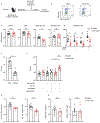
Figure 3:. IL-23 is required for retention of skin-resident memory Th17 cells and TRM17 mediated protection from C. albicans.
(A) Experimental scheme for assessment of memory T cell response to C. albicans. 17Tracker mice were epicutaneously infected with C. albicans and 28 days later, isotype control or anti-IL-23R neutralizing antibody (700 μg) was administered via intraperitoneal injection. 2 weeks later immune cells were isolated from whole skin and secondary lymphoid organs (SLO) and immune cell frequency, number, and phenotype were assessed. (B) Representative flow plots gated on live CD4+ T cells. (C, D) Total numbers of the indicated cell type per cm2 skin after isotype antibody (black circles) or anti-IL-23R treatment (red circles), assessed by flow cytometry at day 42 post-infection. (E) Cohorts of C57/BL6 mice were either uninfected (naïve) or infected on their shaved dorsum with 2x10⁸ CFU C. albicans (memory), and 42 days later rechallenged epicutaneously with C. albicans. CFU per area from homogenized skin 24 hours after infection is shown. (F) C. albicans CFUs isolated from memory mice as in (A) 24 hours after reinfection. Previously infected memory mice (as in E) were injected intraperitoneally with a single dose of isotype (black circles) or anti-IL-23R antibody (red circles) on day 28 post-infection. In addition, mice were given 500μg of TCRγδ T cell depleting antibodies, anti-VCAM-1 and CD49d (500μg), and FTY720 as indicated. CFU/cm2 of skin 24 hours after a second C. albicans infection is shown. (G) Absolute number (left) and frequency (right) of IL-17A-expressing and (H) IFNγ-expressing live PMA/Ionomycin stimulated TCRβ+CD4+ T cells isolated from whole skin of memory mice treated with FTY720, VCAM-1, CD49d, and anti-IL-23R antibody in (F) are shown. All data points represent individual animals. Data are representative of 3 independent experiments with cohorts of 3-5 mice. Significance was calculated using unpaired Student’s t-test, *p<0.05, **p<0.01, ***p<0.001, ****p<0.0001.
Figure 4:
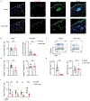
Figure 4:. IL-23 promotes in situ proliferation of TRM17 in skin.
(A) Representative in situ terminal deoxynucleotidyl transferase dUTP nick end labeling (TUNEL) assay of dorsal skin obtained from isotype- and anti-IL-23R antibody-treated 17Tracker mice on day 42 post infection showing incorporation of TUNEL reagent (green), tdTomato (red) and DAPI nuclear stain (blue). Arrows denote cells positive for tdTomato, TUNEL, or merged image as indicated. (B) Quantification of TUNEL+ (left) tdTomato+ cells show in (A), and Annexin V+ (right) viability dye cell quantification of tdTomato+ CD4+ by flow cytometry on day 42 post infection. (C) Analysis of BrdU incorporation by tdTomato+ and tdTomato CD4+ T cells after in vivo administration of BrdU days 28-42 post-infection with C. albicans. Representative flow plots gated on CD4+ TCRβ+ T cells are shown. (D) Quantification showing the percentage of BrdU+ tdTomato+ (left) and tdTomato (right) cells are shown. (E) As in (D) showing total numbers of cells. (F) As in (E) showing number of BrdU+ tdTomato+ cells based on expression of CD69 and CD103. All data points represent individual animals. Data are representative of 3 independent experiments with cohorts of 3-5 mice. Significance was calculated using unpaired Student’s t-test, *p<0.05, **p<0.01, ***p<0.001, ****p<0.0001.
Figure 5:
Figure 5:. IL-23 from CD301b+ myeloid cells is required for maintenance of cutaneous TRM17.
(A) CD45.2+ 17Tracker mice were epicutaneously infected with C. albicans and 36 days later, CD4+ T cells were isolated from pooled spleen and lymph nodes. 1-2 x10⁷ CD4+ T cells were adoptively transferred into naïve congenic (CD45.1+) Mgl2+/DTR-GFP recipients, and epicutaneously infected 1 day later with C. albicans. Mgl2+/DTR-GFP recipients were allowed to rest for 28 days, then 1 μg (loading dose) followed by 100 ng (maintenance dose) of diphtheria toxin (DT) was administered every 3-4 days via intraperitoneal injection to deplete CD301b+ cells. On day 42 immune cells were isolated from whole skin and SLO and T cell frequency, number, and phenotype were assessed. (B) Representative flow cytometry plots gated on live CD90.2+TCRβ+ T cells from skin (top) and SLO (bottom) of Mgl2+/DTR-GFP mice that were sham infected (naïve, left) or infected with C. albicans in the absence (middle, -DT) or presence (right, +DT) of DT treatment are shown. CD45.2+ adoptively transferred 17Tracker cells are gated. (C) Immunofluorescent visualization of GFP (CD301b), CD4, and tdTomato (IL-17A) reporter expression in skin of DT and vehicle-treated mice at day 42 post-infection are shown. Arrows denote tdTomato+ cells and stars denote GFP+ CD301b-expressing cells. (D) Summary data from (B) of total numbers of the indicated cell type in skin per cm2 of skin as assessed by flow cytometry is shown. (E) As in (D) showing total cell numbers from the SLO. (F,G) Lethally irradiated B6.SJL-Ly5.2 (CD45.1) mice were reconstituted with a 1:1 ratio of bone marrow from Mgl2+/DTR-GFP mice and either WT or Il23a−/− mice. After 12 weeks, mice were subject to C. albicans infection and adoptive transfer of CD45.2+ 17Tracker CD4+ T cells as in (A). BrdU and DT or vehicle (PBS) were administered days 28-42 post-infection as in (A). (F) Representative flow plots gated on live CD4+ T cells isolated from skin 42d post-infection. (G) Total numbers of the indicated cell type per cm2 skin, assessed by flow cytometry at day 42 post-infection. All data points represent individual animals. Data are representative of at least 2 independent experiments with cohorts of 2-5 mice. Significance was calculated using unpaired Student’s t-test, *p<0.05, **p<0.01.
Figure 6:
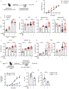
Figure 6:. CD301b+ myeloid cells induce TRM17 proliferation to accelerate development of psoriasiform dermatitis.
(A) Experimental scheme for assessment of psoriasiform dermatitis. 17Tracker mice were epicutaneously infected with C. albicans on the right flank and starting 28 days later, 5% imiquimod cream was applied to shaved skin on the right and left flanks daily for 5 days. Erythema (0-4), scaling (0-4), and thickness (0-4) of the back skin were assessed daily using a modified PASI scoring system as previously described(68). (B) Cumulative inflammation score of 0-12 is depicted for previously infected (red) or naïve (black) mice. (C) Frequency of Thy1.1 (IL-17F)+ CD4+ T cells (left) and dermal TCRγδ cells (right) isolated from back skin previously (red) or naïve (black) mice as determined by flow cytometry. (D) As in (C) showing total numbers of CD4+ T cells, tdTomato+ CD4+ T cells (E, left) and tdTomato CD4+ T cells (E, right) in mice treated with imiquimod or previously infected with C. albicans as indicated. (F) As in C, showing total numbers of dermal TCRγδ+ T cells and tdTomato TCRγδ+ T cells. (G) Frequency of proliferating Ki67+ tdTomato+ CD4+ T cells (left) and TCRγδ+ T cells. (H) Mgl2+/DTR-GFP mice were infected with C. albicans and starting 28 days later, 5% imiquimod cream was applied daily to ear and shaved back skin for 5 consecutive days. On days −1 and +3, diphtheria toxin (DT) was administered to deplete CD301b+ cells. (I) Ear thickness at the indicated time points during imiquimod treatment is shown. (J) Absolute number of CD69+CD103+IL-17A+ CD4+ T cells and (K) Ki67+ expression in tdTomato+ CD4+ T cells present in skin of DT (+DT) or vehicle-treated (−DT) mice on day 3 of imiquimod treatment is shown. All data points represent individual animals. All data are representative of 3 independent experiments with cohorts of 2-6 mice. Significance was calculated using paired (B) or unpaired (C-K) Student’s t-test, *p<0.05, **p<0.01, ***p<0.001.
Figure 7:
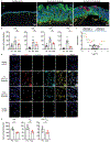
Figure 7:. Clinical anti-IL-23 therapy depletes TRM17 from psoriatic skin.
(A) Representative immunofluorescent staining from healthy control (left), lesional skin from a single psoriatic plaque before (middle) and 1 week after (right) initiation of systemic treatment with an anti-IL-23 monoclonal antibody (150 mg risankizumab or 100 mg guselkumab). Pan-cytokeratin (green), CD3 (red), and nuclear stain (blue). Scale bar 250 μm. (B) Absolute numbers of the indicated cell type or protein assessed by NanoString GeoMX Digital Spatial Profiler (DSP) protein quantification of healthy control (HC), psoriasis pre-anti-IL-23 (Pre), or post anti-IL-23 (Post) treatment skin. (C) Volcano plot displaying proteins expressed by CD3+ cells satisfying threshold criteria of >0.5 fold change (x axis) and −log10 p-value >1 (y-axis). (D) Representative immunofluorescent microscopic images of samples as in (A) showing expression of CD4 (red), IL-17A (green), CD69 (cyan), CD103 (yellow) and DAPI nuclear stain (blue). Scale bar 20 μm. Arrows denote CD4+IL-17A+CD69+CD103+DAPI+ cells. (E) Quantification of numbers of the indicated cells as in (D). Data are representative of 3 independent experiments and include specimens from 5 individual psoriasis patients analyzed pre and post anti-IL-23 treatment. Each data point (B,E) represents an individual patient, with an average of four high powered fields quantified per patient. Significance was calculated using two-way ANOVA with Tukey’s post-t test, *p<0.05, **p<0.001, ***p<0.001.

References

    1. Hirai T, Yang Y, Zenke Y, Li H, Chaudhri VK, Diaz JSDLC, Zhou PY, Nguyen BA-T, Bartholin L, Workman CJ, Griggs DW, Vignali DAA, Singh H, Masopust D, Kaplan DH, Competition for Active TGFβ Cytokine Allows for Selective Retention of Antigen-Specific Tissue- Resident Memory T Cells in the Epidermal Niche. Immunity. 54, 84–98.e5 (2021). - PMC - PubMed
    1. Hirai T, Zenke Y, Yang Y, Bartholin L, Beura LK, Masopust D, Kaplan DH, Keratinocyte-Mediated Activation of the Cytokine TGF-β Maintains Skin Recirculating Memory CD8+ T Cells. Immunity. 50, 1249–1261.e5 (2019). - PMC - PubMed
    1. Hirai T, Whitley SK, Kaplan DH, Migration and Function of Memory CD8+ T Cells in Skin. J Invest Dermatol. 140, 748–755 (2019). - PMC - PubMed
    1. Iijima N, Iwasaki A, A local macrophage chemokine network sustains protective tissue-resident memory CD4 T cells. Science. 346, 93–98 (2014). - PMC - PubMed
    1. Shin H, Kumamoto Y, Gopinath S, Iwasaki A, CD301b+ dendritic cells stimulate tissue-resident memory CD8+ T cells to protect against genital HSV-2. Nat Commun. 7, 13346 (2016). - PMC - PubMed

Publication types