. 2022 Nov 11;13(1):6867.
doi: 10.1038/s41467-022-34589-8.

Molecular identity of proprioceptor subtypes innervating different muscle groups in mice

Affiliations

Molecular identity of proprioceptor subtypes innervating different muscle groups in mice

Stephan Dietrich et al. Nat Commun. .

Abstract

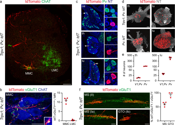
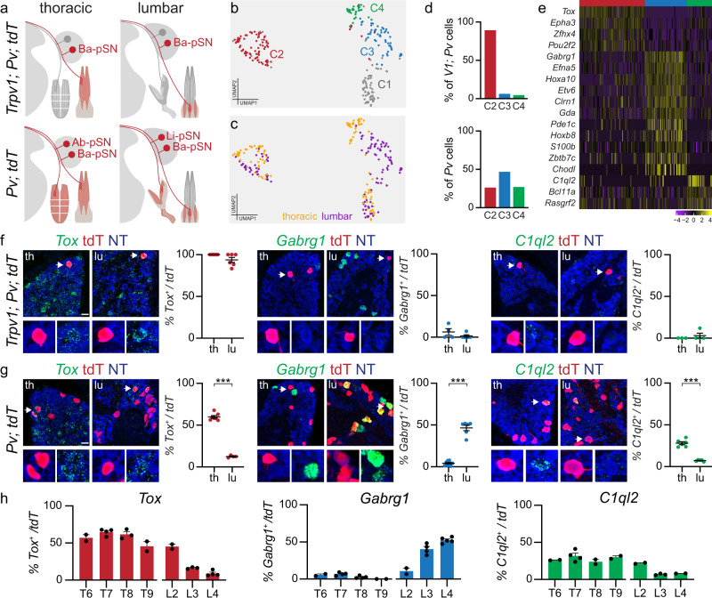
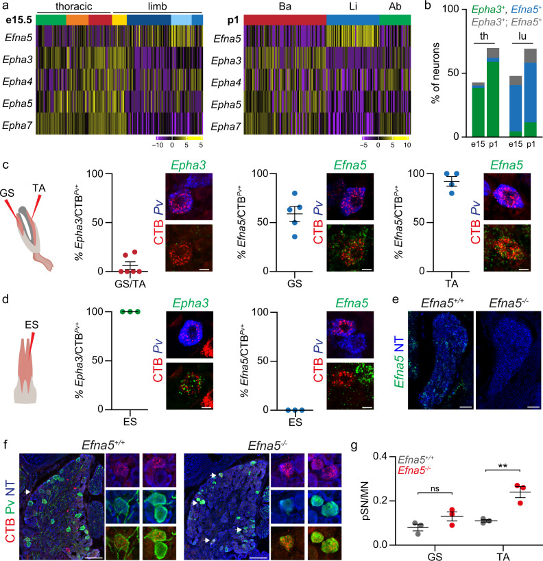
The precise execution of coordinated movements depends on proprioception, the sense of body position in space. However, the molecular underpinnings of proprioceptive neuron subtype identities are not fully understood. Here we used a single-cell transcriptomic approach to define mouse proprioceptor subtypes according to the identity of the muscle they innervate. We identified and validated molecular signatures associated with proprioceptors innervating back (Tox, Epha3), abdominal (C1ql2), and hindlimb (Gabrg1, Efna5) muscles. We also found that proprioceptor muscle identity precedes acquisition of receptor character and comprise programs controlling wiring specificity. These findings indicate that muscle-type identity is a fundamental aspect of proprioceptor subtype differentiation that is acquired during early development and includes molecular programs involved in the control of muscle target specificity.


Conflict of interest statement

The authors declare no competing interests.

Figures

Fig. 1
Fig. 1. Single-cell transcriptome analysis of thoracic and lumbar proprioceptors at e15.5.
a Schematic illustrating central and peripheral connectivity of e15.5 proprioceptors at thoracic (left) and lumbar (right) spinal levels. Ab-pSN, abdominal muscles-connecting proprioceptors; Ba-pSN, back muscles-connecting proprioceptors; Li-pSN, hindlimb muscles-connecting proprioceptors; MMC, median motor column; HMC, hypaxial motor column; LMC, lateral motor column. b UMAP visualization of tdTomato+ neuron clusters from PvtdTom embryos at e15.5. c Gene expression analysis (logcounts) of proprioceptors (Pv, Runx3, Etv1, Ntrk3), mechanoreceptors (Ntrk2, Maf), postmitotic neurons (Avil, Isl1), proliferating neurons (Mki67, Mcm2, Pcna) and glial (Mpz, Apoe) markers. d UMAP visualization of proprioceptor clusters identified from analysis of cluster 1. e UMAP visualization of proprioceptor clusters color-coded according to the thoracic (red) and lumbar (blue) origin of the cells. f Percentage of proprioceptors originating from lumbar (left) and thoracic (right) DRG in different proprioceptor clusters. g Differential gene expression analysis (logcounts) in proprioceptive clusters (pS1, green; pS2, dark blue; pS3, orange; pS4, light blue; pS5, yellow; pS6, red; pS7, blue). h Representative single molecule fluorescent in situ hybridization (smFISH) images of thoracic (top) and lumbar (bottom) e15.5 DRG sections showing proprioceptors (Runx3+; Pv+) expressing Trpv1. Scale bar: 25 µm. i Percentage of proprioceptors (Runx3+; Pv+) expressing Trpv1 in thoracic and lumbar DRG at e15.5 (mean ± SEM, n = 3 animals). Source data are provided as a Source Data file.
Fig. 2
Fig. 2. Genetic targeting of proprioceptors innervating back muscles.
a Representative image of tdTomato+ afferents in a lumbar spinal cord section from p7 Trpv1Cre; PvFlp; Ai65 mice. MMC, median motor column; LMC, lateral motor column. Scale bar: 100 µm. b Representative image (MMC, left) and quantification (right) of tdTomato+; vGluT1+ presynaptic puncta juxtaposed to MMC or LMC neurons cell bodies and proximal dendrites of p7 Trpv1Cre; PvFlp; Ai65 mice (mean ± SEM, n = 4 animals). Scale bar: 25 µm. c Representative images of cervical, thoracic, and lumbar DRG sections showing tdTomato+; Pv+ neurons in p7 Trpv1Cre; PvFlp; Ai65 mice. Scale bar: 100 µm. d Whole mount preparations of thoracic (left) and lumbar (right) DRG showing genetically labeled neurons from p1 Trpv1Cre; PvFlp; Ai65 (top) and PvCre; Ai14 (bottom) mice. Scale bar: 100 µm. e Number of tdTomato+ sensory neurons in DRG from p1 Trpv1Cre; PvFlp; Ai65 and PvCre; Ai14 at thoracic (T1-T12, left) and lumbar (L1-L5, right) levels (mean ± SEM, n = 3 animals). f Representative images (left) and quantification (right) of tdTomato+ group Ia, II, and Ib afferents in muscle spindles (MS) and Golgi tendon organs (GTO) from the erector spinae muscle of Trpv1Cre; PvFlp; Ai65 mice. (mean ± SEM, n = 4 animals). Scale bar: 25 µm. Source data are provided as a Source Data file.
Fig. 3
Fig. 3. Molecular profiles of back-, abdominal- and hindlimb-innervating proprioceptors.
a Schematics illustrating labeling of the Ba-pSN subset captured in Trpv1Cre; PvFlp; Ai65 and all proprioceptors in PvCre; Ai14 at thoracic and lumbar levels. b UMAP visualization of cell clusters after transcriptome analysis of tdTomato+ DRG neurons from p1 Trpv1Cre; PvFlp; Ai65 and PvCre; Ai14 mice at thoracic and lumbar levels. c UMAP visualization of cell clusters color-coded according to the anatomical origin (thoracic, yellow; lumbar, purple) of neurons. d Bar graph illustrating the percentage of Trpv1Cre; PvFlp; Ai65 (top) and PvCre; Ai14 (bottom) cells found in clusters C2 (red), C3 (blue), C4 (green). e Differential gene expression analysis (logcounts) for clusters C2 (red), C3 (blue), and C4 (green). f Representative smFISH images and quantification of Tox (C2), Gabrg1 (C3), and C1ql2 (C4) expression in tdTomato+ thoracic and lumbar DRG neurons from p1 Trpv1Cre; PvFlp; Ai65 mice (mean ± SEM, n ≥ 3 animals). Scale bar: 25 µm. g Representative smFISH images and quantification of Tox (C2), Gabrg1 (C3), and C1ql2 (C4) expression in tdTomato+ thoracic (T6–T9) and lumbar (L3–L4) DRG neurons from p1 PvCre; Ai14 mice (mean ± SEM, n ≥ 4 animals, two-tailed, t-test, ***p < 0.001). Scale bar: 25 µm. h Distribution of Tox (C2), Gabrg1(C3), and C1ql2 (C4) in tdTomato+ neurons from thoracic and lumbar DRG of p1 PvCre; Ai14 mice (mean ± SEM, n ≥ 2 animals). Source data are provided as a Source Data file.
Fig. 4
Fig. 4. Proprioceptors muscle-type identity emerges at early developmental stages.
a, b Representative smFISH images and quantification of Tox (C2), Gabrg1 (C3), and C1ql2 (C4) expression in Pv+ sensory neurons retrogradely labeled after cholera-toxin B (CTB) injection in back (a erector spinae, ES) and hindlimb (b gastrocnemius, GS and tibialis anterior, TA) of p1 wild-type mice (mean ± SEM, n ≥ 4 animals). Scale bar: 10 µm. c, d Heatmaps representing pairwise gene expression correlation values for Ba-pSN (red), Ab-pSN (green), and Li-pSN (blue) molecular signatures at p1 (top) and e15.5 (bottom; Pearson’s r using logcounts). e, f Representative smFISH images and quantification of Tox (C2), and Gabrg1 (C3), expression in Pv+ sensory neurons retrogradely labeled after rhodamine-dextran (RhD) injection in e15.5 back (e) and hindlimb (f) muscles of wild-type mice (mean ± SEM, n ≥ 3 animals). Scale bar: 10 µm. Source data are provided as a Source Data file.
Fig. 5
Fig. 5. Elimination of ephrin-A5 function erodes the specificity of muscle connectivity.
a Gene expression analysis (logcounts) of ephrin-A/EphA family members differentially expressed in proprioceptor clusters identified at e15.5 (thoracic origin: green, orange, red; lumbar origin: yellow, dark blue, light blue, and blue) and p1 (back: red; hindlimb: blue; abdominal: green). b Percentage of Pv+ or tdTomato+ sensory neurons expressing Epha3 (green), Efna5 (blue) or Epha3; Efna5 (gray) at thoracic and lumbar levels in either e15.5 or p1 PvCre; Ai14 mice (mean ± SEM, n ≥ 2 animals). c Representative smFISH images and quantification of Epha3 (left) and Efna5 (center/right) expression in Pv+sensory neurons retrogradely labeled after CTB injection in gastrocnemius (GS), and tibialis anterior (TA) muscles of p1 wild-type mice (mean ± SEM, n ≥ 4 animals). Scale bar: 10 µm. d Representative smFISH images and quantification of Epha3 (left) and Efna5 (right) expression in Pv+ sensory neurons retrogradely labeled after CTB injection in the erector spinae (ES) muscle of p1 wild-type mice (mean ± SEM, n = 3 animals). Scale bar: 10 µm. e Representative smFISH images of Efna5 expression in lumbar DRG of p1 Efna5+/+ and Efna5-/- mice. Scale bar: 25 µm. f Representative images of Pv+; CTB+ sensory neurons retrogradely labeled after CTB injection in the tibialis anterior (TA) muscle of p1 Efna5+/+ (left) and Efna5−/− (right) mice. Scale bar: 100 µm. g Ratio of proprioceptor (Pv+) per motor neuron labeled after CTB injection in the gastrocnemius (GS) and tibialis anterior (TA) muscles of p1 Efna5+/+ (gray) and Efna5−/− (red) mice (mean ± SEM, n = 3 animals, two-tailed t-test, ns p > 0.05, **p < 0.01, p-values 0.12 GS and 0.007308 TA). Source data are provided as a Source Data file.

References

    1. Proske U, Gandevia SC. The proprioceptive senses: their roles in signaling body shape, body position and movement, and muscle force. Physiol. Rev. 2012;92:1651–1697. - PubMed
    1. Balaskas N, Ng D, Zampieri N. The positional logic of sensory-motor reflex circuit assembly. Neuroscience. 2020;450:142–150. - PubMed
    1. Meltzer S, Santiago C, Sharma N, Ginty DD. The cellular and molecular basis of somatosensory neuron development. Neuron. 2021;109:3736–3757. - PMC - PubMed
    1. Zampieri N, de Nooij JC. Regulating muscle spindle and Golgi tendon organ proprioceptor phenotypes. Curr. Opin. Physiol. 2021;19:204–210. - PMC - PubMed
    1. Eccles JC, Eccles RM, Lundberg A. The convergence of monosynaptic excitatory afferents on to many different species of alpha motoneurones. J. Physiol. 1957;137:22–50. - PMC - PubMed

Publication types