. 2022 Dec 31;210(3):309-320.
doi: 10.1093/cei/uxac102.

Exosome-transmitted S100A4 induces immunosuppression and non-small cell lung cancer development by activating STAT3

Affiliations

Exosome-transmitted S100A4 induces immunosuppression and non-small cell lung cancer development by activating STAT3

Xu Wu et al. Clin Exp Immunol. .

Abstract

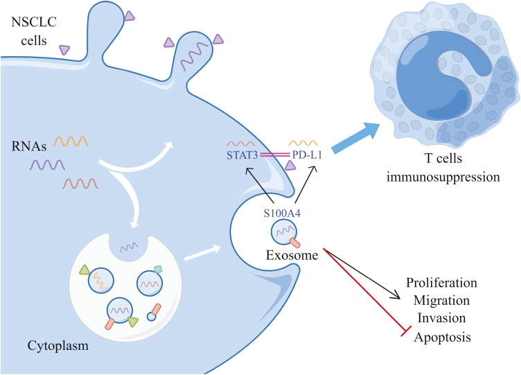
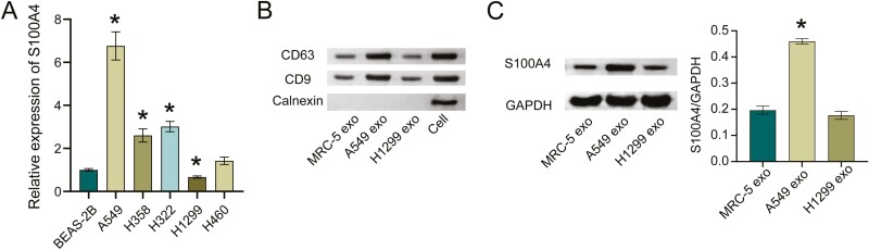
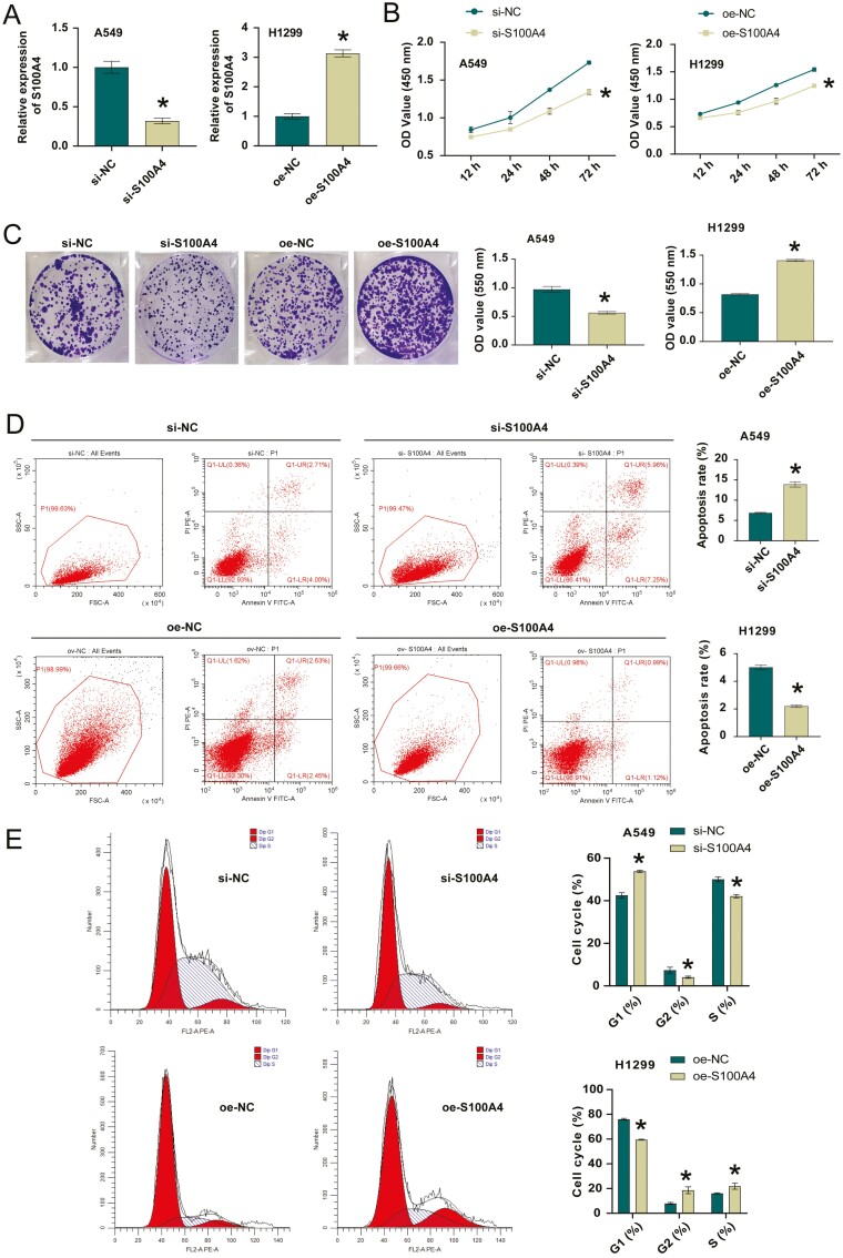
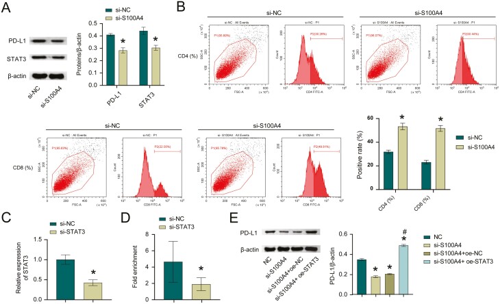
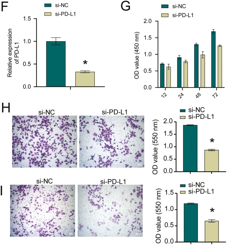
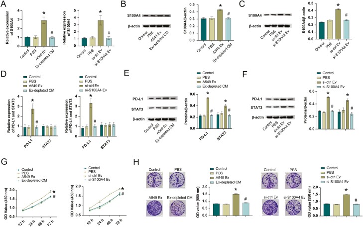
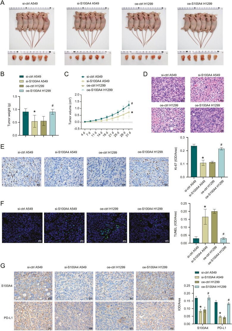
Non-small cell lung cancer (NSCLC) is the primary reason of tumor morbidity and mortality worldwide. We aimed to study the transfer process of S100A4 between cells and whether it affected NSCLC development by affecting STAT3 expression. First, S100A4 expression in NSCLC cells was measured. The exosomes in MRC-5, A549, and H1299 cells were isolated and identified. We constructed si-S100A4 and si-PD-L1 to transfect A549 cells and oe-S100A4 to transfect H1299 cells, and tested the transfection efficiency. Cell function experiments were performed to assess cell proliferation, clone number, apoptosis, cell cycle, migration, and invasion abilities. In addition, ChIP was applied to determine the targeting relationship between S100A4 and STAT3. Next, we explored NSCLC cell-derived exosomes role in NSCLC progress by transmitting S100A4. Finally, we verified the function of exosome-transmitted S100A4 in NSCLC in vivo. High expression of S100A4 was secreted by exosomes. After knocking down S100A4, cell proliferation ability was decreased, clones number was decreased, apoptosis was increased, G1 phase was increased, S phase was repressed, and migration and invasion abilities were also decreased. ChIP validated STAT3 and PD-L1 interaction. After knocking down S100A4, PD-L1 expression was decreased, while ov-STAT3 reversed the effect of S100A4 on PD-L1 expression. Meanwhile, S100A4 inhibited T-cell immune activity by activating STAT3. In addition, knockdown of PD-L1 inhibited cell proliferation, migration, and invasion. NSCLC cell-derived exosomes promoted cancer progression by transmitting S100A4 to activate STAT3 pathway. Finally, in vivo experiments further verified that exosome-transmitted S100A4 promoted NSCLC progression. Exosome-transmitted S100A4 induces immunosuppression and the development of NSCLC by activating STAT3.

Keywords: PD-L1; S100A4; STAT3; exosome; immunosuppression.


Figures

Graphical Abstract
Graphical Abstract
Figure 1:
Figure 1:
the high expression of S100A4 was secreted by exosomes. A. S100A4 mRNA expression in NSCLC cells. *P < 0.05. B. Exosomes marker CD63, CD9, and Calnexin expressions in exosomes. C. Western blot detection of S100A4 level in exosomes. *P < 0.05.
Figure 2:
Figure 2:
S100A4 regulated the proliferation, migration, and invasion of NSCLC cells. A. S100A4 knockdown or overexpression efficiency in NSCLC cells. B. CCK-8 assay for si-NC, si-S100A4 or oe-NC, oe-S100A4 transfected NSCLC cells. C. Colony formation assay for si-NC, si-S100A4 or oe-NC, oe-S100A4 transfected NSCLC cells. D. The percentage of apoptotic cells in si-NC and si-S100A4 groups. E. Cell cycle distribution in si-NC, si-S100A4 or oe-NC, oe-S100A4 transfected NSCLC cells. F and G. Cyclin D1, Bcl-2, and BAX expression in si-NC, si-S100A4 or oe-NC, oe-S100A4 transfected NSCLC cells. H and I. Transwell migration and invasion assays for si-NC, si-S100A4 or oe-NC, oe-S100A4 transfected NSCLC cells. J and K. E-cadherin, N-cadherin, vimentin, twist, slug, snail, β-catenin, and c-Myc expressions in si-NC, si-S100A4 or oe-NC, oe-S100A4 transfected NSCLC cells. *P < 0.05.
Figure 3:
Figure 3:
S100A4 regulated PD-L1 expression and inhibited T-cell immune activity by activating STAT3. A. Western blot measured STAT3 and PD-L1 levels. *P < 0.05. B. The effect of different treatments on CD4+ and CD8+ populations among A549 cells. C. STAT3 knockdown efficiency in A549 cells. *P < 0.05. D. ChIP was utilized to verify STAT3 binding to PD-L1. E. PD-L1 protein level. *P < 0.05 vs NC, #P < 0.05 vs si-S100A4+oe-STAT3. *P < 0.05. F. PD-L1 knockdown efficiency in A549 cells. *P < 0.05. G. CCK-8 assay for si-NC and si-PD-L1 transfected NSCLC cells. H and I. Transwell migration and invasion assays for si-NC and si-PD-L1 transfected NSCLC cells. *P < 0.05.
Figure 3:
Figure 3:
S100A4 regulated PD-L1 expression and inhibited T-cell immune activity by activating STAT3. A. Western blot measured STAT3 and PD-L1 levels. *P < 0.05. B. The effect of different treatments on CD4+ and CD8+ populations among A549 cells. C. STAT3 knockdown efficiency in A549 cells. *P < 0.05. D. ChIP was utilized to verify STAT3 binding to PD-L1. E. PD-L1 protein level. *P < 0.05 vs NC, #P < 0.05 vs si-S100A4+oe-STAT3. *P < 0.05. F. PD-L1 knockdown efficiency in A549 cells. *P < 0.05. G. CCK-8 assay for si-NC and si-PD-L1 transfected NSCLC cells. H and I. Transwell migration and invasion assays for si-NC and si-PD-L1 transfected NSCLC cells. *P < 0.05.
Figure 4:
Figure 4:
exosome-transmitted S100A4 promoted NSCLC progression by targeting STAT3. AC. S100A4 mRNA and protein level. DF. STAT3 and PD-L1 mRNA and protein levels. CCK-8 (G), colony formation (H), transwell migration (I), and invasion (J) assays for A549 cells treated with PBS, Ex-depleted CM, or exosomes from sh-Ctrl and sh-S100A4 transfected A549 cells. *P < 0.05 vs PBS, # P < 0.05 vs A549 Ex or si-ctrl Ev.
Figure 5:
Figure 5:
exosome derived S100A4 promotes NSCLC progression in vivo. AC. Tumor size, weight, and volume of mice injected with A549 cells treated with si-ctrl or si-S100A4 and H1299 cells transfected with oe-ctrl or oe-S100A4. DF. HE, Ki-67, and TUNEL staining in tumor tissues. G. Immunohistochemistry assay for detecting S100A4 and PD-L1 expressions. *P < 0.05 vs PBS, #P < 0.05 vs A549 Ex or si-ctrl Ev.

References

    1. Tan AC. Targeting the PI3K/Akt/mTOR pathway in non-small cell lung cancer (NSCLC). Thorac Cancer 2020, 11, 511–8. doi:10.1111/1759-7714.13328. - DOI - PMC - PubMed
    1. Suster DI, Mino-Kenudson M.. Molecular pathology of primary non-small cell lung cancer. Arch Med Res 2020, 51, 784–98. doi:10.1016/j.arcmed.2020.08.004. - DOI - PubMed
    1. Azghadi S, Daly ME.. Radiation and immunotherapy combinations in non-small cell lung cancer. Cancer Treat Res Commun 2021, 26, 100298. doi:10.1016/j.ctarc.2020.100298. - DOI - PubMed
    1. Marmarelis ME, Langer CJ.. Treatment of patients with non-small-cell lung cancer harboring rare Oncogenic mutations. Clin Lung Cancer 2020, 21, 395–406. doi:10.1016/j.cllc.2020.01.010. - DOI - PubMed
    1. Baran K, Brzeziańska-Lasota E.. Proteomic biomarkers of non-small cell lung cancer patients. Adv Respir Med 2021, 89, 419–26. doi:10.5603/ARM.a2021.0089. - DOI - PubMed

Publication types

MeSH terms