. 2022 Nov 12;79(12):587.
doi: 10.1007/s00018-022-04615-5.

Compound A attenuates proinflammatory cytokine-induced endoplasmic reticulum stress in beta cells and displays beneficial therapeutic effects in a mouse model of autoimmune diabetes

Affiliations

Compound A attenuates proinflammatory cytokine-induced endoplasmic reticulum stress in beta cells and displays beneficial therapeutic effects in a mouse model of autoimmune diabetes

Luz Andreone et al. Cell Mol Life Sci. .

Abstract

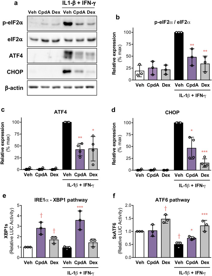
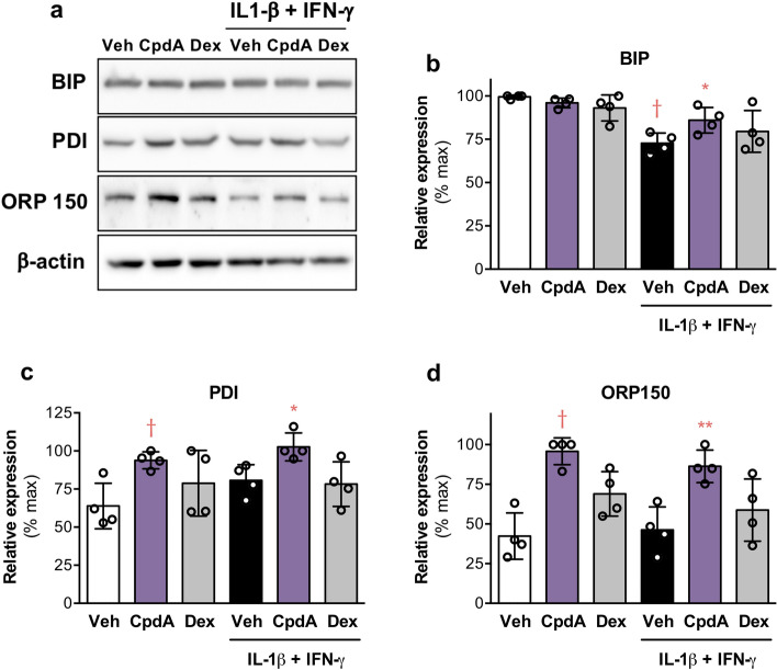
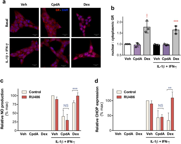
Type 1 diabetes (T1D) is characterized by an immune-mediated progressive destruction of the insulin-producing β-cells. Proinflammatory cytokines trigger endoplasmic reticulum (ER) stress and subsequent insulin secretory deficiency in cultured β-cells, mimicking the islet microenvironment in T1D. β-cells undergo physiologic ER stress due to the high rate of insulin production and secretion under stimulated conditions. Severe and uncompensated ER stress in β-cells is induced by several pathological mechanisms before onset and during T1D. We previously described that the small drug Compound A (CpdA), a selective glucocorticoid receptor (GR/NR3C1, nuclear receptor subfamily 3, group C, member 1) ligand with demonstrated inflammation-suppressive activity in vivo, is an effective modulator of effector T and dendritic cells and of macrophages, yet, in a GR-independent manner. Here, we focus on CpdA's therapeutic potential in T1D cellular and animal models. We demonstrate that CpdA improves the unfolded protein response (UPR) by attenuating ER stress and favoring the survival and function of β-cells exposed to an environment of proinflammatory cytokines. CpdA administration to NODscid mice adoptively transferred with diabetogenic splenocytes (from diabetic NOD mice) led to a delay of disease onset and reduction of diabetes incidence. Histological analysis of the pancreas showed a reduction in islet leukocyte infiltration (insulitis) and preservation of insulin expression in CpdA-treated normoglycemic mice in comparison with control group. These new findings together with our previous reports justify further studies on the administration of this small molecule as a novel therapeutic strategy with dual targets (effector immune and β-cells) during autoimmune diabetes.

Keywords: Inflammation; Islets; SEGRAM; Small-molecule; Type 1 diabetes.


Conflict of interest statement

The authors have no relevant financial or non-financial interests to disclose.

Figures

Fig. 1
Fig. 1
CpdA inhibits cytokine-triggered NF-κB pathway activation and reduces nitric oxide (NO) production in INS-1E cells. ac INS-1E cells were pretreated with vehicle, CpdA 10 μM or dexamethasone (Dex) 0.1 μM for 1 h and then challenged or not with IL-1β 100 pg/mL and IFN-γ 5 ng/mL (CYT). After indicated time, levels of phospho-IkBα and total IkBα were analyzed by Western blot. Representative blots (a) and quantitative analysis of phospho-IkBα (b) and IkBα (c) protein expression expressed as mean ± SEM of n = 4 independent experiments; β-actin was used as loading control. (*) p < 0.05 vs. vehicle. d INS-1 cells were treated as described in (a) with 30 min CYT stimulation. NF-κB (RelA) cellular localization was analyzed by immunofluorescence staining. A representative confocal microscopy pictures of INS-1E cells immunostained for NF-κB (red) in different experimental conditions as indicated; nuclei were stained with DAPI (blue); scale bars 10 μm. e Quantification of nuclear:cytoplasmic ratio of NF-kB staining from analysis of 5 separate high-power field images for each experimental condition. Data are shown as mean ± SD of n = 3 independent experiments. f INS-1E were treated as described in (a) with 16 h CYT stimulation. NO secretion was assessed by Griess reaction. Data are shown as mean ± SD of n = 5 independent experiments. g–i INS-1E were treated as described in (a) with 6 h CYT stimulation, iNOS mRNA and protein expression were analyzed by RT-qPCR and Western blot, respectively. Relative iNOS mRNA levels (g) normalized to HPRT expressed as mean ± SD of n = 3 independent experiments. Representative blots (h) and quantitative analysis of iNOS (i) protein expression expressed as mean ± SD of n = 3 independent experiments; β-actin was used as loading control. di (†) p < 0.05 vs. vehicle; (*) p < 0.05, (**) p < 0.01, (***) p < 0.001 vs. vehicle + CYT
Fig. 2
Fig. 2
CpdA hampers the cytokine-induced activation of ER stress related pathways and favors unfolded protein response (UPR) pathways in INS-1E cells. ad INS-1E cells were pretreated with vehicle, CpdA 10 μM or dexamethasone (Dex) 0.1 μM for 1 h and then challenged or not with IL-1β 100 pg/mL and IFN-γ 5 ng/mL (CYT). After 16 h, levels of phospho- and total eIF2α, ATF4 and CHOP were analyzed by Western blot. Representative blots (a) and quantitative analysis of phospho- and total eIF2α (b), ATF4 (c) and CHOP (d) protein expression expressed as mean ± SD of n = 3/4 independent experiments; β-actin was used as loading control. ef INS-1 cells were transiently transfected with XBP1u-LUC (e) or 5xATF6-LUC (f) and RSV-βGal reporter plasmids. At 24 h post-transfection, cells were treated as described in (a). After 16 h, cells were collected and firefly luciferase (LUC) activity was measured and normalized against β-galactosidase (β-Gal) activity for transfection efficiency. Relative LUC activity is expressed as mean ± SD of n = 3 independent experiments; (†) p < 0.05 vs. vehicle; (*) p < 0.05, (**) p < 0.01, (***) p < 0.001 vs. vehicle + CYT
Fig. 3
Fig. 3
CpdA enhances the expression of ER chaperones in INS-1E cells. a–d INS-1E cells were pretreated with vehicle, CpdA 10 μM or dexamethasone (Dex) 0.1 μM for 1 h and then challenged or not with IL-1β 100 pg/mL and IFN-γ 5 ng/mL (CYT). After 16 h, levels of BIP, PDI and ORP150 were analyzed by Western blot. Representative blots (a) and quantitative analysis of BIP (b), PDI (c) and ORP150 (d) protein expression expressed as mean ± SD of n = 4 independent experiments; β-actin was used as loading control. (†) p < 0.05 vs. vehicle; (*) p < 0.05, (**) p < 0.01 vs. vehicle + CYT
Fig. 4
Fig. 4
CpdA acts independently of the GR-complex. ab INS-1E cells were pretreated with vehicle, CpdA 10 μM or dexamethasone (Dex) 0.1 μM for 1 h and then challenged or not with IL-1β 100 pg/mL and IFN-γ 5 ng/mL (CYT). After 30 min, glucocorticoid receptor (GR) expression was analyzed by immunofluorescence staining. a Representative confocal microscopy pictures of INS-1E cells immunostained for GR (red) in the different experimental conditions as indicated; nuclei were stained with DAPI (blue); scale bars 10 μm. b Quantification of nuclear:cytoplasmic ratio of GR staining was performed from analysis of 5 separate high-power field images for each experimental condition. Data are shown as mean ± SD of n = 3 independent experiments. (†) p < 0.05 vs. vehicle; (***) p < 0.001 vs. vehicle + CYT. cd INS-1E were treated as described in (a) with or without the GCs antagonist RU486 (1 μM) and 16 h of CYT stimulation. c NO secretion was assessed by Griess reaction. Data are shown as mean ± SD of n = 3 independent experiments. d Levels of CHOP protein expression were analyzed by Western blot; quantitative analysis of blots is expressed as mean ± SD of n = 3 independent experiments; β-actin was used as loading control. cd (**) p < 0.01, (***) p < 0.001 between indicated experimental conditions; NS  not significant differences
Fig. 5
Fig. 5
CpdA attenuates CYT-induced apoptosis and preserves glucose stimulated insulin secretion in β-cells. af INS-1E cells were pretreated with vehicle, CpdA 10 μM or dexamethasone (Dex) 0.1 μM for 1 h and then challenged or not with IL-1β 100 pg/mL and IFN-γ 5 ng/mL (CYT) for 16 h. a Cell viability was assessed by MTT assay. Viability in control group (Veh) has been considered as 100%. Data are shown as mean ± SD of n = 3 independent experiments. b Bax mRNA to Bcl-2 mRNA ratio, c DP5 mRNA and d TNF-α mRNA expression were analyzed by RT-qPCR. Relative mRNA levels normalized to HPRT are expressed as mean ± SD of n = 3/4 independent experiments. e Cumulative insulin secretion was determined in the conditioned media (11 mM glucose) by specific ELISA. Values were normalized to total protein content measured by BCA. Relative insulin secretion is shown as mean ± SD of n = 3 independent experiments. f Glucose-Stimulated Insulin Secretion (GSIS) was assessed by ELISA in the conditioned media of cells cultured in the presence of low (2 mM) or high (20 mM) glucose. Insulin secretion index (20 mM/2 mM) is shown as mean ± SD of n = 4 independent experiments. g–h Murine islets (5 IEQ/well) were pretreated with vehicle, CpdA 10 μM or dexamethasone (Dex) 0.1 μM for 1 h and then challenged or not with IL1-β 100 pg/mL + IFN-γ 5 ng/mL + TNF-α 8 ng/mL for 16 h. (g) Apoptosis was assessed by Hoechst and PI double fluorescence staining. Percentages of apoptotic cells in islets are expressed as mean ± SD of n = 3 independent experiments. h GSIS was assessed in isolated mice islets as described in (f). Insulin secretion index (20 mM/2 mM) is shown as mean ± SD of n = 4 independent experiments. (†) p < 0.05 vs. vehicle; (*) p < 0.05, (**) p < 0.01, (***) p < 0.001 vs. vehicle + CYT
Fig. 6
Fig. 6
CpdA improves glucose response in cytokine-challenged isolated human islets. ah Isolated human islets were pretreated with vehicle, CpdA 10 μM or dexamethasone (Dex) 0.1 μM for 1 h and then challenged or not with IL1-β 250 pg/mL + IFN-γ 50 ng/mL (CYT) for 16 h. ab Glucose-Stimulated Insulin Secretion (GSIS) was assessed by ELISA in the conditioned media of islets (5 IEQ/well) cultured in the presence of low (2 mM) or high (20 mM) glucose. Values were normalized to total protein content measured by BCA. Islets from two non-diabetic donors were tested in independent experiments with similar results. Data are shown as insulin secretion index (20 mM/2 mM). c IL-6 mRNA d IL-1β mRNA and e TNF-α mRNA expression were analyzed by RT-qPCR. Islets (50 IEQ/well) from three non-diabetic donors were tested in independent experiments (n = 3); relative mRNA levels normalized to RPL19 are expressed as mean ± SD. (†) p < 0.05 vs. vehicle; (**) p < 0.01, (***) p < 0.001 vs. vehicle + CYT
Fig. 7
Fig. 7
Beneficial effects of CpdA administration in the adoptive transfer of autoimmune diabetes in mice. a Experimental scheme. Non-obese diabetic (NODscid) mice were adoptively transferred with diabetogenic splenocytes (at day 0 i.p. 5 × 106 cells/mice) and treated with CpdA (i.p. 100μg, n = 21) or vehicle (Control, n = 10) three times a week from day  – 1 to day 50. b Kaplan–Meier plot of cumulative diabetes incidence. p < 0.0001 vs. vehicle, by log-rank (Mantel–Cox) test. c Graph representing the classification of pancreatic islets according to the severity of leukocyte infiltration (insulitis) in each experimental group. Grade: 0, no insulitis; 1: < 25% infiltrate; 2: 25–50% infiltrate; 3: 50–75% infiltrate; and 4: > 75% infiltrate. Bars show mean ± SEM of independent individuals. Control d28 after adoptive transfer (n = 2), CpdA-treated d28 (n = 3), CpdA-treated d100 (n = 3), NODscid sham control (n = 2). d Islet immunostaining for insulin expression from each experimental group. Representative islets are shown. Scale bar 50 μm. Arrows (➤) indicate the infiltrating leukocytes. Insulitis: NODscid sham, grade 0; Control d28, grade 4; CpdA-treated d28, grade 2; CpdA-treated d100, grade 1
Fig. 8
Fig. 8
Compound A impacts several cell targets with potential therapeutic effects on autoimmune diabetes. Schematic outline of results. We previously reported that CpdA is an effective modulator of effector T and dendritic cells, and macrophages in vitro and in vivo. In this study, we found that CpdA improves UPR and attenuates ER stress-related apoptotic pathways, favoring the survival and function of β-cells exposed to an environment of proinflammatory cytokines. CpdA administration to NODscid mice adoptively transferred with diabetogenic splenocytes attenuated the progress of the autoimmune attack leading to a delay of disease onset and reduction of diabetes incidence. These findings together with our previous reports justify further studies on the administration of this small molecule as a novel therapeutic strategy with dual targets (effector immune and β-cells) during autoimmune diabetes

References

    1. Clark AL, Urano F (2016) Endoplasmic reticulum stress in β-cells and autoimmune diabetes. Curr Opin Immunol 43:60–66. 10.1016/j.coi.2016.09.006 - PMC - PubMed
    1. Tersey SA, Nishiki Y, Templin AT et al (2012) Islet β-cell endoplasmic reticulum stress precedes the onset of type 1 diabetes in the nonobese diabetic mouse model. Diabetes 61(4):818–827. 10.2337/db11-1293 - PMC - PubMed
    1. Cardozo AK, Ortis F, Storling J et al (2005) Cytokines downregulate the sarcoendoplasmic reticulum pump Ca2+ ATPase 2b and deplete endoplasmic reticulum Ca2+, leading to induction of endoplasmic reticulum stress in pancreatic b-cells. Diabetes 54:452–461. 10.2337/diabetes.54.2.452 - PubMed
    1. Miani M, Colli ML, Ladrière L, Cnop M, Eizirik DL (2012) Mild endoplasmic reticulum stress augments the proinflammatory effect of IL-1β in pancreatic rat β-cells via the IRE1α/XBP1s pathway. Endocrinology 153(7):3017–3028. 10.1210/en.2011-2090 - PubMed
    1. Mandrup-Poulsen T, Pickersgill L, Donath MY (2010) Blockade of interleukin 1 in type 1 diabetes mellitus. Nat Rev Endocrinol 6(3):158–166. 10.1038/nrendo.2009.271 (PMID: 20173777) - PubMed

LinkOut - more resources