Comparative Study
. 2023 Mar;111(3):684-700.
doi: 10.1002/jbm.b.35186. Epub 2022 Nov 12.

An in vitro comparison of human corneal epithelial cell activity and inflammatory response on differently designed ocular amniotic membranes and a clinical case study

Affiliations
Comparative Study

An in vitro comparison of human corneal epithelial cell activity and inflammatory response on differently designed ocular amniotic membranes and a clinical case study

Yong Mao et al. J Biomed Mater Res B Appl Biomater. 2023 Mar.

Abstract

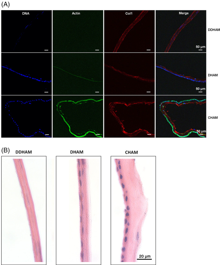
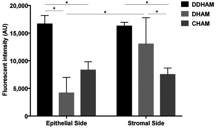
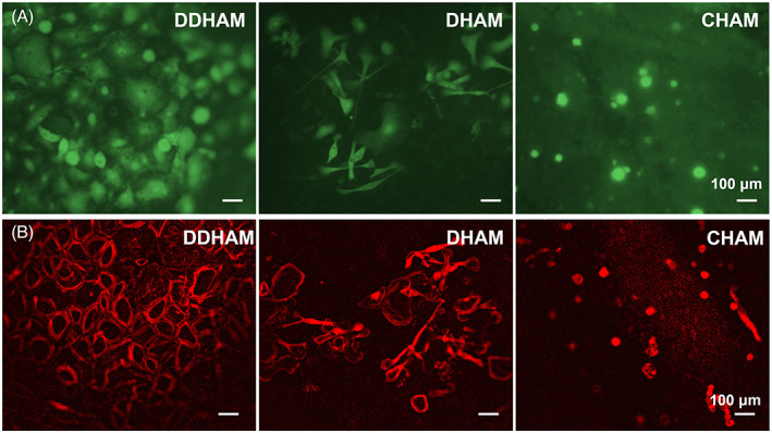
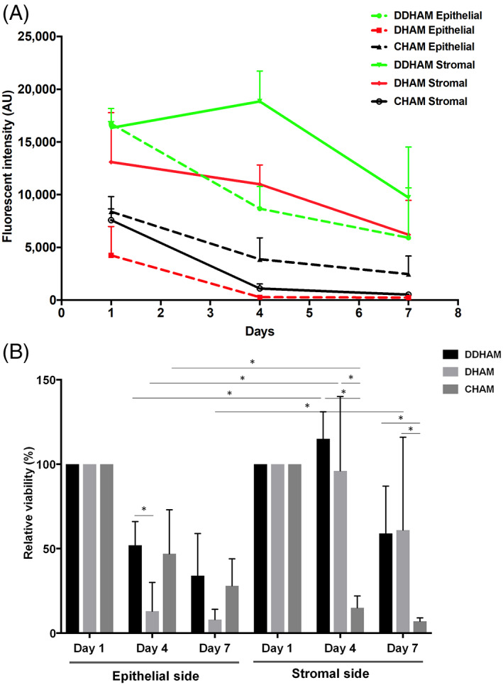
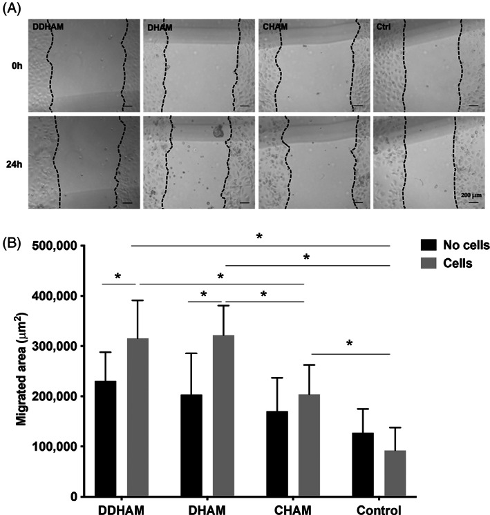
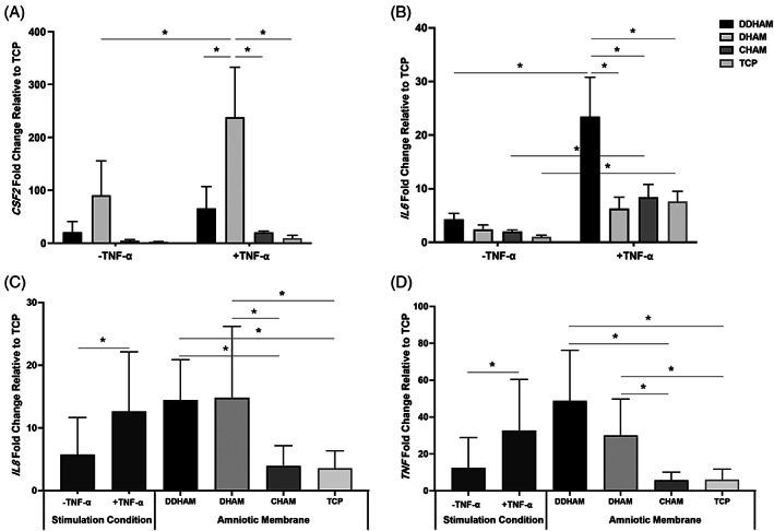
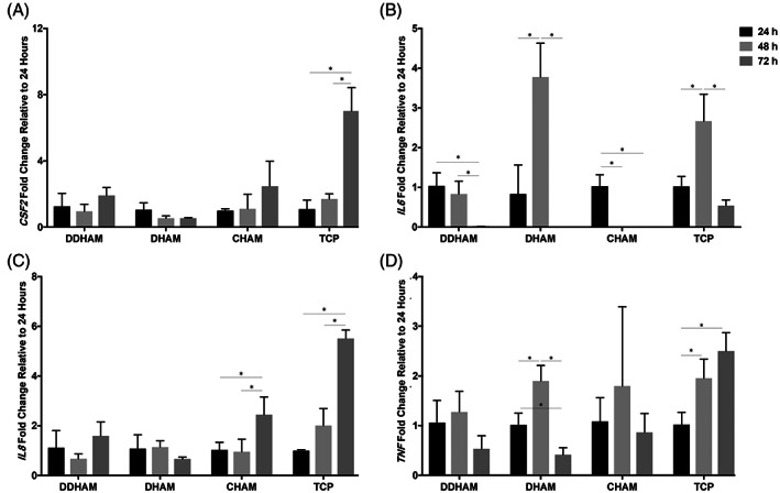
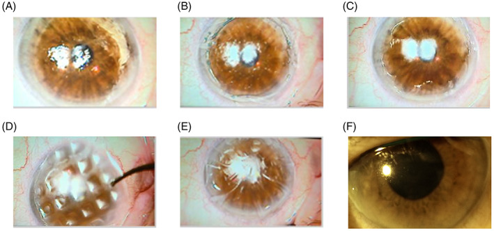
Amniotic membrane (AM) is a naturally derived biomaterial with biological and mechanical properties important to Ophthalmology. The epithelial side of the AM promotes epithelialization, while the stromal side regulates inflammation. However, not all AMs are equal. AMs undergo different processing with resultant changes in cellular content and structure. This study evaluates the effects of sidedness and processing on human corneal epithelial cell (HCEC) activity, the effect of processing on HCEC inflammatory response, and then a case study is presented. Three differently processed, commercially available ocular AMs were selected: (1) Biovance®3L Ocular, a decellularized, dehydrated human AM (DDHAM), (2) AMBIO2®, a dehydrated human AM (DHAM), and (3) AmnioGraft®, a cryopreserved human AM (CHAM). HCECs were seeded onto the AMs and incubated for 1, 4 and 7 days. Cell adhesion and viability were evaluated using alamarBlue assay. HCEC migration was evaluated using a scratch wound assay. An inflammatory response was induced by TNF-α treatment. The effect of AM on the expression of pro-inflammatory genes in HCECs was compared using quantitative polymerase chain reaction (qPCR). Staining confirmed complete decellularization and the absence of nuclei in DDHAM. HCEC activity was best supported on the stromal side of DDHAM. Under inflammatory stimulation, DDHAM promoted a higher initial inflammatory response with a declining trend across time. Clinically, DDHAM was used to successfully treat anterior basement membrane dystrophy. Compared with DHAM and CHAM, DDHAM had significant positive effects on the cellular activities of HCECs in vitro, which may suggest greater ocular cell compatibility in vivo.

Keywords: anterior basement membrane dystrophy; biomaterial; decellularized scaffold; human amniotic membrane; human ocular epithelial cells; ocular surface.


Conflict of interest statement

Adam Kuehn, Desiree Long, Raja Sivalenka, Radoslaw A. Junka, Anna Gosiewska, Stephen A. Brigido, and Robert J. Hariri are salaried employees at Celularity Inc. Anish Shah functions as a key opinion leader for Celularity Inc. Yong Mao reports a grant from Celularity Inc. during the study. Nicole M. Protzman serves as an independent contractor for Celularity Inc. and reports personal fees from Celularity Inc. during the study. Nikita John has nothing to disclose.

Figures

FIGURE 1
FIGURE 1
Staining confirms the absence of cells and nuclei in DDHAM. Immunofluorescent staining of DDHAM, DHAM, and CHAM is shown (A). The cross‐sections of the membranes were stained with Hoechst Dye (DNA in blue), phalloidin (Actin in green) and anti‐human type I collagen antibodies (Col1 in red). Representative images are shown and the scale bar = 50 μm. H&E staining (nuclei in blue and cytoplasm in red) of DDHAM, DHAM, and CHAM is shown (B). Representative images are shown and the scale bar = 20 μm. Abbreviations: CHAM, cryopreserved human amniotic membrane; DDHAM, decellularized dehydrated human amniotic membrane; DHAM, dehydrated human amniotic membrane
FIGURE 2
FIGURE 2
The epithelial and stromal sides of DDHAM best support cell adhesion. Human corneal epithelial cells were seeded onto the epithelial and stromal sides of amniotic membranes and incubated for 24 h. A two‐way analysis of variance with Tukey post‐hoc tests was conducted to evaluate the effects of sidedness and amniotic membrane on cell adhesion. Significant interactions were evaluated with simple main effects analysis with Sidak correction for multiple comparisons. Comparisons between the epithelial and stromal sides of each amniotic membrane are shown, and comparisons between amniotic membranes for each side are shown. Fluorescent intensity is expressed in arbitrary units (AU). Data shown are mean ± SD (n = 4). *p ≤ .05. Abbreviations: CHAM, cryopreserved human amniotic membrane; DDHAM, decellularized dehydrated human amniotic membrane; DHAM, dehydrated human amniotic membrane
FIGURE 3
FIGURE 3
Staining confirms human corneal epithelial cell viability and morphology on the stromal side of DDHAM and DHAM on day 4. Human corneal epithelial cells were seeded onto the stromal side of the three amniotic membranes, cultured, and stained with Calcein AM to visualize live cells at day 4 (A). The morphology of human corneal epithelial cells on amniotic membranes was monitored by Actin staining on day 4 and pseudo‐colored red (B). Images were captured using epi‐fluorescent microscope. Scale bar = 100 μm. Abbreviations: CHAM, cryopreserved human amniotic membrane; DDHAM, decellularized dehydrated human amniotic membrane; DHAM, dehydrated human amniotic membrane
FIGURE 4
FIGURE 4
Stromal side of DDHAM best supports human corneal epithelial cell viability over time. Human corneal epithelial cells were seeded onto the epithelial and stromal sides of amniotic membranes and incubated for 1, 4 and 7 days. Absolute cell viability is plotted over time for each side of the amniotic membranes (A). The relative cell viability, expressed as a percentage of day 1, is plotted over time for each side of the amniotic membranes (B). A three‐way analysis of variance with Tukey post hoc tests was conducted to examine the effects of sidedness, amniotic membrane, and time on relative cell viability. Significant interactions were evaluated with simple main effects analysis with Sidak correction for multiple comparisons. Comparisons between the epithelial and stromal sides of each amniotic membrane are shown and comparisons between the amniotic membranes for each side are shown. Data shown are mean ± SD (n = 4). *p ≤ .05. Abbreviations: CHAM, cryopreserved human amniotic membrane; DDHAM, decellularized dehydrated human amniotic membrane; DHAM, dehydrated human amniotic membrane
FIGURE 5
FIGURE 5
Migration is greatest on DDHAM and DHAM in the presence of human corneal epithelial cells. Representative scratch wound images are shown to demonstrate the effects of conditioned media on the migration of human corneal epithelial cells at 0 and 24 h. Scale bar = 200 μm (A). The conditioned media from different amniotic membranes (with and without cells) were tested to evaluate the effect of amniotic membranes alone on the migration of human corneal epithelial cells (B). A two‐way analysis of variance with Tukey post‐hoc tests was conducted to evaluate the effects of cell presence and amniotic membrane on cell migration. Significant interactions were evaluated with simple main effects analysis with Sidak correction for multiple comparisons. The wound areas (μm2) were measured using Image J. The migrated area = Area0h − Area24h. Data shown are mean ± SD (n = 4). *p ≤ .05. Abbreviations: CHAM, cryopreserved human amniotic membrane; DDHAM, decellularized dehydrated human amniotic membrane; DHAM, dehydrated human amniotic membrane; Medium Ctrl, medium control
FIGURE 6
FIGURE 6
DDHAM and DHAM support an inflammatory response at 24 h. Relative mRNA expressions of CSF2 (A), IL6 (B), IL8 (C), and TNF (D) at 24 h are shown. Relative mRNA expressions at 24 h are normalized to tissue culture treated plastic in the resting condition. A two‐way analysis of variance with Tukey post‐hoc tests was conducted to evaluate the effects of stimulation condition and amniotic membrane on mRNA expressions at 24 h. Significant interactions were evaluated with simple main effects analysis with Sidak correction for multiple comparisons. Data shown are mean ± SD (n = 3). *p ≤ .05. Abbreviations: CHAM, cryopreserved human amniotic membrane; CSF2, colony‐stimulating factor 2; DDHAM, decellularized dehydrated human amniotic membrane; DHAM, dehydrated human amniotic membrane; IL6, interleukin 6; IL8, interleukin 8; TCP, tissue culture treated plastic; TNF, tumor necrosis factor
FIGURE 7
FIGURE 7
DDHAM supports a declining inflammatory response across time. Relative mRNA expressions of CSF2 (A), IL6 (B), IL8 (C), and TNF (D) across time in the stimulated condition (+TNF‐α) are shown. Relative mRNA expressions across time are normalized to expression at 24 h. A one‐way analysis of variance with Tukey post‐hoc tests was conducted to examine the effect of time on mRNA expressions. Statistical comparisons are between time points for each amniotic membrane in the stimulated condition (+TNF‐α). Data shown are mean ± SD (n = 3). *p ≤ .05. Abbreviations: CHAM, cryopreserved human amniotic membrane; CSF2, colony‐stimulating factor 2; DDHAM, decellularized dehydrated human amniotic membrane; DHAM, dehydrated human amniotic membrane; IL6, interleukin 6; IL8, interleukin 8; TCP, tissue culture treated plastic; TNF, tumor necrosis factor
FIGURE 8
FIGURE 8
Successful clinical application of DDHAM to treat a case of anterior basement membrane dystrophy. Images of the epithelial surface were taken to illustrate the clinical course: pre‐operatively, showing the poor irregular surface of the epithelium (A), post removal of poor epithelium with visible sub epithelial debris from Anterior Basement Membrane Dystrophy (B), post burring of all sub‐epithelial scarring and Anterior Basement Membrane Dystrophy debris (C), placement of DDHAM (D), placement of bandage contact lens over DDHAM (E), and two months postoperatively, showing a clear surface (F)

Similar articles

Cited by

References

    1. Leal‐Marin S, Kern T, Hofmann N, et al. Human amniotic membrane: a review on tissue engineering, application, and storage. J Biomed Mater Res B Appl Biomater. 2021;109:1198‐1215. - PubMed
    1. Liu J, Li L, Li X. Effectiveness of cryopreserved amniotic membrane transplantation in corneal ulceration: a meta‐analysis. Cornea. 2019;38:454‐462. - PubMed
    1. Malhotra C, Jain AK. Human amniotic membrane transplantation: different modalities of its use in ophthalmology. World J Transplant. 2014;4:111‐121. - PMC - PubMed
    1. Fernandes M, Sridhar MS, Sangwan VS, Rao GN. Amniotic membrane transplantation for ocular surface reconstruction. Cornea. 2005;24:643‐653. - PubMed
    1. Walkden A. Amniotic membrane transplantation in ophthalmology: an updated perspective. Clin Ophthalmol. 2020;14:2057‐2072. - PMC - PubMed

Publication types