. 2022 Dec:175:105923.
doi: 10.1016/j.nbd.2022.105923. Epub 2022 Nov 10.

Neonatal ketamine exposure impairs infrapyramidal bundle pruning and causes lasting increase in excitatory synaptic transmission in hippocampal CA3 neurons

Affiliations

Neonatal ketamine exposure impairs infrapyramidal bundle pruning and causes lasting increase in excitatory synaptic transmission in hippocampal CA3 neurons

Omar Hoseá Cabrera et al. Neurobiol Dis. 2022 Dec.

Abstract

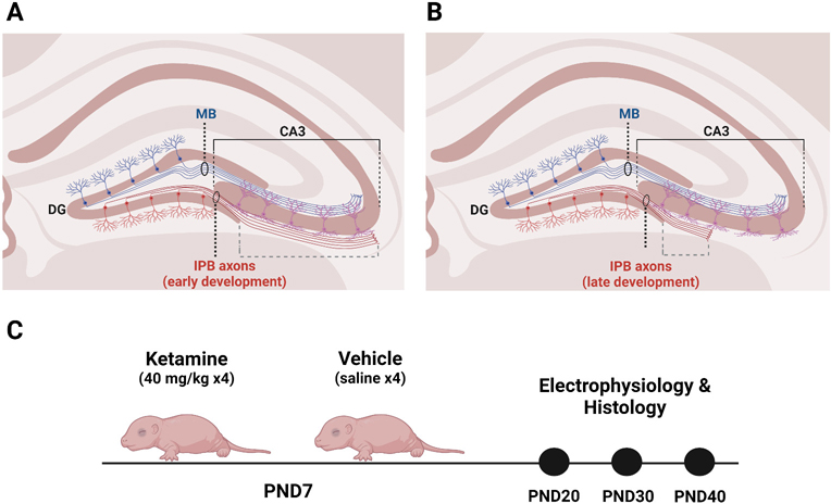
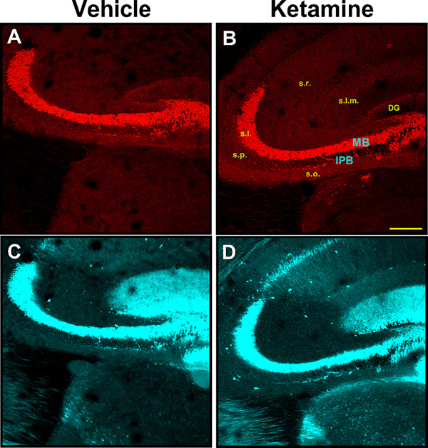
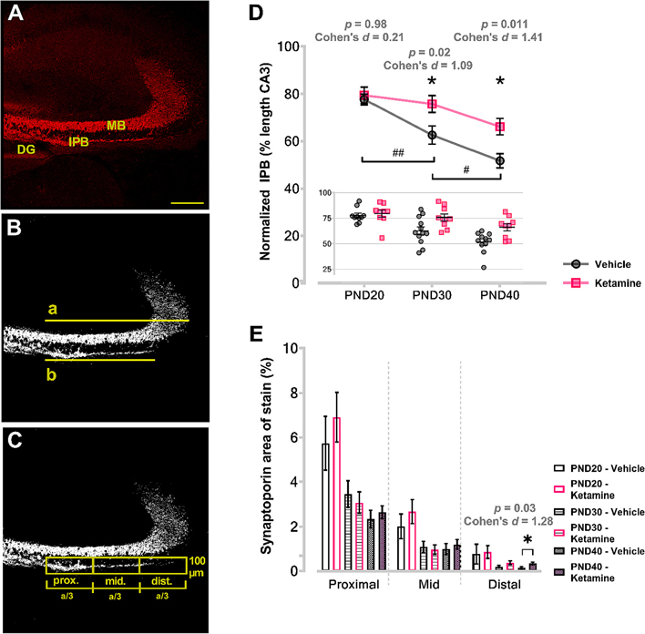
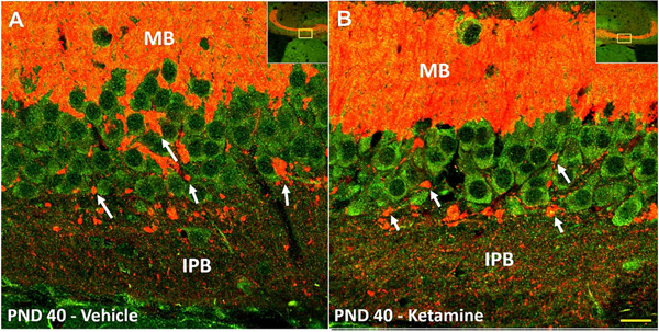
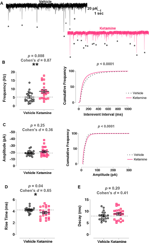
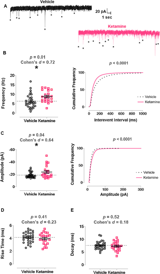
Preclinical models demonstrate that nearly all anesthetics cause widespread neuroapoptosis in the developing brains of infant rodents and non-human primates. Anesthesia-induced developmental apoptosis is succeeded by prolonged neuropathology in the surviving neurons and lasting cognitive impairments, suggesting that anesthetics interfere with the normal developmental trajectory of the brain. However, little is known about effects of anesthetics on stereotyped axonal pruning, an important developmental algorithm that sculpts neural circuits for proper function. Here, we proposed that neonatal ketamine exposure may interfere with stereotyped axonal pruning of the infrapyramidal bundle (IPB) of the hippocampal mossy fiber system and that impaired pruning may be associated with alterations in the synaptic transmission of CA3 neurons. To test this hypothesis, we injected postnatal day 7 (PND7) mouse pups with ketamine or vehicle over 6 h and then studied them at different developmental stages corresponding to IPB pruning (PND20-40). Immunohistochemistry with synaptoporin (a marker of mossy fibers) revealed that in juvenile mice treated with ketamine at PND7, but not in vehicle-treated controls, positive IPB fibers extended farther into the stratum pyramidale of CA3 region. Furthermore, immunofluorescent double labeling for synaptoporin and PSD-95 strongly suggested that the unpruned IPB caused by neonatal ketamine exposure makes functional synapses. Importantly, patch-clamp electrophysiology for miniature excitatory postsynaptic currents (mEPSCs) in acute brain slices ex vivo revealed increased frequency and amplitudes of mEPSCs in hippocampal CA3 neurons in ketamine-treated groups when compared to vehicle controls. We conclude that neonatal ketamine exposure interferes with normal neural circuit development and that this interference leads to lasting increase in excitatory synaptic transmission in hippocampus.

Keywords: Glutamate; Neonate; Neurotoxicity; Neurotransmission; hippocampus.


Conflict of interest statement

Declaration of Competing Interest The authors declare that the research was conducted in the absence of any commercial or financial relationships that could be construed as a potential conflict of interest.

Figures

Fig. 1.
Fig. 1.. Diagrams of stereotyped axonal pruning in the IPB of hippocampus and of experimental design.
A. In early rodent postnatal development, the IPB of the mossy fiber system arises from the axons of DG granule neurons. IPB fibers send axon collaterals that innervate the basal dendrites of neurons in CA3. B. Starting around PND20, the IPB undergoes stereotyped axonal pruning that continues until at least PND40. IPB axons distal to DG are pruned hundreds of microns towards source neurons in DG. When the pruning window closes, the IPB is short, innervating neurons more proximal to the DG. C. PND7 mice were injected with 40 mg/kg ketamine or vehicle then subjected to electrophysiological and histological analyses at three developmental stages coinciding with the window of IPB pruning early pruning (PND20), active pruning (PND30), and late pruning (PND40). Normalized IPB length was the measure of IPB pruning and calculated as the length of the IPB (dashed gray bracket in A. and B.) divided by the length of CA3 (solid black bracket in A and B). Created with BioRender.com. DG, dentate gyrus; IPB, infrapyramidal bundle; MB, main bundle; PND, postnatal day.
Fig. 2.
Fig. 2.. Dual immunofluorescence staining indicates that synaptoporin more specifically labels mossy fiber system in mouse hippocampus compared to calbindin.
Dual immunofluorescence staining of synaptoporin (top panels – red) and calbindin (bottom panels – cyan) of representative PND40 animals following neonatal administration of vehicle (A, C) or ketamine (B, D). Synaptoporin brightly stained mossy fibers of IPB and MB in stratum oriens and stratum lucidum, respectively; other areas were virtually devoid of the stain. Conversely, calbindin more broadly labeled hippocampal structures, extending into the stratum radiatum, stratum lacunosum-moleculare and dentate gyrus, in addition to areas that were also positive for synaptoporin. Virtually all synaptoporin-labeled structures showed almost complete colocalization of calbindin-stain; the reverse was not true. In all cases, IPB is readily observable as a bundle of fibers emanating from the tip of the inferior blade of the dentate gyrus, traversing the stratum oriens beneath the main bundle. Scale bar – 200 μm. (For interpretation of the references to colour in this figure legend, the reader is referred to the web version of this article.) DG, dentate gyrus; IPB, infrapyramidal bundle; MB, main bundle; s.l.m., stratum lacunosum-moleculare; s.l., stratum lucidum; s.o., stratum oriens; s.p., stratum pyramidale; s.r. stratum radiatum.
Fig. 3.
Fig. 3.. Neonatal ketamine treatment impairs stereotyped axonal pruning in hippocampus.
Left panels show examples of measurements performed on a PND20 ketamine-treated animal. Panel A represents compressed 12 μM thickness stack of synaptoporin-stained coronal hippocampal section obtained with 10× objective and very high resolution (2048 × 2048). The compressed stack was thresholded and size-filtered for particles <0.5 μM diameter to eliminate the background noise (B). Measurements of CA3 MB (a) and IPB length (b) were then taken from the tip of the inferior blade of the DG, and normalized IPB ratio was obtained by dividing the two measurements (b/a). Next, the area that IPB traverses was divided into three rectangular areas (C) spanning exactly the distance measured with “a”. The long axis of the rectangle equaled one-third of the MB length (a/3), whereas the short axis was constant and set at 100 μm for all images. The results were expressed as percent area positive for synaptoporin. Analysis of normalized IPB length (D) revealed that animals subjected to ketamine as neonates had significantly longer IPB at both PND30 and PND40 compared to vehicle counterparts (asterisks). Furthermore, IPB lengths between consecutive timepoints were significantly different in vehicle controls (pounds); in ketamine, they were not. Further analysis of IPB pruning revealed that percent area positive for synaptoporin immunolabeling was significantly higher in the most distal portions relative to DG in PND40 ketamine-treated animals compared to age and area-matched vehicle counterparts (E), indicating that distal sections of IPB were most affected by the process of stereotyped pruning. Scale bar – 200 μm. DG, dentate gyrus; IPB, infrapyramidal bundle; MB, main bundle; PND, postnatal day.
Fig. 4.
Fig. 4.. Remaining IPB fibers likely make functional synapses with pyramidal layer neurons at PND40.
Immunofluorescence co-labeling of PSD-95 (green) and synaptoporin (red) in PND40 animal following neonatal vehicle (left panel) or ketamine (right panel), visualized with 60× objective. Multiple collaterals were observed emanating from the remaining IPB fibers as they migrate towards the MB. As these collaterals crossed the pyramidal layer, multiple large nodules (arrows) were detected in close proximity to PSD-95 positive pyramidal neurons, possibly en passant synapses, and also numerous small puncta around cell soma and basal dendrites. These histomorphological features of putative connections are indicative of functional synaptic connections between the presynaptic IPB terminals and CA3 pyramidal neurons. Insert is the 10× magnification image showing the location from which the 60× images were obtained. Scale bar – 20 μm. (For interpretation of the references to colour in this figure legend, the reader is referred to the web version of this article.) IPB, infrapyramidal bundle; MB, main bundle; PND, postnatal day.
Fig. 5.
Fig. 5.. Neonatal ketamine exposure increases excitatory synaptic transmission in CA3 at PND20.
A. Left panel shows representative trace of mEPSCs recorded from a juvenile mouse treated with vehicle as a neonate. Right panel displays representative trace of mEPSCs recorded from a mouse treated with ketamine as a neonate. Asterisks mark mEPSCs. B. Mice treated with ketamine as neonates (n = 9 mice, n = 19 neurons) had significantly increased frequency of mEPSCs in CA3 neurons compared to those treated with vehicle (n = 8 mice, n = 22 neurons) (left panel) and a significant leftward shift in the cumulative frequency of the interevent interval (right panel). C. There was no change in the average amplitudes of mEPSCs between groups (left panel), but there was a significant rightward shift in cumulative frequency of mEPSC amplitude (right panel). D. Neonatal ketamine also significantly shortened rise time of mEPSC events compared to vehicle. E. There was no change in decay of mEPSCs between groups. mEPSC, miniature excitatory postsynaptic current; PND, postnatal day.
Fig. 6.
Fig. 6.. Neonatal ketamine exposure significantly increased frequency and amplitudes of mEPSCs at later (PND30 and PND40) developmental stages.
A. Left panel shows representative trace of mEPSCs recorded from a vehicle treated mouse in later juvenile development. Right panel shows representative trace of mEPSCs from a mouse treated with ketamine as a neonate. Asterisks mark mEPSCs. B. Mice treated with ketamine as neonates (n = 12 mice, n = 22 neurons) had higher average frequency of mEPSCs compared to mice that received vehicle (n = 11 mice, n = 30 neurons) (left panel) and a leftward shift in mEPSC interevent interval (right panel). C. Neonatal ketamine caused an increase in average amplitude of mEPSCs compared to vehicle (left panel) and a rightward shift in cumulative frequency of mEPSC amplitude (right panel). D. There was no change in mEPSC rise time between groups. E. There was no change in mEPSC decay between groups. mEPSC, miniature excitatory postsynaptic current; PND, postnatal day.

References

    1. Amaral DG, Scharfman HE, Lavenex P, 2007. The dentate gyrus: fundamental neuroanatomical organization (dentate gyrus for dummies). Prog. Brain Res. 163. - PMC - PubMed
    1. Andreae LC, Burrone J, 2018. The role of spontaneous neurotransmission in synapse and circuit development. J. Neurosci. Res. 96, 354–359. Available at: https://onlinelibrary.wiley.com/doi/full/10.1002/jnr.24154 [Accessed December 28, 2021]. - DOI - PMC - PubMed
    1. Bagri A, Cheng HJ, Yaron A, Pleasure SJ, Tessier-Lavigne M, 2003. Stereotyped pruning of long hippocampal axon branches triggered by retraction inducers of the semaphorin family. Cell 113, 285–299. - PubMed
    1. Bischofberger J, Engel D, Frotscher M, Jonas P, 2006a. Timing and efficacy of transmitter release at mossy fiber synapses in the hippocampal network. Pflugers Arch. - Eur. J. Physiol. 453, 361–372. Available at: https://link.springer.com/article/10.1007/s00424-006-0093-2 [Accessed December 28, 2021]. - DOI - PubMed
    1. Bischofberger J, Engel D, Li L, Geiger JRP, Jonas P, 2006b. Patch-clamp recording from mossy fiber terminals in hippocampal slices. Nat. Protoc. 1, 2075–2081. - PubMed

Publication types