. 2022 Dec:86:104343.
doi: 10.1016/j.ebiom.2022.104343. Epub 2022 Nov 11.

Comprehensive genetic and functional analyses of Fc gamma receptors influence on response to rituximab therapy for autoimmunity

Affiliations

Comprehensive genetic and functional analyses of Fc gamma receptors influence on response to rituximab therapy for autoimmunity

James I Robinson et al. EBioMedicine. 2022 Dec.

Abstract

Background: Rituximab is widely used to treat autoimmunity but clinical response varies. Efficacy is determined by the efficiency of B-cell depletion, which may depend on various Fc gamma receptor (FcγR)-dependent mechanisms. Study of FcγR is challenging due to the complexity of the FCGR genetic locus. We sought to assess the effect of FCGR variants on clinical response, B-cell depletion and NK-cell-mediated killing in rheumatoid arthritis (RA) and systemic lupus erythematosus (SLE).

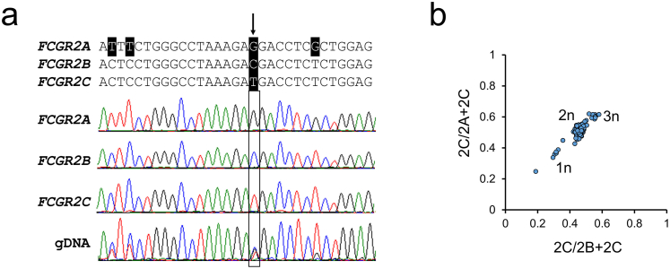
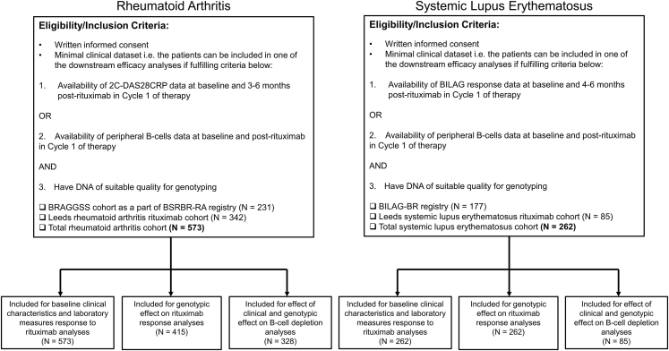
Methods: A longitudinal cohort study was conducted in 835 patients [RA = 573; SLE = 262]. Clinical outcome measures were two-component disease activity score in 28-joints (2C-DAS28CRP) for RA and British Isles Lupus Assessment Group (BILAG)-2004 major clinical response (MCR) for SLE at 6 months. B-cells were evaluated by highly-sensitive flow cytometry. Single nucleotide polymorphism and copy number variation for genes encoding five FcγRs were measured using multiplex ligation-dependent probe amplification. Ex vivo studies assessed NK-cell antibody-dependent cellular cytotoxicity (ADCC) and FcγR expression.

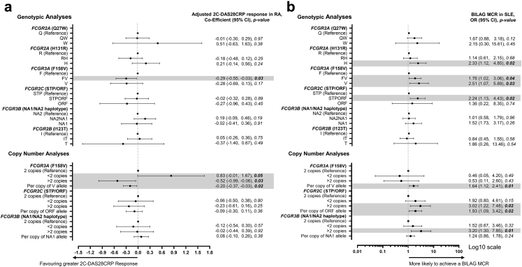
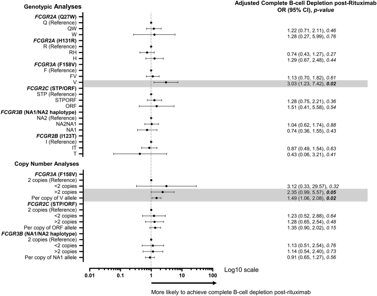
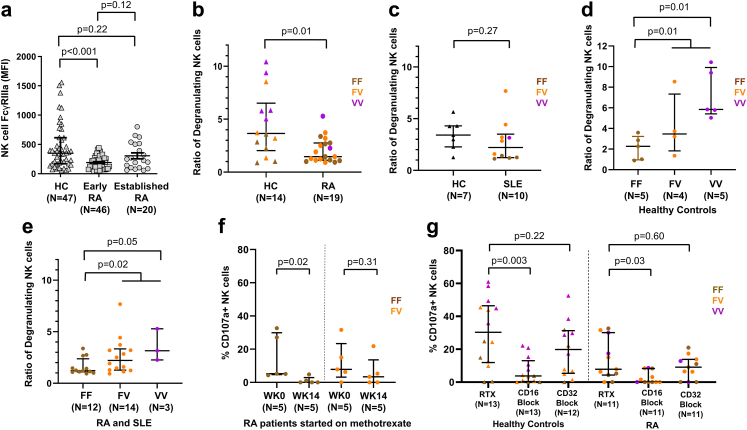
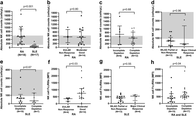
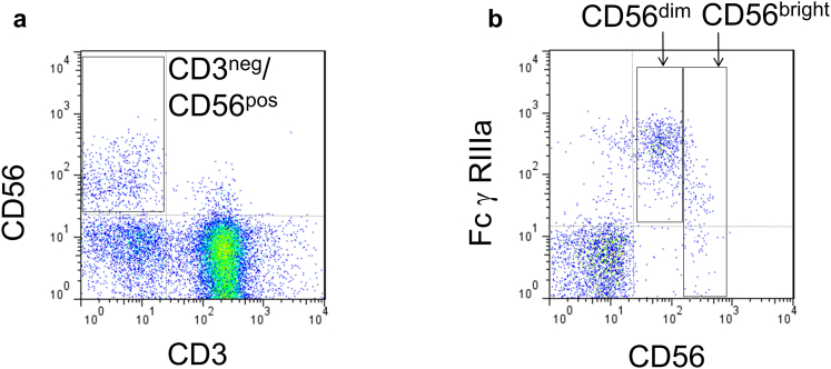
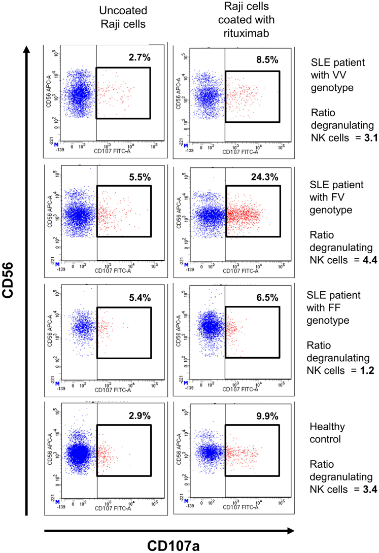
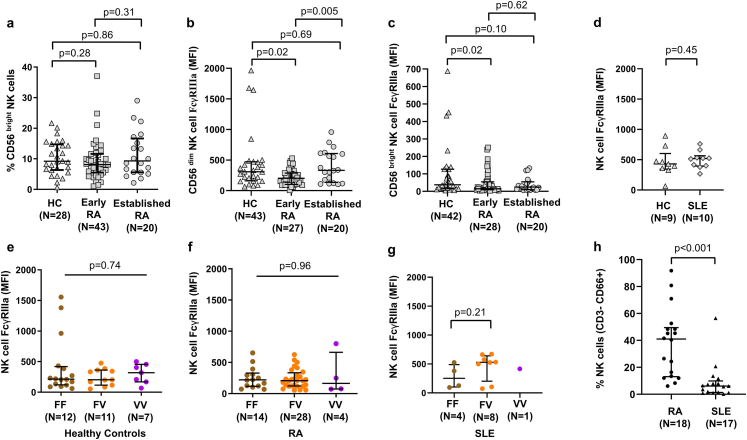
Findings: In RA, carriage of FCGR3A-158V and increased FCGR3A-158V copies were associated with greater 2C-DAS28CRP response (adjusted for baseline 2C-DAS28CRP). In SLE, MCR was associated with increased FCGR3A-158V, OR 1.64 (95% CI 1.12-2.41) and FCGR2C-ORF OR 1.93 (95% CI 1.09-3.40) copies. 236/413 (57%) patients with B-cell data achieved complete depletion. Homozygosity for FCGR3A-158V and increased FCGR3A-158V copies were associated with complete depletion in combined analyses. FCGR3A genotype was associated with rituximab-induced ADCC, and increased NK-cell FcγRIIIa expression was associated with improved clinical response and depletion in vivo. Furthermore, disease status and concomitant therapies impacted both NK-cell FcγRIIIa expression and ADCC.

Interpretation: FcγRIIIa is the major low affinity FcγR associated with rituximab response. Increased copies of the FCGR3A-158V allele (higher affinity for IgG1), influences clinical and biological responses to rituximab in autoimmunity. Enhancing FcγR-effector functions could improve the next generation of CD20-depleting therapies and genotyping may stratify patients for optimal treatment protocols.

Funding: Medical Research Council, National Institute for Health and Care Research, Versus Arthritis.

Keywords: Autoimmune diseases; B-lymphocytes; Genetics; Rheumatoid arthritis; Rituximab; Systemic lupus erythematosus.


Conflict of interest statement

Declaration of interests Prof Bruce has received research grants from GSK, consulting fees from GSK, UCB, Eli Lilly & Co, BMS, Aurinia, IL-TOO and AstraZeneca and speaker fees from AstraZeneca, GSK and UCB within the last 3 years. Dr Vital has received honoraria and consulting fees from Roche within the last 3 years. All other authors declare no competing interest related to the work described in this manuscript.

Figures

Fig. 1
Fig. 1
FCGR2C QSV assay is associated with CD32 expression on NK-cells.(a) Schematic of the two main copy number regions (CNR1 and 2) and the relative positions of the functionally relevant nonsynonymous variants affecting FCGR3A and FCGR2C. (b) Expression of CD32 on NK-cells in treatment-naïve, early RA patients (n = 59) stratified by FCGR2C open reading frame (ORF) copy number. Data were summarised as median and the error bars denoted interquartile range. Using our FCGR2C QSV assay (Supplementary Fig. S1), in combination with multiplex ligation dependent assay probe values for rs10917661, copies of FCGR2C-ORF were genotyped where matched NK-cell CD32 (clone KB61) expression data were available. It was not possible to distinguish classical and non-classical FCGR2C-ORF in our study. The latter variant also contains a premature STP codon, precluding FcγRIIc translation. A particular locus rearrangement including a deletion of one copy of FCGR2C and one copy of FCGR3B (CNR1 del), has been described to lead to expression of FcγRIIb on NK-cells. The rearrangement was observed in conjunction with FCGR2C-STP in 5/6 individuals, where we would anticipate FcγRIIc would be the only class II FcγR expressed. (c) The relationship between % positive CD32 expression on NK-cells, and geometric mean fluorescence intensity (MFI) in treatment-naïve, early RA patients (n = 59). P-values calculated using non-parametric Mann–Whitney test. Data are summarised using median and interquartile range.
Fig. 2
Fig. 2
Association of FCGR genotype and copy number with clinical response in RA and SLE.(a) Co-efficient, 95% confidence intervals (CI) and p-value for the effect of the indicated genotype or copy number on 2C-DAS28CRP response (adjusted for baseline 2C-DAS28CRP) at 6 months post-rituximab, compared with reference genotype. All tests were performed using univariable linear regression; negative coefficients for clinical response outcomes indicate a favourable outcome. (b) Odds ratio (OR), 95% CI and p-value for the effect of the indicated genotype or copy number on BILAG Major Clinical Response (MCR) at 6 months post-rituximab, compared with reference genotype. All tests were performed using logistic regression. The x-axis was transformed to log10 scale. For both figures, the dots represent either the co-efficient or the OR and the error bars denote the 95% CI. The vertical broken lines denote lines of no effect.
Fig. 3
Fig. 3
Association of FCGR genotype and copy number with complete B-cell depletion. Odds ratio (OR), 95% confidence intervals (CI) and p-value for the effect of the indicated genotype or copy number on complete B-cell depletion post-rituximab, compared with reference genotype. The x-axis was transformed to log10 scale. The dots represent the OR and the error bars denote the 95% CI. All tests were performed using logistic regression, adjusted for age, concomitant disease-modifying anti-rheumatic drug use including hydroxychloroquine, and baseline plasmablast count.
Fig. 4
Fig. 4
Effect of disease, genotype and methotrexate on NK-cell degranulation.(a) Natural Killer (NK)-cell (CD3-CD56+CD16+) FcγRIIIa (CD16; clone 3G8) geometric mean fluorescence intensity (MFI) using flow cytometry for healthy controls (HC) (n = 47), early (symptom onset <1 year and treatment naïve) (n = 46) and established (>2 years) rheumatoid arthritis (RA) (n = 20). Comparison of NK-cell degranulation following incubation with B-cell lineage, Daudi cells between HC (n = 14) and RA (n = 19) (b) and Raji cells between HC (n = 7) and systemic lupus erythematosus (SLE) (n = 10) (c) using an effector:target (E:T) ratio of 1:1 and rituximab. Ratio of degranulating NK-cells were compared in individuals with two copies of FCGR3A between the three F158V genotypes in HC (n = 14) (d) and RA and SLE combined (n = 29) (e). (f) NK-cell degranulation (%CD107a positive NK-cells) was compared before and 14 weeks after RA patients (n = 10) started on methotrexate according to their FCGR3A genotype. (g) %CD107a positive NK-cells was assessed after incubation with rituximab and subsequent inclusion of CD16 (clone 3G8) and CD32 (clone KB61) blocking antibodies in HC (n = 13) and RA (n = 11). All p-values calculated using non-parametric Mann–Whitney test. Data were summarised as median and the error bars denoted interquartile range.
Fig. 5
Fig. 5
Peripheral blood NK-cell abundance and FcγRIIIa expression in rituximab-treated RA and SLE patients. Comparison of absolute natural killer (NK)-cell (CD3-CD56+) counts between (a) rituximab-treated rheumatoid arthritis (RA) (n = 18) and systemic lupus erythematosus (SLE) (n = 17) patients; (b) rituximab-treated RA patients exhibiting no European League Against Rheumatism (EULAR) clinical response (n = 4) and moderate/good clinical response (n = 14); (c) rituximab-treated RA patients exhibiting incomplete (n = 6) and complete B-cell depletion (n = 12); (d) rituximab-treated SLE patients exhibiting British Isles Lupus Assessment Group (BILAG) partial clinical response or no clinical response (n = 13) and major clinical response (n = 4); and (e) rituximab-treated SLE patients with incomplete (n = 10) and complete B-cell depletion (n = 7). The shaded grey areas represent adult reference ranges of the absolute NK-cell counts (90–600 cells/μL). Expression of FcγRIIIa (CD16; clone 3G8) on NK-cells of (f) RA patients exhibiting EULAR non-response (n = 4) and moderate/good clinical response (n = 14) to rituximab; (g) SLE patients exhibiting BILAG partial clinical response/non-response (n = 13) and major clinical response (n = 4) to rituximab; and (h) RA and SLE patients exhibiting incomplete (n = 15) and complete B-cell depletion (n = 17) in response to rituximab. All p-values calculated using non-parametric Mann–Whitney test. Data were summarised as median and the error bars denoted interquartile range.
None
Fig. S1
None
Fig. S2
Fig. S3
Fig. S3
Fig. S4
Fig. S4
Fig. S5
Fig. S5

References

    1. Buch M.H., Smolen J.S., Betteridge N., et al. Updated consensus statement on the use of rituximab in patients with rheumatoid arthritis. Ann Rheum Dis. 2011;70(6):909–920. - PMC - PubMed
    1. Garcia-Montoya L., Villota-Eraso C., Yusof M.Y.M., Vital E.M., Emery P. Lessons for rituximab therapy in patients with rheumatoid arthritis. Lancet Rheumatol. 2020;2(8):e497–e509. - PubMed
    1. Fanouriakis A., Kostopoulou M., Alunno A., et al. 2019 Update of the EULAR recommendations for the management of systemic lupus erythematosus. Ann Rheum Dis. 2019;78(6):736–745. - PubMed
    1. Ramos-Casals M., Soto M.J., Cuadrado M.J., Khamashta M.A. Rituximab in systemic lupus erythematosus: a systematic review of off-label use in 188 cases. Lupus. 2009;18(9):767–776. - PubMed
    1. Dass S., Rawstron A.C., Vital E.M., Henshaw K., McGonagle D., Emery P. Highly sensitive B cell analysis predicts response to rituximab therapy in rheumatoid arthritis. Arthritis Rheum. 2008;58(10):2993–2999. - PubMed

MeSH terms