. 2023 Jan:226:109306.
doi: 10.1016/j.exer.2022.109306. Epub 2022 Nov 11.

A tamoxifen-inducible Cre knock-in mouse for lens-specific gene manipulation

Affiliations

A tamoxifen-inducible Cre knock-in mouse for lens-specific gene manipulation

Zongbo Wei et al. Exp Eye Res. 2023 Jan.

Abstract

Mouse models are valuable tools in studying lens biology and biochemistry, and the Cre-loxP system is the most used technology for gene targeting in the lens. However, numerous genes are indispensable in lens development. The conventional knockout method either prevents lens formation or causes simultaneous cataract formation, hindering the studies of their roles in lens structure, growth, metabolism, and cataractogenesis during lens aging. An inducible Cre-loxP mouse line is an excellent way to achieve such a purpose. We established a lens-specific Cre ERT2 knock-in mouse (LCEK), an inducible mouse model for lens-specific gene targeting in a spatiotemporal manner. LCEK mice were created by in-frame infusion of a P2A-CreERT2 at the C-terminus of the last coding exon of the gene alpha A crystallin (Cryaa). LCEK mice express tamoxifen-inducible Cre recombinase uniquely in the lens. Through ROSAmT/mG and two endogenous genes (Gclc and Rbpj) targeting, we found no Cre recombinase leakage in the lens epithelium, but 50-80% leakage was observed in the lens cortex and nucleus. Administration of tamoxifen almost completely abolished target gene expression in both lens epithelium and cortex but only mildly enhanced gene deletion in the lens nucleus. Notably, no overt leakage of Cre activity was detected in developing LCEK lens when bred with mice carrying loxP floxed genes that are essential for lens development. This newly generated LCEK line will be a powerful tool to target genes in the lens for gene functions study in lens aging, posterior capsule opacification (PCO), and other areas requiring precision gene targeting.


Conflict of interest statement

Declaration of competing interest ZW, CH, JC, LG, and XF declare they have no conflict of interest.

Figures
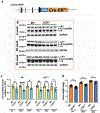
Figure 1.
Figure 1.
LCEK knock-in mouse design and αA crystallin expression. (A) Cyaa-2A-CreERT2 knock-in allele design. (B) Lens αA crystallin protein expression at 9, 20, and 28 weeks of age. The lower band close to the 15KD marker is native αA crystallin, and the upper band close to 25KD is the αA crystallin insertion isoform. GAPDH serves as a loading control. (C) Semi-quantitative results of αA crystallin protein expression based on densitometry (n=4). (D) Lens wet weight (n≥8). Results are expressed as mean ± SD. αA crystallin expression was analyzed using one-way ANOVA with Sidak’s multiple comparison test, and lens wet weight was analyzed using unpaired two-tailed Student’s t-test. Only p<0.05 is considered significant. *<0.05, **<0.01, ***<0.001, ****<0.0001, n.s, not significant.
Figure 2.
Figure 2.
LCEK knock-in mouse and WT mice lens phenotypic exam. (A) Slit lamp image of WT and heterozygous LCEK mice at 2, 4, 6, and 12 months of age. (B) Darkfield image of WT and heterozygous LCEK mice lenses at 2, 4, 6, and 12 months of age. (C) H&E staining of WT and heterozygous LCEK mice lenses at 2, 4, 6, and 12 months of age. N=5 in each group.
Figure 3.
Figure 3.
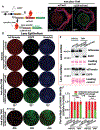
Cre recombinase expression patterns and induction efficiency in ROSAmT/mG/LCEK+/− mice. (A) Breeding strategy for ROSAmT/mG/LCEK+/− production. (B, C) Four weeks after tamoxifen induction, Cre-loxP recombination manifested with EGFP expression only occurs in the lens. (B) mT/mG mice, (C) LCEK+/−/mT/mG mice. (D) Lens epithelium Cre-loxP recombination with or without tamoxifen induction. Upper panel: mT/mG and LCEK+/−/mT/mG mice lens epithelia without tamoxifen induction. Lower panel: mT/mG and LCEK+/−/mT/mG mice lens epithelia with tamoxifen induction (4 weeks). EGFP was only seen in tamoxifen-induced epithelia. Hoechst 33342 was used for nuclei staining. (E) EGFP and tdTomato protein expression in lens cortex and nucleus with and without tamoxifen induction. Ponceau S staining served as a loading control. (F) The ratio between EGFP and tdTomato proteins in the lens cortex and nucleus. The protein level was determined by semi-quantitative measurement of protein immuno-blot densitometry from (E) images. N=6 in each group. TAM: tamoxifen. Results are expressed as mean ± SD. Two-Way ANOVA with Sidak’s multiple comparison test was used to compare each group in (F). Only p<0.05 is considered significant. *<0.05, **<0.01, ***<0.001, ****<0.0001, n.s, not significant.
Figure 4.
Figure 4.
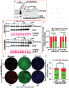
Age-related changes of Cre recombinase expression and induction efficiency in ROSAmT/mG/LCEK+/− mice. (A) Lens epithelium has no Cre recombinase leakage in 1- and 5-months old mice without tamoxifen induction. tdTomato was measured by immuno-blot assay, and GAPDH served as a loading control. Lane 1: mT/mG/Cre lens fiber homogenate without tamoxifen induction. We found several tdTomato bands from fiber homogenate (also see in Fig 3F); Lane 2: Mty lane: empty lane (avoid spillover from lane 1); Lane 3: mT/mG lens epithelium; lanes 4-7 are 1-month-old mT/mG/CreERT2 lens epithelium, and lanes 8-12 are 5-month-old mT/mG/CreERT2 lens epithelium. (B) EGFP and tdTomato protein expression in the lens cortex and nucleus region of 1- and 5-month-old mice without tamoxifen induction. Ponceau S staining served as a loading control. (C) The ratio between EGFP and tdTomato proteins in the lens cortex and nucleus without tamoxifen induction. The protein level was determined by semi-quantitative measurement of protein immuno-blot densitometry from (B) images, n=4/group. (D) The tamoxifen-induced EGFP expression in the lens epithelium of 1- and 8-month-old mice. The EGFP expression was recorded 4 weeks after tamoxifen administration. Hoechst 33342 was used for nuclei staining. (E) The ratio of green (EGFP) and red (tdTomato) expression was estimated by the number of green and red fluorescent cell counts. 500 cells were counted in each lens epithelium from 5 sites. Results are expressed as mean ± SD. Two-Way ANOVA with Sidak’s multiple comparison test was used to compare each group in (E). Only p<0.05 is considered significant. *<0.05, **<0.01, ***<0.001, ****<0.0001, n.s, not significant.
Figure 5.
Figure 5.
LCEK mediated Gclc gene targeting in the lens. (A) Breeding strategy for LCEK+/−/Gclcfl/fl production. (B) Level of GCLC protein expression in corneal, retina, and lens after tamoxifen induction. Only lens GCLC was targeted. GAPDH was used as a loading control. (C) Lens glutathione (GSH) levels in WT, LEGSKO, and tamoxifen-induced LCEK+/−/Gclcfl/fl mice lenses. (D) Almost complete GCLC deletion was seen in the lens epithelium after tamoxifen induction. GCLC was determined by an immuno-blot assay. (E) Semi-quantitative results of GCLC protein expression based on densitometry (n=4). (F) About 70% and 80% reduced GCLC protein levels were seen in the lens cortex and nucleus without tamoxifen induction, respectively. After tamoxifen induction, almost complete GCLC deletion was seen in the lens cortex, but no significant changes were seen in the lens nucleus. (G) Semi-quantitative results of GCLC protein expression based on densitometry (n=4). Results are expressed as mean ± SD. GCLC expression was analyzed using one-way ANOVA with Sidak’s multiple comparison test. Only p<0.05 is considered significant. *<0.05, **<0.01, ***<0.001, ****<0.0001, n.s, not significant.
Figure 6.
Figure 6.
LCEK mediated Rbpj gene targeting in the lens. (A) Breeding strategy for LCEK+/−/Rbpjfl/fl production. (B, C) Slit lamp image of 1-month-old LCEK+/− and LCEK+/−/Rbpjfl/fl mice eyes. Both mice showed normal lens morphology (n=5). (D, E) Lens darkfield image of 1-month-old LCEK+/− and LCEK+/−/Rbpjfl/fl mice lenses. Both mice showed normal lens morphology (n=5). (F) Near complete RBPJ protein deletion was seen in LCEK+/−/Rbpjfl/fl mice lens epithelium after tamoxifen induction (n=4). GAPDH served as a loading control.

Similar articles

References

    1. Andley UP, 2008. The lens epithelium: focus on the expression and function of the alpha-crystallin chaperones. Int J Biochem Cell Biol 40, 317–323. - PMC - PubMed
    1. Asbell PA, Dualan I, Mindel J, Brocks D, Ahmad M, Epstein S, 2005. Age-related cataract. Lancet 365, 599–609. - PubMed
    1. Ashery-Padan R, Marquardt T, Zhou X, Gruss P, 2000. Pax6 activity in the lens primordium is required for lens formation and for correct placement of a single retina in the eye. Genes Dev 14, 2701–2711. - PMC - PubMed
    1. Audette DS, Anand D, So T, Rubenstein TB, Lachke SA, Lovicu FJ, Duncan MK, 2016. Prox1 and fibroblast growth factor receptors form a novel regulatory loop controlling lens fiber differentiation and gene expression. Development 143, 318–328. - PMC - PubMed
    1. Augusteyn RC, 2010. On the growth and internal structure of the human lens. Exp Eye Res 90, 643–654. - PMC - PubMed

Publication types