Review
. 2023 Apr;53(4):640-648.
doi: 10.1007/s00247-022-05539-9. Epub 2022 Nov 14.

Chest magnetic resonance imaging in cystic fibrosis: technique and clinical benefits

Affiliations
Review

Chest magnetic resonance imaging in cystic fibrosis: technique and clinical benefits

Daniel Gräfe et al. Pediatr Radiol. 2023 Apr.

Abstract

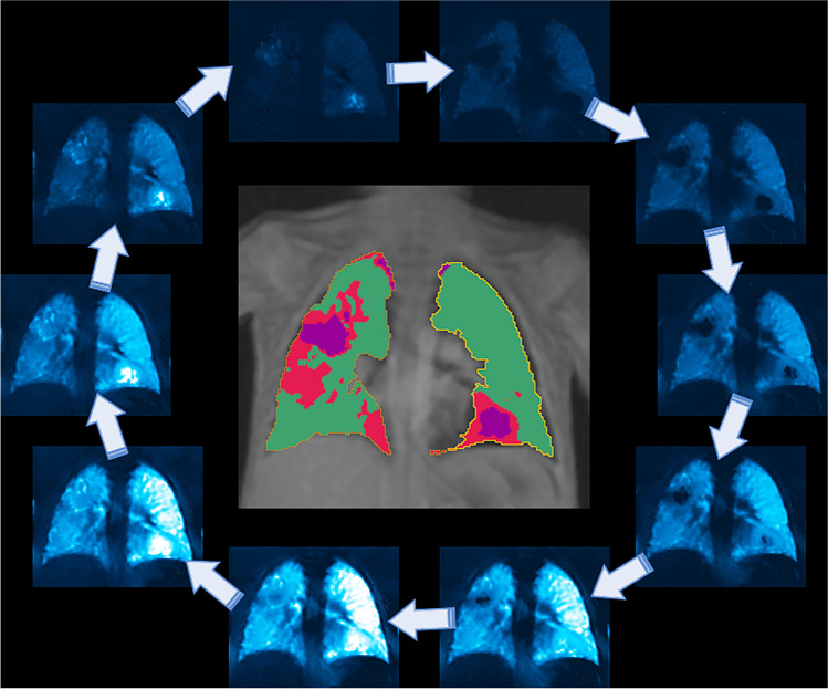
Cystic fibrosis (CF) is one of the most common inherited and life-shortening pulmonary diseases in the Caucasian population. With the widespread introduction of newborn screening and the development of modulator therapy, tremendous advances have been made in recent years both in diagnosis and therapy. Since paediatric CF patients tend to be younger and have lower morbidity, the type of imaging modality that should be used to monitor the disease is often debated. Computed tomography (CT) is sensitive to many pulmonary pathologies, but radiation exposure limits its use, especially in children and adolescents. Conventional pulmonary magnetic resonance imaging (MRI) is a valid alternative to CT and, in most cases, provides sufficient information to guide treatment. Given the expected widespread availability of sequences with ultra-short echo times, there will be even fewer reasons to perform CT for follow-up of patients with CF. This review aims to provide an overview of the process and results of monitoring CF with MRI, particularly for centres not specialising in the disease.

Keywords: Bronchiectasis; Chest; Children; Computed tomography; Cystic fibrosis; Eichinger score; Fourier decomposition; Magnetic resonance imaging; Pulmonary; Ultra-short echo times.


Conflict of interest statement

None

Figures

Fig. 1
Fig. 1
Magnetic resonance images in a 16-year-old girl with bronchiectasis in the upper lobes (arrows). a Coronal respiratory-triggered T2 fast spin echo and self-gated ultra-short echo time sequences with (b) coronal, (c) axial and (d) sagittal multiplanar reconstructions. Please note that air trapping distal to the dilated bronchi is only depicted in the latter
Fig. 2
Fig. 2
Coronal (a) and axial (b) T2 fast spin echo images in a 16-year-old girl with severe cystic fibrosis and infection with Staphylococcus aureus and Pseudomonas aeruginosa show signs of bronchiectasis in the right upper lobe (arrows) and small mucus plugs in the left upper lobe (arrowheads)
Fig. 3
Fig. 3
A coronal ultra-short echo time sequence in a 10-year-old boy with cystic fibrosis and good pulmonary function. Self-gating (a) with an acquisition time of 6:15 min. Single breath-hold technique (b) with an acquisition time of 17 s. Peripheral mucus is found in both upper lobes (arrows)
Fig. 4
Fig. 4
An axial T2 fast spin echo image with fat saturation in a 12-year-old girl with severe pulmonary involvement in cystic fibrosis. Marked bronchiectasis and bronchial wall thickening affect both upper lobes (arrows). In addition, minor consolidations are seen in the right upper lobe (arrowhead)
Fig. 5
Fig. 5
A 16-year-old boy with cystic fibrosis. A T2 fast spin echo magnetic resonance (MR) image (a). The mucus in the bronchiectasis in the basal right upper lobe is hypointense on T2 (arrow). An axial unenhanced computed tomography (CT) image (b). The mucus in the bronchiectasis in the basal right upper lobe is hyperdense on CT (arrow). This combination of MR and CT findings is characteristic of allergic bronchopulmonary aspergillosis and aspergilloma
Fig. 6
Fig. 6
An example of an unenhanced functional proton magnetic resonance image (MRI) (Fourier decomposition) for the estimation of ventilation by means of phase-resolved functional lung MRI (PREFUL) [16] in a free-breathing 6-year-old boy after severe pneumonia (acquisition time of 60 s). Bullae can be observed in the left lower lobe (basal) and the right upper lobe, which ventilate with delayed filling and emptying of air (ventilation circle with arrows around the image). The automated computation map (within the ventilation circle) reveals healthy lung (green) as well as extensive damage to the lungs with perfusion deficits (red) and ventilation-perfusion mismatch (purple)

References

    1. Scotet V, L’Hostis C, Férec C. The changing epidemiology of cystic fibrosis: Incidence, survival and impact of the CFTRGene discovery. Genes (Basel) 2020;11:589. doi: 10.3390/genes11060589. - DOI - PMC - PubMed
    1. Farrell PM. The prevalence of cystic fibrosis in the European Union. J Cyst Fibros. 2008;7:450–453. doi: 10.1016/j.jcf.2008.03.007. - DOI - PubMed
    1. Elborn JS. Cystic fibrosis. Lancet. 2016;388:2519–2531. doi: 10.1016/S0140-6736(16)00576-6. - DOI - PubMed
    1. Balfour-Lynn IM, King JA. CFTR modulator therapies – Effect on life expectancy in people with cystic fibrosis. Paediatr Respir Rev. 2020;42:3–8. - PMC - PubMed
    1. Southern KW, Mérelle MM, Dankert-Roelse JE, Nagelkerke AD. Newborn screening for cystic fibrosis. Cochrane Database Syst Rev. 2009;2009:CD001402. - PMC - PubMed

LinkOut - more resources