Observational Study
. 2023 Feb;93(2):297-302.
doi: 10.1002/ana.26549. Epub 2022 Dec 2.

Cerebral Cortical Encephalitis in Myelin Oligodendrocyte Glycoprotein Antibody-Associated Disease

Affiliations
Observational Study

Cerebral Cortical Encephalitis in Myelin Oligodendrocyte Glycoprotein Antibody-Associated Disease

Cristina Valencia-Sanchez et al. Ann Neurol. 2023 Feb.

Abstract

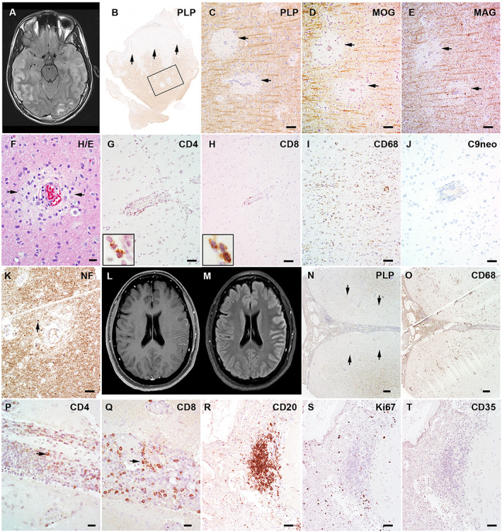
Cerebral cortical encephalitis (CCE) is a recently described myelin oligodendrocyte glycoprotein antibody-associated disease (MOGAD) phenotype. In this observational retrospective study, we characterized 19 CCE patients (6.7% of our MOGAD cohort). Headache (n = 15, 79%), seizures (n = 13, 68%), and encephalopathy (n = 12, 63%) were frequent. Magnetic resonance imaging revealed unilateral (n = 12, 63%) or bilateral (n = 7, 37%) cortical T2 hyperintensity and leptomeningeal enhancement (n = 17, 89%). N-Methyl-D-aspartate receptor autoantibodies coexisted in 2 of 15 tested (13%). CCE pathology (n = 2) showed extensive subpial cortical demyelination (n = 2), microglial reactivity (n = 2), and inflammatory infiltrates (perivascular, n = 1; meningeal, n = 1). Most received high-dose steroids (n = 17, 89%), and all improved, but 3 had CCE relapses. This study highlights the CCE spectrum and provides insight into its pathogenesis. ANN NEUROL 2023;93:297-302.


Conflict of interest statement

C.V.‐S., Y.G., E.S., M.M., K.N.K., P.M.E., V.R., J.‐M.T., A.B., C.F.L., report no conflicts of interest. J.J.C. has served as a consultant for Roche and UCB, which have upcoming treatment trials in MOGAD. S.L.‐C. and A.K. have served on an advisory board for Genentech/Roche, which has an upcoming treatment trial in MOGAD. D.D. has consulted for UCB, which has an upcoming treatment trial in MOGAD. S.J.P. reports grants, personal fees, and nonfinancial support from Roche/Genentech, and personal fees for consulting services from UCB, which have upcoming treatment trials in MOGAD. E.P.F. has participated in advisory boards for Roche and UCB who have upcoming treatment trials in MOGAD. E.P.F. has received funding from the NIH (R01NS113828). Mayo Clinic Laboratories offer commercial testing for MOG‐IgG, but none of the authors receive financial compensation for this.

Figures

FIGURE 1
FIGURE 1
The magnetic resonance imaging (MRI) and pathology of 2 patients with myelin oligodendrocyte glycoprotein (MOG) antibody‐associated disease cerebral cortical encephalitis. (A–K) Patient 12 (Table S1). (A) The axial MRI fluid‐attenuated inversion recovery (FLAIR) image shows swelling and T2 hyperintensity in the right more than left cortex and in subcortical white matter. (B) Cortical biopsy of the right temporal lobe in this patient shows diffuse subpial cortical demyelination (arrows) and small perivascular intracortical demyelination within the frame (proteolipid protein [PLP] immunohistochemistry). (C–E) The enlarged views of framed region in B show perivascular myelin loss to an equal extent in PLP (C), MOG (D), and myelin‐associated glycoprotein (MAG; E). (F) Hematoxylin and eosin (H&E) stain shows mild perivascular infiltration and edema. (G, H) Immunohistochemistry reveals perivascular infiltration of both CD4+ (G) and CD8+ (H) T lymphocytes. (I) Extensive microglial reactions are present beyond the demyelination in the cortex. (J) No C9 neoantigen (terminal complement activation product) is present. (K) Axons are preserved with mild perivascular spheroid formation (indicated with arrow). (L–Q) Patient 18 (Table S1). (L) Axial T1‐weighted postgadolinium MRI shows cortical and leptomeningeal enhancement. (M) Axial FLAIR images reveal cortical T2 hyperintensity and swelling in the left hemispheric cortex. (N) Left hemispheric cortical biopsy using the PLP stain shows extensive subpial cortical demyelination (indicated with arrows) with marked meningeal inflammatory infiltration. (O) CD68 stain highlights extensive macrophage/microglial reactivity in both meninges and subpial lesions. (P, Q) CD4+ (P) and CD8+ (Q) lymphocytes are both present in the subarachnoid space. (R) CD20 immunohistochemistry shows B‐cell abundant aggregates present in the leptomeninges. (S) The staining for the proliferation marker, Ki67, reveals no proliferating activity in the B‐cell aggregates. (T) CD35 immunohistochemistry shows absence of follicle dendritic cells. Scale bars: 100μm (C–E, I, K), 20μm (F, P, Q), 50μm (G, H, J, R‐T), 200μm (N, O). [Color figure can be viewed at www.annalsofneurology.org]
FIGURE 2
FIGURE 2
Additional magnetic resonance imaging examples of myelin oligodendrocyte glycoprotein antibody‐associated disease cerebral cortical encephalitis. (A) Axial fluid‐attenuated inversion recovery (FLAIR) image reveals right posterior temporo‐occipital cortical T2 hyperintensity and swelling (A1, yellow bracket), and accompanying T1‐weighted postgadolinium image reveals associated cortical and leptomeningeal enhancement (A2, yellow bracket [inset shows higher magnification]). (B) Axial FLAIR image reveals left parietal cortical T2 hyperintensity and swelling (B1, yellow bracket), and T1‐weighted postgadolinium image reveals concomitant subtle leptomeningeal enhancement (B2 [inset shows higher magnification]). (C) Axial FLAIR image reveals right temporal lobe cortical T2 hyperintensity and swelling (C1, yellow bracket) with T1‐weighted postgadolinium image revealing accompanying leptomeningeal enhancement (C2, yellow bracket [inset shows higher magnification]). Follow‐up imaging 18 months later reveals resolution of right temporal lobe cortical swelling, T2 hyperintensity, and enhancement with new left temporal T2 hyperintensity (C3, yellow bracket) with subtle leptomeningeal enhancement although slightly limited by motion artifact (C4, yellow bracket [inset shows higher magnification]) in a patient with recurrent cerebral cortical encephalitis attacks. [Color figure can be viewed at www.annalsofneurology.org]

References

    1. Ogawa R, Nakashima I, Takahashi T, et al. MOG antibody‐positive, benign, unilateral, cerebral cortical encephalitis with epilepsy. Neurol Neuroimmunol Neuroinflamm 2017;4:e322. - PMC - PubMed
    1. Budhram A, Mirian A, Le C, et al. Unilateral cortical FLAIR‐hyperintense lesions in anti‐MOG‐associated encephalitis with seizures (FLAMES): characterization of a distinct clinico‐radiographic syndrome. J Neurol 2019;266:2481–2487. - PubMed
    1. Yao T, Zeng Q, Xie Y, et al. Clinical analysis of adult MOG antibody‐associated cortical encephalitis. Mult Scler Relat Disord 2022;5:103727. - PubMed
    1. Shu H, Ding M, Shang P, et al. Myelin oligodendrocyte glycoprotein antibody associated cerebral cortical encephalitis: case reports and review of literature. Front Hum Neurosci 2021;15:782490. - PMC - PubMed
    1. Fujimori J, Ogawa R, Murata T, et al. Decreased subcortical T2 FLAIR signal with cortical T2 FLAIR hyperintense lesions in unilateral cerebral cortical encephalitis with myelin oligodendrocyte glycoprotein antibody. Neuroimmunol Rep 2022;2:100096.

Publication types

Substances