. 2022 Dec 21;10(6):e0364622.
doi: 10.1128/spectrum.03646-22. Epub 2022 Nov 14.

Diverse Sensory Repertoire of Paralogous Chemoreceptors Tlp2, Tlp3, and Tlp4 in Campylobacter jejuni

Affiliations

Diverse Sensory Repertoire of Paralogous Chemoreceptors Tlp2, Tlp3, and Tlp4 in Campylobacter jejuni

Taha et al. Microbiol Spectr. .

Abstract

Campylobacter jejuni responds to extracellular stimuli via transducer-like chemoreceptors (Tlps). Here, we describe receptor-ligand interactions of a unique paralogue family of dCache_1 (double Calcium channels and chemotaxis) chemoreceptors: Tlp2, Tlp3, and Tlp4. Phylogenetic analysis revealed that Tlp2, Tlp3, and Tlp4 receptors may have arisen through domain duplications, followed by a divergent evolutionary drift, with Tlp3 emerging more recently, and unexpectedly, responded to glycans, as well as multiple organic and amino acids with overlapping specificities. All three Tlps interacted with five monosaccharides and complex glycans, including Lewis's antigens, P antigens, and fucosyl GM1 ganglioside, indicating a potential role in host-pathogen interactions. Analysis of chemotactic motility of single, double, and triple mutants indicated that these chemoreceptors are likely to work together to balance responses to attractants and repellents to modulate chemotaxis in C. jejuni. Molecular docking experiments, in combination with saturation transfer difference nuclear magnetic resonance spectroscopy and competition surface plasmon resonance analysis, illustrated that the ligand-binding domain of Tlp3 possess one major binding pocket with two overlapping, but distinct binding sites able to interact with multiple ligands. A diverse sensory repertoire could provide C. jejuni with the ability to modulate responses to attractant and repellent signals and allow for adaptation in host-pathogen interactions. IMPORTANCE Campylobacter jejuni responds to extracellular stimuli via transducer-like chemoreceptors (Tlps). This remarkable sensory perception mechanism allows bacteria to sense environmental changes and avoid unfavorable conditions or to maneuver toward nutrient sources and host cells. Here, we describe receptor-ligand interactions of a unique paralogue family of chemoreceptors, Tlp2, Tlp3, and Tlp4, that may have arisen through domain duplications, followed by a divergent evolutionary drift, with Tlp3 emerging more recently. Unlike previous reports of ligands interacting with sensory proteins, Tlp2, Tlp3, and Tlp4 responded to many types of chemical compounds, including simple and complex sugars such as those present on human blood group antigens and gangliosides, indicating a potential role in host-pathogen interactions. Diverse sensory repertoire could provide C. jejuni with the ability to modulate responses to attractant and repellent signals and allow for adaptation in host-pathogen interactions.

Keywords: Campylobacter jejuni; chemoreceptor; chemotaxis; ligand discovery.


Conflict of interest statement

The authors declare no conflict of interest.

Figures

FIG 1
FIG 1
Phyletic distribution of dCache_1 domains from Campylobacterota chemoreceptors. The taxonomy tree, based on 120 protein sequences, was retrieved from AnnoTree (24). C. jejuni 11168 belongs to Campylobacter_D jejuni and is indicated in boldface. Tlp2,3,4 were assigned to the 28H MCP based on 28 helical heptads in the signaling domain by using hidden Markov models (23). Tlp1 homologs were assigned based on the sequence similarity of the LBD (see Fig. S3); differences in the MCP signaling domain heptad class might be due either domain swap or insertions/deletions.
FIG 2
FIG 2
(A) Phylogenetic distribution of Tlp2, Tlp3, and Tlp4 homologues among Campylobacter_D genus. (B) Fragment of dCache_1 sequence alignment of Tlp2, Tlp3, and Tlp4 from C. jejuni 11168 and PctA, PctB, and PctC from P. aeruginosa. Conserved amino acid recognition motif is highlighted in gray. Amino acid numeration is based on the tlp3 gene sequence.
FIG 3
FIG 3
Structural analysis of arginine binding to Tlp3 using molecular docking experiments and saturation transfer difference (STD) NMR experiments. (A) Ribbon structure of dimeric Tlp3 (PDB 4XMR) (15) (left) and surface structure (right). (B) A monomeric Tlp3 structure was used for an unbiased global docking experiments using glucose and arginine as ligands with all clusters A to D shown, with cluster A being the predominant high-occupancy cluster for both ligands. (C) Zoomed view of cluster A that exhibits a main and an extended binding site with arginine bound to the main binding site. (D) STD (blue) and 1H NMR spectrum of arginine in complex with Tlp3 acquired at 283K and 600.13 MHz. Strong saturation transfer could be detected for H3/H3′ and H5/H5′ with H4/H4′ overlapped with protein background. The epitope is consistent with the docked structure shown in panel C.
FIG 4
FIG 4
Representative sensograms from SPR analysis of Tlp3peri, Tlp2peri, and Tlp4peri. Single-cycle SPR plots are shown as concentration-dependent interactions between proteins and ligands, as illustrated.
FIG 5
FIG 5
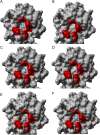
Molecular docking results of glucose (Glc) and arginine (Arg) bound to cluster A of Tlp3. Glc bound to cluster A’s main (A) and extended (B) sites; Arg bound to cluster A’s main (C) and extended (D) sites; Arg bound to cluster A’s main site and Glc bound to the extended site (E); Glc bound to cluster A’s main site and Arg bound to the extended site (F). Structures E and F were submitted to 50-ns Molecular Dynamics (MD) simulations using the AMBER force field (see the supplemental material).
FIG 6
FIG 6
SPR competition analysis illustrates one binding pocket with two distinct binding sites for Tlp3 ligands. SPR competition analysis of the binding of glucose (glc), fucose (fuc), thiamine (thia), arginine (arg), and isoleucine (iso) to WT Tlp3LBD was performed. The experimental RU value is the actual RU value, as follows. (A) “glc” indicates responses to glucose only, “fuc” indicates responses to fucose only, “glc+fuc” indicates glucose responses following saturation with fucose, and “fuc+glc” indicates fucose responses following saturation with glucose (see Fig. S6 for the representative ABA sensorgram for this analysis). (B) “iso” indicates responses to isoleucine only, “glc” indicates responses to glucose only, “iso+ glc” indicates isoleucine responses following saturation with glucose, and “glc+iso” indicates glucose responses following saturation with isoleucine. (C) “iso” indicates responses to isoleucine only, “thia” indicates responses to thiamine only, “iso+thia” indicates isoleucine responses following saturation with thiamine, and “thia+iso” indicates thiamine responses following saturation with isoleucine. (D) “glc” indicates responses to glucose only, “arg” indicates responses to arginine only, “glc+arg” indicates glucose responses following saturation with arginine, and “arg+glc” indicates arginine responses following saturation with glucose. (E) “glc” indicates responses to glucose only, “thia” indicates responses to thiamine only, “glc+thia” indicates glucose responses following saturation with thiamine, and “thia+glc” indicates thiamine responses following saturation with glucose. (F) “glc” indicates responses to glucose only, “malic” indicates responses to malic acid only, “glc+malic” indicates glucose responses following saturation with malic acid, and “malic +glc” indicates malic acid responses following saturation with glucose. The theoretical value is RU values based on mathematical theory. The binding status of the ligands to protein are classified as follows: independent site (additive/accumulative effect), ligands binding to different binding sites; shared site, ligands binding/sharing same binding site; or preferential shared site, ligands binding/sharing same binding site, but the protein binds to one ligand better than the other when in equilibrium. All response data were normalized to a 100-Da molecular weight for each analyte, allowing direct comparison of responses.
FIG 7
FIG 7
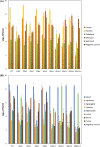
Nutrient depletion chemotaxis assay. The agarose plugs contained the indicated ligands. (A) fucose, glucose, galactose, mannose, and sialic acid. (B) Serine, methionine, asparagine, lysine, cysteine, purine, and aspartate. The C. jejuni 81116 ΔflaA ΔflaB isogenic mutant was used as a nonmotile, nonchemotactic control; agar plugs containing no added ligand were used as a negative control, and mucin was used as a positive control. Standard errors are shown as bars above the means of three replicates. Viable counts of C. jejuni from the assays are shown on a log scale. The asterisk (*) indicates a statistically significant difference compared to the WT strain (P < 0.05).

Similar articles

Cited by

References

    1. Kramer JM, Frost JA, Bolton FJ, Wareing DR. 2000. Campylobacter contamination of raw meat and poultry at retail sale: identification of multiple types and comparison with isolates from human infection. J Food Prot 63:1654–1659. doi:10.4315/0362-028x-63.12.1654. - DOI - PubMed
    1. Altekruse SF, Stern NJ, Fields PI, Swerdlow DL. 1999. Campylobacter jejuni–an emerging foodborne pathogen. Emerg Infect Dis 5:28–35. doi:10.3201/eid0501.990104. - DOI - PMC - PubMed
    1. Shane S. 2000. Campylobacter infection of commercial poultry. Rev Sci Tech 19:376–395. doi:10.20506/rst.19.2.1224. - DOI - PubMed
    1. Humphrey S, Chaloner G, Kemmett K, Davidson N, Williams N, Kipar A, Humphrey T, Wigley P. 2014. Campylobacter jejuni is not merely a commensal in commercial broiler chickens and affects bird welfare. mBio 5:e01364-14–e01314. doi:10.1128/mBio.01364-14. - DOI - PMC - PubMed
    1. Acheson D, Allos BM. 2001. Campylobacter jejuni infections: update on emerging issues and trends. Clin Infect Dis 32:1201–1206. doi:10.1086/319760. - DOI - PubMed

Publication types

LinkOut - more resources