

The mast cell reaction in premalignant and malignant lesions of the head and neck
- PMID: 36374145
- PMCID: PMC9804062
- DOI: 10.47162/RJME.63.2.11
The mast cell reaction in premalignant and malignant lesions of the head and neck
Abstract
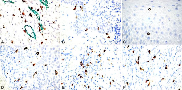
Head and neck squamous cell carcinoma (HNSCC) is one of the most frequent and aggressive neoplasms of this anatomical region. Many studies evaluated the neoplastic cells, but few works focused on the tumor microenvironment. In the present study, we investigated the distribution and mast cell density (MCD) in malignant and premalignant lesions of the oral cavity, tongue, pharynx, and larynx. There were analyzed 52 specimens of HNSCC, and 15 biopsies taken from patients with dysplasia. Results were compared with those found in a control group of 10 biopsies of oral mucosa from patients with inflammatory diseases. Slides stained with Hematoxylin-Eosin were used for the histopathological diagnosis and grade, and mast cells (MCs) were identified by immunohistochemistry, using anti-MC tryptase. MCs were counted using a method similar to that proposed for microvessel density. We found a significant increase in the number of MCs from the normal oral mucosa until overt carcinoma. Unlike normal tissues, in HNSCC, many MCs were found between tumor cells. We found no relationship between MCs and blood vessels in the tumor area. A significant statistical correlation was found between dysplastic and malignant tumors, but not between tumors with a different grade. Also, it was not found relationship between MCD and the anatomical location of the tumor. Based on these results, we believe that MCD evaluated by anti-MC tryptase is an independent factor of prognosis and reflects an unfavorable outcome.
Conflict of interest statement
None to declare.
Figures
Similar articles
-
Low Mast Cell Density Predicts Poor Prognosis in Oral Squamous Cell Carcinoma and Reduces Survival in Head and Neck Squamous Cell Carcinoma.Anticancer Res. 2016 Oct;36(10):5499-5506. doi: 10.21873/anticanres.11131. Anticancer Res. 2016. PMID: 27798921
-
The role of mast cell tryptase in neoangiogenesis of premalignant and malignant lesions of the uterine cervix.J Histochem Cytochem. 2001 Aug;49(8):1061-2. doi: 10.1177/002215540104900816. J Histochem Cytochem. 2001. PMID: 11457936
-
Increase of mast cells and tumor angiogenesis in oral squamous cell carcinoma.J Oral Pathol Med. 2003 Apr;32(4):195-9. doi: 10.1034/j.1600-0714.2003.00128.x. J Oral Pathol Med. 2003. PMID: 12653857
-
Prognostic and clinicopathological significance of tumor-stroma ratio in head and neck squamous cell carcinoma: A systematic review.Med Oral Patol Oral Cir Bucal. 2022 Jul 1;27(4):e301-e309. doi: 10.4317/medoral.24922. Med Oral Patol Oral Cir Bucal. 2022. PMID: 35717622 Free PMC article.
-
[Morphology of non cutaneous head and neck squamous cell carcinoma].Pathologe. 2018 Feb;39(1):3-10. doi: 10.1007/s00292-017-0395-5. Pathologe. 2018. PMID: 29209797 Review. German.
Cited by
-
The Complex Role of Mast Cells in Head and Neck Squamous Cell Carcinoma: A Systematic Review.Medicina (Kaunas). 2024 Jul 19;60(7):1173. doi: 10.3390/medicina60071173. Medicina (Kaunas). 2024. PMID: 39064602 Free PMC article.
-
Assessment of Expression of Mast Cells Density in Oral Premalignant and Malignant Lesions by Histochemical Analysis.J Pharm Bioallied Sci. 2024 Dec;16(Suppl 5):S4765-S4768. doi: 10.4103/jpbs.jpbs_920_24. Epub 2025 Jan 30. J Pharm Bioallied Sci. 2024. PMID: 40061668 Free PMC article.
-
Type 2 Diabetes Mellitus in Patients with Different Types of Thyroid Nodular Lesions Among Western Romanian Patients: A Comprehensive Clinical, Biochemical, and Hormonal Analysis.Medicina (Kaunas). 2025 Jul 14;61(7):1270. doi: 10.3390/medicina61071270. Medicina (Kaunas). 2025. PMID: 40731899 Free PMC article.
-
A novel mast cell marker gene-related prognostic signature to predict prognosis and reveal the immune landscape in head and neck squamous cell carcinoma.Front Immunol. 2025 Jul 9;16:1538641. doi: 10.3389/fimmu.2025.1538641. eCollection 2025. Front Immunol. 2025. PMID: 40703522 Free PMC article.
References
-
- Balica N, Raica M, Cotulbea S, Doros C. Mast cell reaction in malignant laryngeal neoplasm. Rom J Morphol Embryol. 2007;48(4):395–401. - PubMed
MeSH terms
Substances
LinkOut - more resources
Full Text Sources
Medical
