. 2022 Nov 11;14(22):9000-9019.
doi: 10.18632/aging.204377. Epub 2022 Nov 11.

NAP1L5 targeting combined with MYH9 Inhibit HCC progression through PI3K/AKT/mTOR signaling pathway

Affiliations

NAP1L5 targeting combined with MYH9 Inhibit HCC progression through PI3K/AKT/mTOR signaling pathway

Rui Zhao et al. Aging (Albany NY). .

Abstract

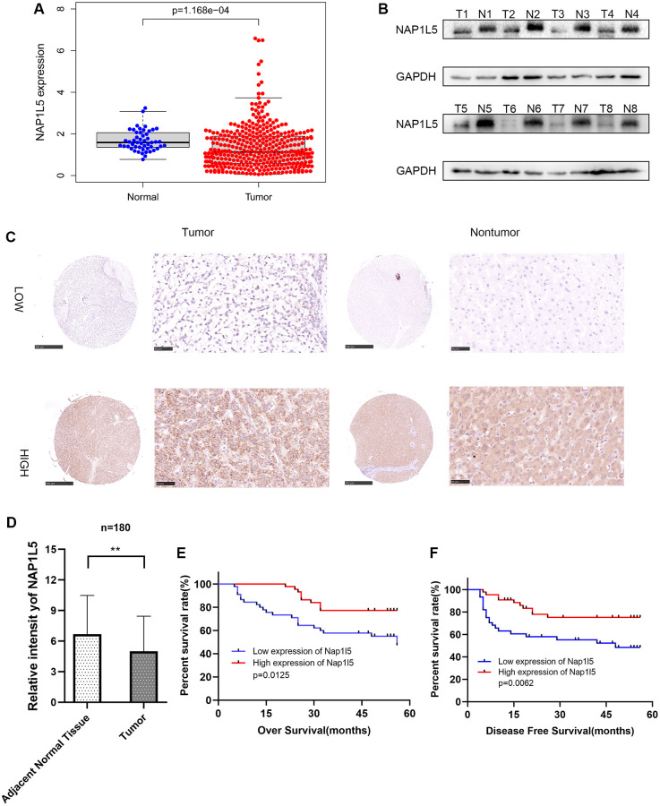
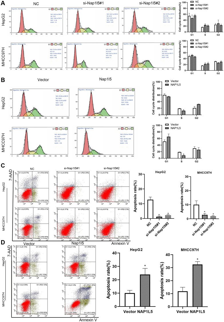
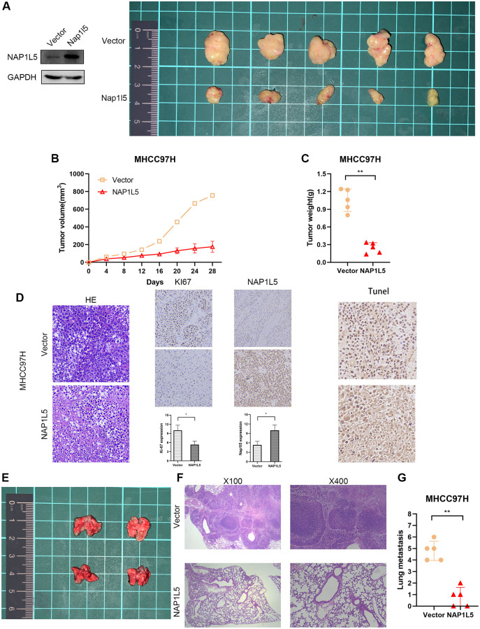
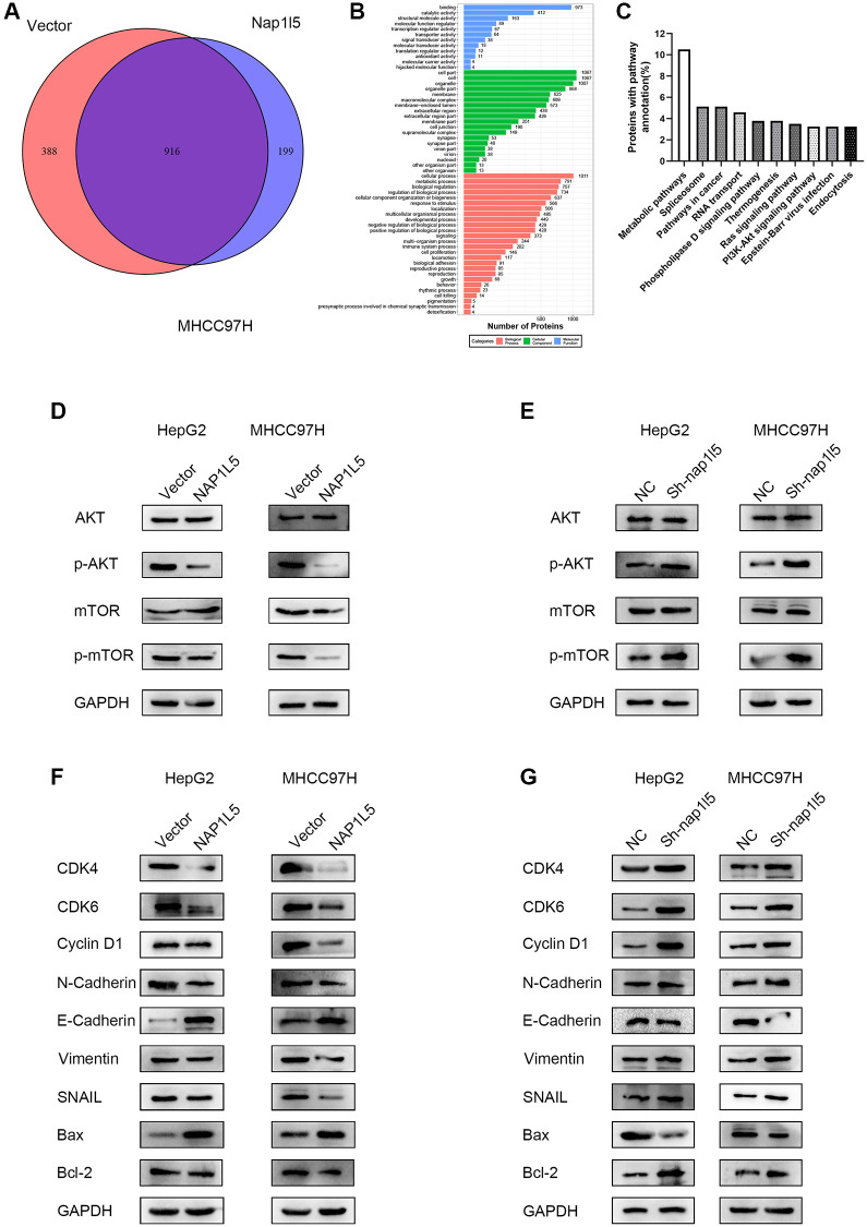
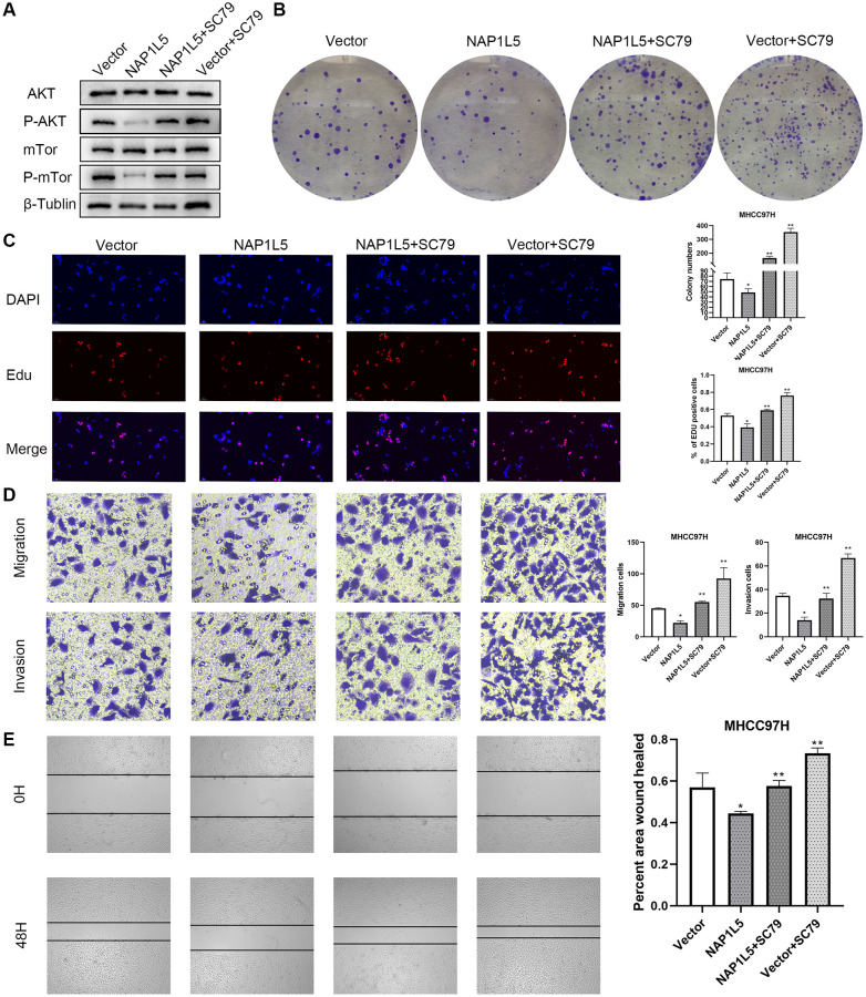
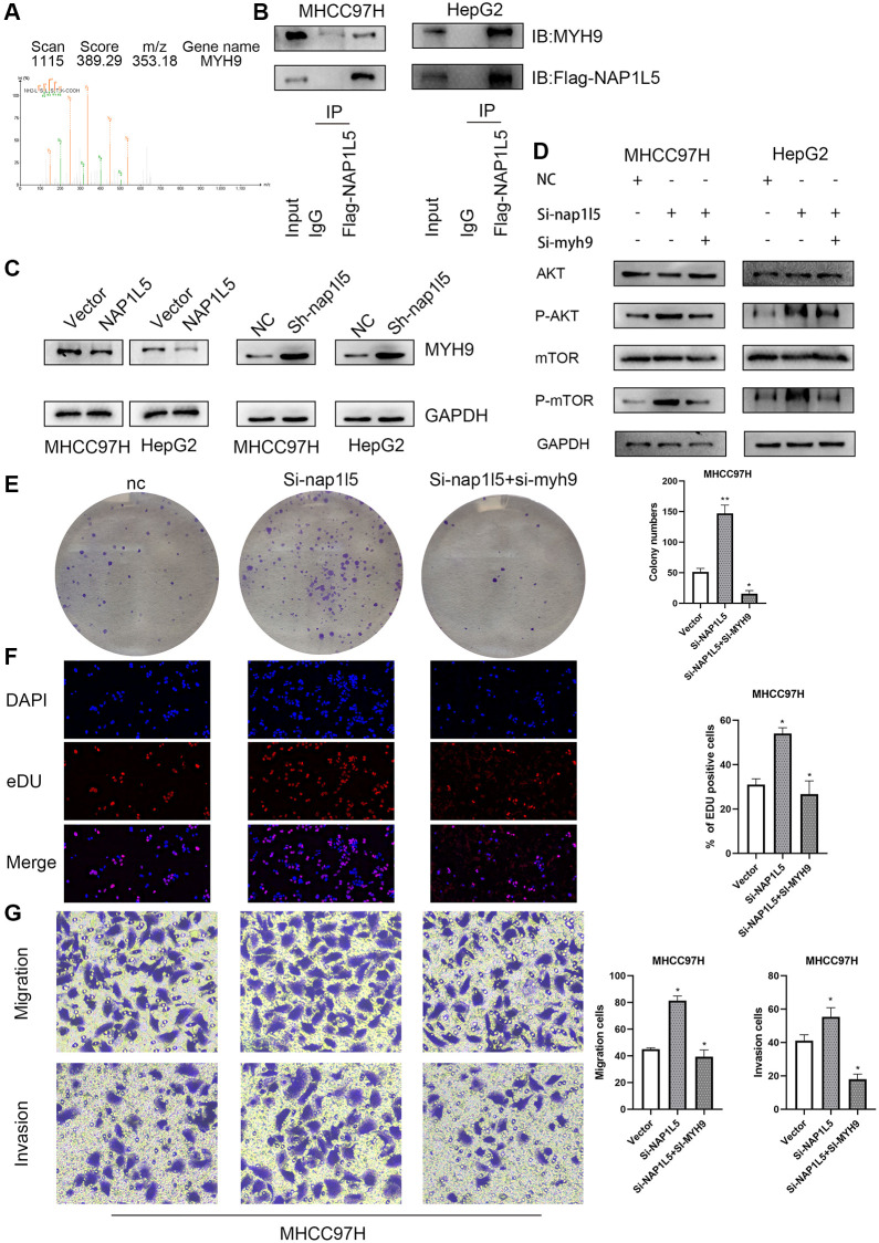
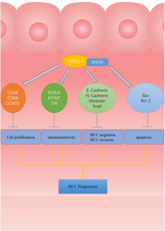
Hepatocellular carcinoma (HCC) is one of the leading causes of cancer death worldwide. Nucleosome assembly protein 1-like 5 (NAP1L5) is a protein-coding gene that encodes a protein similar to nucleosome assembly protein 1 (NAP1). It is a histone chaperone that plays an important role in gene transcription in organisms. However, the role of NAP1L5 in the pathogenesis of hepatocellular carcinoma remains to be elucidated. In this study, low expression of NAP1L5 was found in hepatocellular carcinoma, and the downregulation of NAP1L5 was related to shorter survival and disease-free survival. In addition, its expression is also related to the tumor size and recurrence of hepatocellular carcinoma. The overexpression and knockdown of NAP1L5 by plasmid and siRNA showed that NAP1L5 inhibited the proliferation, migration and invasion and induced apoptosis of hepatoma cells. In vivo experiments confirmed that NAP1L5 can inhibit the growth and metastasis of hepatocellular carcinoma cells. In the mechanistic study, we found that NAP1L5 affects the occurrence and development of hepatocellular carcinoma by regulating MYH9 to inhibit the PI3K/AKT/mTOR signaling pathway. As a functional tumor suppressor, NAP1L5 is expressed at low levels in HCC. NAP1L5 inhibits the PI3K/AKT/mTOR signaling pathway in hepatocellular carcinoma by regulating MYH9. It may be a new potential target for liver cancer treatment.

Keywords: MYH9; Nap1L5; hepatocellular carcinoma; invasion; migration; proliferation.


Conflict of interest statement

CONFLICTS OF INTEREST: The authors declare no conflicts of interest related to this study.

Figures

Figure 1
Figure 1
The expression of NAP1L5 in hepatocellular carcinoma is decreased and is related to poor prognosis. (A) The expression of the NAP1L5 gene in the TCGA database: 50 paracancerous tissues and 374 cancerous tissues. (B) The expression of NAP1L5 protein in 8 pairs of hepatocellular carcinoma (Tumor) and paracancerous tissues (Normal) was assessed. (C) Immunohistochemistry was used to detect the expression of NAP1L5 in the HCC tissue microarray. (D) The immunohistochemical score of 90 samples of HCC and paracancerous tissues in the tissue microarray. (E, F) Kaplan–Meier survival analysis was performed to evaluate the effect of NAP1L5 on disease-free survival and overall survival. *p < 0.05; **p < 0.01;***p < 0.001.
Figure 2
Figure 2
Downregulation of NAP1L5 can promote the proliferation, migration and invasion of hepatocellular carcinoma cells in vitro. (A) NAP1L5 mRNA expression in the LO2 and hepatoma cell lines. (B) The expression of NAP1L5 protein in the LO2 and hepatoma cell lines. (C) Validation of siRNaA transfection efficiency. (D) The effect of the downregulation of NAP1L5 on the proliferation of HepG2 and MHCC97H cells was detected by EdU staining. (E) A colony formation assay was used to detect the effect of NAP1L5 downregulation on the proliferation of HepG2 and MHCC97H cells. (F) Transwell assays were used to detect the effect of NAP1L5 downregulation on the migration and invasion of HepG2 and MHCC97H cells. (G) The effect of the downregulation of NAP1L5 on the migration of HepG2 and MHCC97H cells was detected by a wound healing migration assay. *p < 0.05; **p < 0.01; ***p < 0.001.
Figure 3
Figure 3
In vitro, NAP1L5 overexpression inhibited cell proliferation, migration and invasion. (A) The mRNA expression of NAP1L5 in HepG2 and MHCC97H cell lines after plasmid transfer into vector and NAP1L5. (B) The expression of NAP1L5 protein in HepG2 and MHCC97H cell lines after plasmid transfer into vector and NAP1L5. (C) EdU staining was used to detect the effect of NAP1L5 overexpression on the proliferation of HepG2 and MHCC97H cells. (D) The effect of NAP1L5 overexpression on the proliferation of HepG2 and MHCC97H cells was detected by a colony formation assay. (E) Transwell assays were performed to detect the effect of NAP1L5 overexpression on the migration and invasion of HepG2 and MHCC97H cells. (F) The effects of NAP1L5 overexpression on the migration and invasion of HepG2 and MHCC97H cells were detected by a wound healing migration assay. *p < 0.05; **p < 0.01; ***p < 0.001.
Figure 4
Figure 4
NAP1L5 induces cell cycle S phase cell reduction and promotes apoptosis in vitro. (A) The effect of NAP1L5 downregulation on the cell cycle distribution of HepG2 and MHCC97H cells. (B) The effect of NAP1L5 overexpression on the cell cycle distribution of HepG2 and MHCC97H cells. (C) The effect of the downregulation of NAP1L5 on the apoptosis of HepG2 and MHCC97H cells. (D) The effect of NAP1L5 overexpression on HepG2 and MHCC97H cell apoptosis. *p < 0.05; **p < 0.01; ***p < 0.001.
Figure 5
Figure 5
NAP1L5 inhibits tumor growth and metastasis in vivo. (AC) Xenografted tumors were produced by injection of MHCC97H cells overexpressing NAP1L5 (experimental group) or carrying control vector (control group). The growth of the transplanted tumor was measured by volume and weight. (D) HE, IHC, Ki-67 and TUNEL staining were performed on the transplanted tumor. (E) The lung metastasis model was established by injecting MHCC97H cells overexpressing NAP1L5 (experimental group) or control vector (control group) through the tail vein. (F, G)The number of metastatic foci in lung tissue was observed, and HE staining was performed on lung tissue. *p < 0.05; **p < 0.01; ***p < 0.001.
Figure 6
Figure 6
NAP1L5 regulates the PI3K/AKT/MTOR signaling pathway in hepatoma cells. (A) Venn diagram of the mass spectrometry analysis of MHCC97H cells overexpressing NAP1L5 and transferred into the control vector. (B) Mass spectrometry analysis of the GO functional enrichment map of MHCC97H cells overexpressing NAP1L5. (C) Enrichment of differentially expressed gene pathways in NAP1L5-overexpressing and MHCC97H cells transfected with the control vector. (D) Western blotting was used to detect the expression of p-AKT and p-mTOR in HepG2 and MHCC97H cells overexpressing NAP1L5. (E) Western blotting was used to detect the expression of p-AKT and p-mTOR in HepG2 and MHCC97H cells downregulated by NAP1L5. (F) Western blotting was used to analyze the expression of key molecules involved in cell cycle regulation, EMT and apoptosis in HepG2 and MHCC97H cells overexpressing NAP1L5. (G) Western blotting was used to analyze the expression of key molecules involved in cell cycle regulation, EMT and apoptosis in HepG2 and MHCC97H cells downregulated by NAP1L5.
Figure 7
Figure 7
NAP1L5 inhibits the progression of hepatocellular carcinoma by regulating PI3K/AKT/MTOR. (A) The expression of p-AKT and p-mTOR in MHCC97H-Vector cells and MHCC97H-NAP1L5 cells treated with SC79 (100 ng/mL) and the two types of cells without SC79 treatment was detected by Western blotting. (B) A colony formation assay was used to detect the effect of SC79 (100 ng/mL) on the proliferation of MHCC97H-Vector and MHCC97H-NAP1L5 cells. (C) EdU staining was used to detect the effect of SC79 (100 ng/mL) on the proliferation of MHCC97H-Vector and MHCC97H-NAP1L5 cells. (D) The effect of SC79 (100 ng/mL) on the migration and invasion of MHCC97H-Vector and MHCC97H-NAP1L5 cells was detected by a Transwell assay. (E) The effect of SC79 (100 ng/mL) on the migration of MHCC97H-Vector and MHCC97H-NAP1L5 cells was detected by a wound healing migration assay. *p < 0.05; **p < 0.01; ***p < 0.001.
Figure 8
Figure 8
Effects of NAP1L5 binding to MYH9 on downstream signaling pathways and cell function in hepatocellular carcinoma. (A) The possible interacting proteins were analyzed by mass spectrometry. Trypsin digestion fragments were detected by mass spectrometry, and B and Y represent N-terminal and C-terminal collision-induced dissociation fragment ions. (B) Co-IP confirmed the combination of NAP1L5 and MYH9. (C) Western blotting showed that the expression of NAP1L5 protein was negatively correlated with that of MYH9 protein. (D) Both NAP1L5 and MYH9 were downregulated in the MHCC97H cell line. Western blotting showed that the phosphorylation levels of AKT and mTOR were lower than those in the NAP1L5-downregulated cell line. (E) A colony formation assay verified the change in the proliferation ability of MHCC97H cells with simultaneous downregulation of NAP1L5 and MYH9. (F) EdU staining verified the change in the proliferation ability of MHCC97H cells with simultaneous downregulation of NAP1L5 and MYH9. (G) A Transwell assay verified the change in the migration and invasion ability of MHCC97H cells with simultaneous downregulation of NAP1L5 and MYH9. *p < 0.05; **p < 0.01; ***p < 0.001.
Figure 9
Figure 9
The flow chart of NAP1L5 targeting combined with MYH9 Inhibit HCC progression. NAP1L5 targets and binds MYH9 to regulate cycle, apoptosis, EMT-related protein expression and PI3K/AKT/mTOR signal pathway activity, thus inhibiting the progression of hepatocellular carcinoma.

Similar articles

Cited by

References

    1. Sung H, Ferlay J, Siegel RL, Laversanne M, Soerjomataram I, Jemal A, Bray F. Global Cancer Statistics 2020: GLOBOCAN Estimates of Incidence and Mortality Worldwide for 36 Cancers in 185 Countries. CA Cancer J Clin. 2021; 71:209–49. 10.3322/caac.21660 - DOI - PubMed
    1. Andrews AJ, Chen X, Zevin A, Stargell LA, Luger K. The histone chaperone Nap1 promotes nucleosome assembly by eliminating nonnucleosomal histone DNA interactions. Mol Cell. 2010; 37:834–42. 10.1016/j.molcel.2010.01.037 - DOI - PMC - PubMed
    1. Akishina AA, Kuvaeva EE, Vorontsova YE, Simonova OB. NAP Family Histone Chaperones: Characterization and Role in Ontogenesis. Russ J Dev Biol. 2020; 51:343–55. 10.1134/S1062360420060028 - DOI
    1. Attia M, Förster A, Rachez C, Freemont P, Avner P, Rogner UC. Interaction between nucleosome assembly protein 1-like family members. J Mol Biol. 2011; 407:647–60. 10.1016/j.jmb.2011.02.016 - DOI - PubMed
    1. Rogner UC, Spyropoulos DD, Le Novère N, Changeux JP, Avner P. Control of neurulation by the nucleosome assembly protein-1-like 2. Nat Genet. 2000; 25:431–5. 10.1038/78124 - DOI - PubMed

Publication types

MeSH terms