

Development and external validation of automated detection, classification, and localization of ankle fractures: inside the black box of a convolutional neural network (CNN)
- PMID: 36374292
- PMCID: PMC10175446
- DOI: 10.1007/s00068-022-02136-1
Development and external validation of automated detection, classification, and localization of ankle fractures: inside the black box of a convolutional neural network (CNN)
Abstract
Purpose: Convolutional neural networks (CNNs) are increasingly being developed for automated fracture detection in orthopaedic trauma surgery. Studies to date, however, are limited to providing classification based on the entire image-and only produce heatmaps for approximate fracture localization instead of delineating exact fracture morphology. Therefore, we aimed to answer (1) what is the performance of a CNN that detects, classifies, localizes, and segments an ankle fracture, and (2) would this be externally valid?
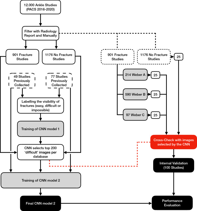
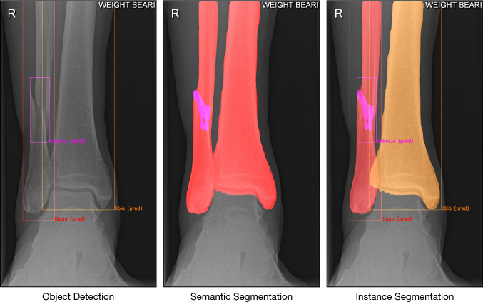
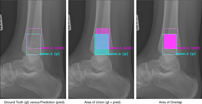
Methods: The training set included 326 isolated fibula fractures and 423 non-fracture radiographs. The Detectron2 implementation of the Mask R-CNN was trained with labelled and annotated radiographs. The internal validation (or 'test set') and external validation sets consisted of 300 and 334 radiographs, respectively. Consensus agreement between three experienced fellowship-trained trauma surgeons was defined as the ground truth label. Diagnostic accuracy and area under the receiver operator characteristic curve (AUC) were used to assess classification performance. The Intersection over Union (IoU) was used to quantify accuracy of the segmentation predictions by the CNN, where a value of 0.5 is generally considered an adequate segmentation.
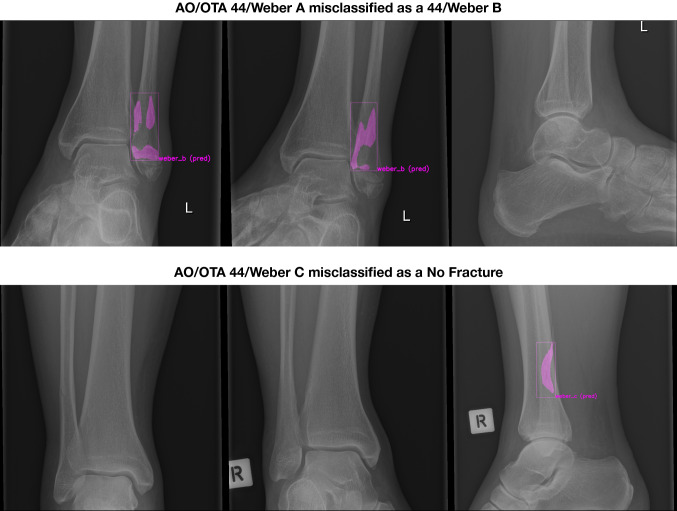
Results: The final CNN was able to classify fibula fractures according to four classes (Danis-Weber A, B, C and No Fracture) with AUC values ranging from 0.93 to 0.99. Diagnostic accuracy was 89% on the test set with average sensitivity of 89% and specificity of 96%. External validity was 89-90% accurate on a set of radiographs from a different hospital. Accuracies/AUCs observed were 100/0.99 for the 'No Fracture' class, 92/0.99 for 'Weber B', 88/0.93 for 'Weber C', and 76/0.97 for 'Weber A'. For the fracture bounding box prediction by the CNN, a mean IoU of 0.65 (SD ± 0.16) was observed. The fracture segmentation predictions by the CNN resulted in a mean IoU of 0.47 (SD ± 0.17).
Conclusions: This study presents a look into the 'black box' of CNNs and represents the first automated delineation (segmentation) of fracture lines on (ankle) radiographs. The AUC values presented in this paper indicate good discriminatory capability of the CNN and substantiate further study of CNNs in detecting and classifying ankle fractures.
Level of evidence: II, Diagnostic imaging study.
Keywords: Ankle; Artificial Intelligence; CNN; Lateral Malleolus.
© 2022. The Author(s).
Conflict of interest statement
One author (JP) certifies that he has received, an amount less than USD 15,000 from the Michael van Vloten Foundation (Rotterdam, The Netherlands), an amount less than USD 10,000 from ZonMw (Den Haag, The Netherlands), and an amount less than USD 10,000 from the Prins Bernhard Cultuur Fonds (Amsterdam, The Netherlands). One author (JND) certifies that he has received an unrestricted Postdoc Research Grant from the Marti-Keuning-Eckhardt Foundation.
Figures


References
-
- Oliveira ECL, van den Merkhof A, Olczak J, Gordon M, Jutte PC, Jaarsma RL, Ijpma FFA, Doornberg JN, Prijs J. An increasing number of convolutional neural networks for fracture recognition and classification in orthopaedics: are these externally validated and ready for clinical application? Bone Jt Open. 2021;2(10):879–885. doi: 10.1302/2633-1462.210.BJO-2021-0133. - DOI - PMC - PubMed
MeSH terms
LinkOut - more resources
Full Text Sources
Medical
Research Materials
