. 2023 Jan 2;20(1):183-193.
doi: 10.1021/acs.molpharmaceut.2c00548. Epub 2022 Nov 14.

Structure-Based Discovery of Small-Molecule Inhibitors of the Autocatalytic Proliferation of α-Synuclein Aggregates

Affiliations

Structure-Based Discovery of Small-Molecule Inhibitors of the Autocatalytic Proliferation of α-Synuclein Aggregates

Sean Chia et al. Mol Pharm. .

Abstract

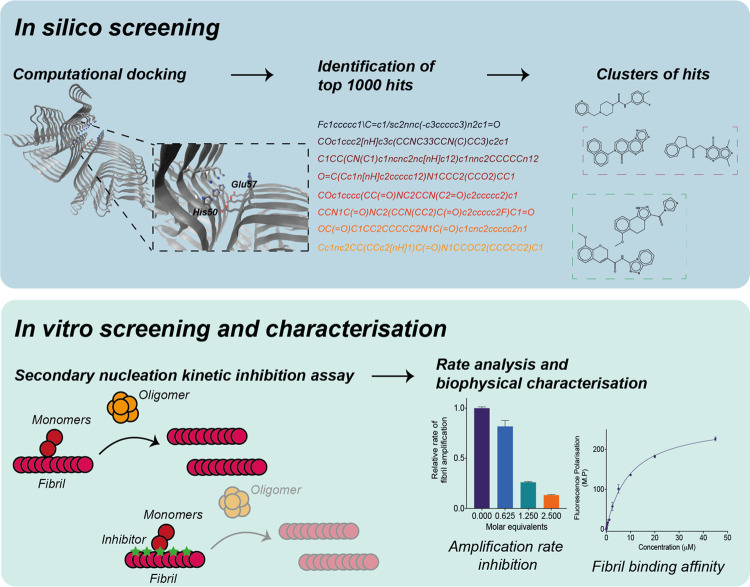
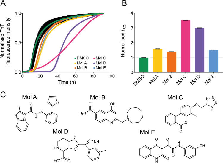
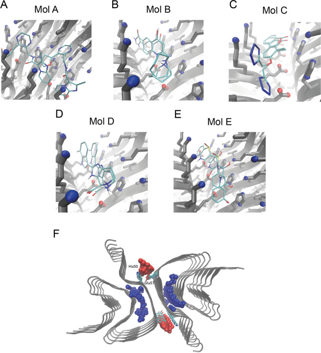
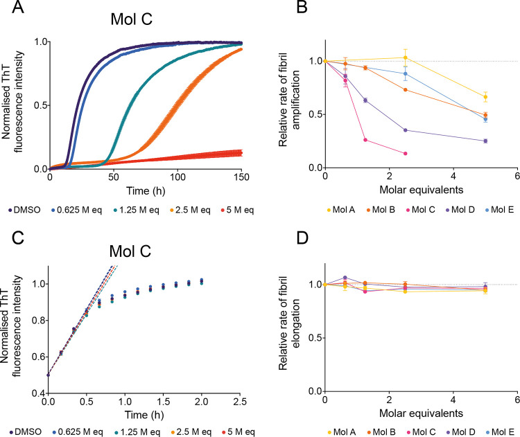
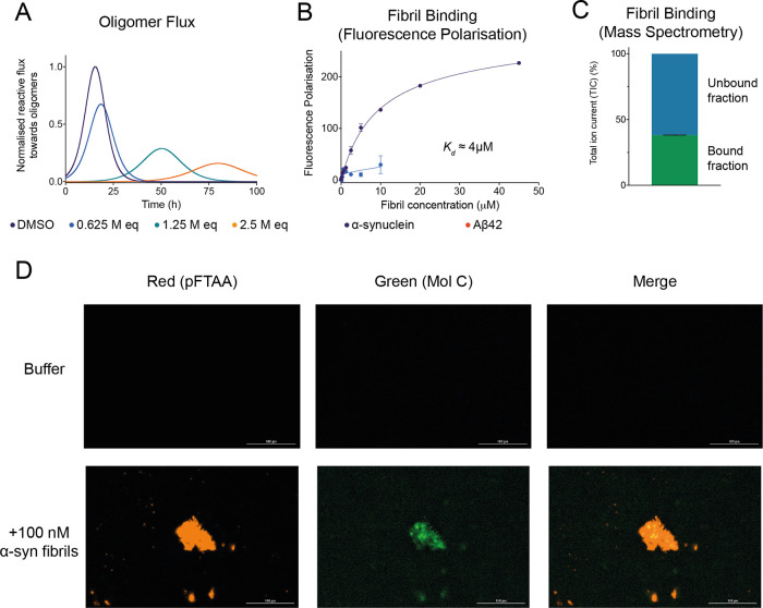
The presence of amyloid fibrils of α-synuclein is closely associated with Parkinson's disease and related synucleinopathies. It is still very challenging, however, to systematically discover small molecules that prevent the formation of these aberrant aggregates. Here, we describe a structure-based approach to identify small molecules that specifically inhibit the surface-catalyzed secondary nucleation step in the aggregation of α-synuclein by binding to the surface of the amyloid fibrils. The resulting small molecules are screened using a range of kinetic and thermodynamic assays for their ability to bind α-synuclein fibrils and prevent the further generation of α-synuclein oligomers. This study demonstrates that the combination of structure-based and kinetic-based drug discovery methods can lead to the identification of small molecules that selectively inhibit the autocatalytic proliferation of α-synuclein aggregates.

Keywords: Parkinson’s disease, α-synuclein; computational docking; kinetic-based small-molecule discovery; protein aggregation; structure-based small-molecule discovery.


Conflict of interest statement

The authors declare the following competing financial interest(s): Sara Linse, Tuomas P. J. Knowles, Johnny Habchi and Michele Vendruscolo are founders of Wren Therapeutics. Andrea Possenti, Benedetta Mannini, Roxine Staats are currently employees of Wren Therapeutics. Sean Chia and Rodrigo Cataldi have been employees of Wren Therapeutics. Robert I. Horne is a consultant for Wren Therapeutics Magdalena Nowinska has been a consultant for Wren Therapeutics.

Figures

Figure 1
Figure 1
Combined structure-based and kinetic-based approach to identify small molecules that bind α-synuclein fibrils and inhibit its aggregation. In the first step, computational docking is performed on a large library of small molecules. The top candidates are then clustered to identify a subset of chemically diverse compounds that exhibit high predicted binding scores for α-synuclein fibrils. Subsequently, these compounds are experimentally validated through a kinetic assay for their ability to inhibit the secondary nucleation aggregation of α-synuclein by binding to the surface of fibrils. Further rate constant analysis and fibril-binding experiments allow for the positive compounds to be characterized based on both their inhibition of the kinetic assay, as well as their binding affinity toward α-synuclein fibrils.
Figure 2
Figure 2
Five compounds selected from the docking library inhibit the aggregation of α-synuclein. (A) Kinetic profiles of a 10 μM solution of α-synuclein in the presence of 25 nM seeds at pH 4.8 and 37 °C, in the presence of 1% DMSO alone (beige), in the presence of 10 molar equivalents of compounds A–E (represented in different colors), or in the presence of 10 molar equivalents of other compounds in the docking library that did not affect significantly α-synuclein aggregation (black). (B) Relative t1/2 of the aggregation of α-synuclein in the presence of compounds A–E as shown in (A), normalized to the DMSO control. (C) Chemical structures of compounds A–E. Throughout, error bars represent mean ± SEM of two replicates.
Figure 3
Figure 3
Computational docking of compounds to α-synuclein fibrils. (A–E) Binding poses of compounds A–E to the selected binding pocket in α-synuclein fibrils (centered between residues His50 and Glu57), determined either through FRED or AutoDock Vina. (F) Representation of possible binding pockets in the fibril structure (PDB: 6cu7, cyan) identified by Fpocket, with pockets in the fibril core (blue spheres), and at the fibril surface (red spheres). Key binding site residues His50 and Glu57 are shown in licorice representation.
Figure 4
Figure 4
Compounds identified by docking specifically inhibit the proliferation of α-synuclein aggregates by secondary nucleation. (A) Kinetic profiles of a 10 μM solution of α-synuclein in the presence of 25 nM seeds at pH 4.8, 37 °C, in the presence of either 1% DMSO alone (purple) or increasing molar equivalents of compound C (represented in different colors). (B) Relative rate of fibril amplification of α-synuclein in the presence of compounds A–E as shown in (A) and Figure S3, normalized to the DMSO control. (C) Kinetic profiles of a 10 μM solution of α-synuclein in the presence of 5 μM seeds at pH 4.8, 37 °C, in the presence of either 1% DMSO alone (purple) or increasing molar equivalents of compound C (represented in different colors). Dotted lines indicate the vmax of the reaction which is used to extract the elongation rate of the aggregation process. (D) Relative rate of fibril elongation of α-synuclein in the presence of compounds A–E as shown in (C) and Figure S5,6, normalized to the DMSO control. Throughout, error bars represent mean ± SEM of three replicates.
Figure 5
Figure 5
Compound C inhibits the reactive flux toward α-synuclein oligomers and displays binding affinity and specificity toward α-synuclein fibrils. (A) Time dependence of the reactive flux toward α-synuclein oligomers either in the presence of 1% DMSO alone (purple) or in the presence of increasing molar equivalents of compound C (represented in different colors), normalized to the DMSO control. (B) Change in fluorescence polarization (in mP units) of 10 μM compound C with increasing concentrations of either α-synuclein fibrils (purple) or Aβ42 fibrils (red). The solid lines are fits to the points using a one-step binding curve, estimating a Kd of 4 μM for compound C toward α-synuclein fibrils. (C) Total ion current (TIC) of 10 μM compound C bound and unbound to 10 μM α-synuclein fibrils detected by mass spectrometry (see the Materials and Methods section). (D) Representative images indicating either the fluorescence of the red channel (amyloid-specific dye pFTAA) or the green channel (compound C) following incubation in the absence (top) or presence (bottom) of 100 nM α-synuclein fibrils. Throughout, error bars represent mean ± SEM of two replicates.

References

    1. Poewe W.; Seppi K.; Tanner C. M.; Halliday G. M.; Brundin P.; Volkmann J.; Schrag A. E.; Lang A. E. Parkinson Disease. Nat. Rev. Dis. Primers 2017, 3, 17013.10.1038/nrdp.2017.13. - DOI - PubMed
    1. Nichols E.; Szoeke C. E. I.; Vollset S. E.; Abbasi N.; Abd-Allah F.; Abdela J.; Aichour M. T. E.; Akinyemi R. O.; Alahdab F.; Asgedom S. W.; Awasthi A.; Barker-Collo S. L.; Baune B. T.; Béjot Y.; Belachew A. B.; Bennett D. A.; Biadgo B.; Bijani A.; Bin Sayeed M. S.; Brayne C.; Carpenter D. O.; Carvalho F.; Catalá-López F.; Cerin E.; Choi J. Y. J.; Dang A. K.; Degefa M. G.; Djalalinia S.; Dubey M.; Duken E. E.; Edvardsson D.; Endres M.; Eskandarieh S.; Faro A.; Farzadfar F.; Fereshtehnejad S. M.; Fernandes E.; Filip I.; Fischer F.; Gebre A. K.; Geremew D.; Ghasemi-Kasman M.; Gnedovskaya E. V.; Gupta R.; Hachinski V.; Hagos T. B.; Hamidi S.; Hankey G. J.; Haro J. M.; Hay S. I.; Irvani S. S. N.; Jha R. P.; Jonas J. B.; Kalani R.; Karch A.; Kasaeian A.; Khader Y. S.; Khalil I. A.; Khan E. A.; Khanna T.; Khoja T. A. M.; Khubchandani J.; Kisa A.; Kissimova-Skarbek K.; Kivimäki M.; Koyanagi A.; Krohn K. J.; Logroscino G.; Lorkowski S.; Majdan M.; Malekzadeh R.; März W.; Massano J.; Mengistu G.; Meretoja A.; Mohammadi M.; Mohammadi-Khanaposhtani M.; Mokdad A. H.; Mondello S.; Moradi G.; Nagel G.; Naghavi M.; Naik G.; Nguyen L. H.; Nguyen T. H.; Nirayo Y. L.; Nixon M. R.; Ofori-Asenso R.; Ogbo F. A.; Olagunju A. T.; Owolabi M. O.; Panda-Jonas S.; Passos V. M. d. A.; Pereira D. M.; Pinilla-Monsalve G. D.; Piradov M. A.; Pond C. D.; Poustchi H.; Qorbani M.; Radfar A.; Reiner R. C.; Robinson S. R.; Roshandel G.; Rostami A.; Russ T. C.; Sachdev P. S.; Safari H.; Safiri S.; Sahathevan R.; Salimi Y.; Satpathy M.; Sawhney M.; Saylan M.; Sepanlou S. G.; Shafieesabet A.; Shaikh M. A.; Sahraian M. A.; Shigematsu M.; Shiri R.; Shiue I.; Silva J. P.; Smith M.; Sobhani S.; Stein D. J.; Tabarés-Seisdedos R.; Tovani-Palone M. R.; Tran B. X.; Tran T. T.; Tsegay A. T.; Ullah I.; Venketasubramanian N.; Vlassov V.; Wang Y. P.; Weiss J.; Westerman R.; Wijeratne T.; Wyper G. M. A.; Yano Y.; Yimer E. M.; Yonemoto N.; Yousefifard M.; Zaidi Z.; Zare Z.; Vos T.; Feigin V. L.; Murray C. J. L. Global, Regional, and National Burden of Alzheimer’s Disease and Other Dementias, 1990–2016: A Systematic Analysis for the Global Burden of Disease Study 2016. Lancet Neurol. 2019, 18, 88–106. 10.1016/S1474-4422(18)30403-4. - DOI - PMC - PubMed
    1. Balestrino R.; Schapira A. H. V. Parkinson Disease. Eur. J. Neurol. 2020, 27, 27–42. 10.1111/ene.14108. - DOI - PubMed
    1. Pringsheim T.; Jette N.; Frolkis A.; Steeves T. D. L. The Prevalence of Parkinson’s Disease: A Systematic Review and Meta-Analysis. Mov. Disord. 2014, 29, 1583–1590. 10.1002/mds.25945. - DOI - PubMed
    1. Spillantini M. G.; Schmidt M. L.; Lee V. M.; Trojanowski J. Q.; Jakes R.; Goedert M. Alpha-Synuclein in Lewy Bodies. Nature 1997, 388, 839–840. 10.1038/42166. - DOI - PubMed

Publication types