. 2022 Nov 22;119(47):e2208886119.
doi: 10.1073/pnas.2208886119. Epub 2022 Nov 14.

Mono-(2-ethyl-5-hydroxyhexyl) phthalate promotes uterine leiomyoma cell survival through tryptophan-kynurenine-AHR pathway activation

Affiliations

Mono-(2-ethyl-5-hydroxyhexyl) phthalate promotes uterine leiomyoma cell survival through tryptophan-kynurenine-AHR pathway activation

Takashi Iizuka et al. Proc Natl Acad Sci U S A. .

Abstract

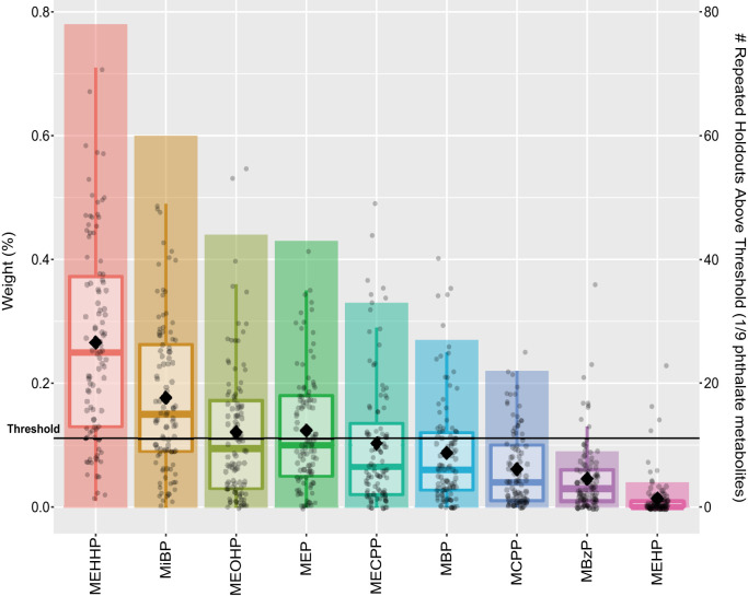
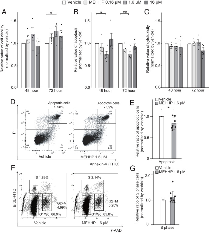
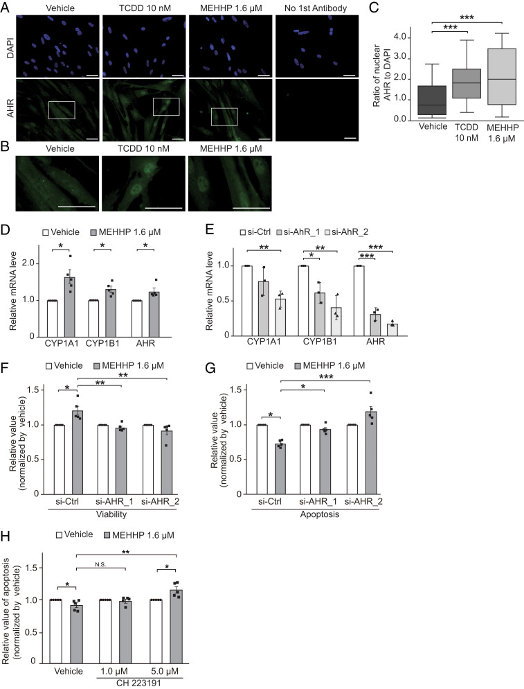
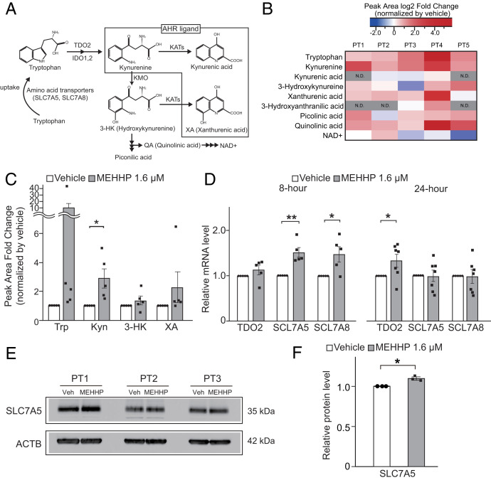
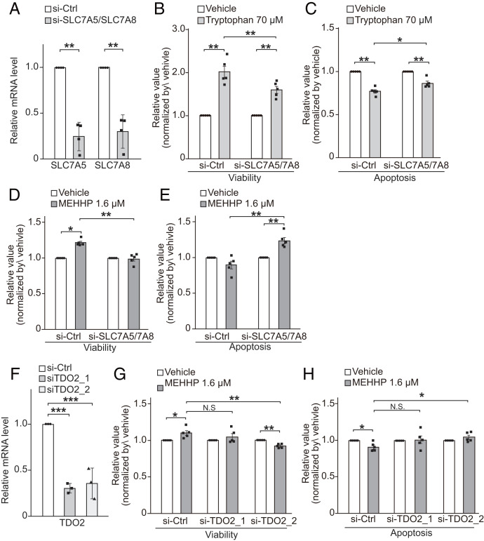
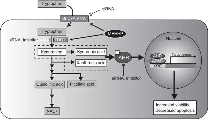
Uterine leiomyoma is the most common tumor in women and causes severe morbidity in 15 to 30% of reproductive-age women. Epidemiological studies consistently indicate a correlation between leiomyoma development and exposure to endocrine-disrupting chemical phthalates, especially di-(2-ethylhexyl) phthalate (DEHP); however, the underlying mechanisms are unknown. Here, among the most commonly encountered phthalate metabolites, we found the strongest association between the urine levels of mono(2-ethyl-5-hydroxyhexyl) phthalate (MEHHP), the principal DEHP metabolite, and the risk of uterine leiomyoma diagnosis (n = 712 patients). The treatment of primary leiomyoma and smooth muscle cells (n = 29) with various mixtures of phthalate metabolites, at concentrations equivalent to those detected in urine samples, significantly increased cell viability and decreased apoptosis. MEHHP had the strongest effects on both cell viability and apoptosis. MEHHP increased cellular tryptophan and kynurenine levels strikingly and induced the expression of the tryptophan transporters SLC7A5 and SLC7A8, as well as, tryptophan 2,3-dioxygenase (TDO2), the key enzyme catalyzing the conversion of tryptophan to kynurenine that is the endogenous ligand of aryl hydrocarbon receptor (AHR). MEHHP stimulated nuclear localization of AHR and up-regulated the expression of CYP1A1 and CYP1B1, two prototype targets of AHR. siRNA knockdown or pharmacological inhibition of SLC7A5/SLC7A8, TDO2, or AHR abolished MEHHP-mediated effects on leiomyoma cell survival. These findings indicate that MEHHP promotes leiomyoma cell survival by activating the tryptophan-kynurenine-AHR pathway. This study pinpoints MEHHP exposure as a high-risk factor for leiomyoma growth, uncovers a mechanism by which exposure to environmental phthalate impacts leiomyoma pathogenesis, and may lead to the development of novel druggable targets.

Keywords: aryl hydrocarbon receptor; endocrine-disrupting chemicals; leiomyoma; phthalate; tryptophan.


Conflict of interest statement

The authors declare no competing interest.

Figures

Fig. 1.
Fig. 1.
Relative importance of urinary phthalate metabolites to the overall association of the metabolite mixture with prior LM diagnosis. WQSR models, which were constrained in the positive direction, evaluated associations between the mixture and the risk of having (n = 193) compared to not having (n = 519) a prior LM diagnosis (RR = 1.06; 95% CI = 1.00, 1.12; P = 0.04). Boxplots present the distributions of relative weights for each metabolite from 100 repeated holdouts where each dot is a weight from one repeated holdout, the diamonds indicate the mean, and bar height indicates the proportion of repeated holdouts with weights above the threshold (1 of 9 phthalate metabolites or 11.1%). Notable metabolite contributors to the overall mixture association are those with diamonds above the threshold.
Fig. 2.
Fig. 2.
MEHHP increases viability and decreases apoptosis in LM cells. (A–C) LM cells were treated with vehicle or various concentrations of MEHHP (0.16, 1.6, 16 μM) for 48 or 72 h. Cell viability (A), apoptosis (B), and cytotoxicity (C) were assessed using the ApoTox-Glo Triplex Assay kit. Values are presented as mean ± SEM (n = 5). (D) Representative flow cytometry plots showing the percentage of apoptotic cells. LM cells were treated with vehicle or MEHHP (1.6 μM) for 48 h followed by Annexin-V and PI staining and flow cytometry analysis. (E) Bar graph showing the relative ratio of apoptotic LM cells after treatment with MEHHP vs. vehicle. Values are presented as mean ± SEM (n = 7). (F) Representative flow cytometry plots showing the distribution of LM cells in different phases of the cell cycle. Cells were treated with vehicle or MEHHP (1.6 μM) for 72 h and BrdU was added for the last 24 h. The cells were analyzed by flow cytometry. (G) Bar graph showing the relative ratio of LM cells in S phase after treatment with MEHHP vs. vehicle. Values are presented as mean ± SEM (n = 8). Statistical analysis was performed using Student’s t test or Dennett’s multiple comparison test compared with vehicle control. *P < 0.05, **P < 0.01.
Fig. 3.
Fig. 3.
MEHHP promotes LM cell survival through activation of the AHR pathway. (A) Representative images of immunocytochemistry staining for AHR (green) in LM cells treated with vehicle (DMSO), TCDD (10 nM), or MEHHP (1.6 μM) for 24 h. DAPI was used for nuclear staining (blue). (Scale bars, 100 μm.) (B) High-magnification images of cells highlighted in A. (Scale bars, 100 μm.) (C) The relative ratio of the AHR-positive nuclear area normalized to the DAPI-stained nuclear area quantified by ImageJ. Statistical analysis was performed using the Kruskal–Wallis test compared with vehicle control (n = 3). ***P < 0.001. (D) Bar graph showing RT-qPCR quantification of CYP1A1, CYP1B1, and AHR gene expression levels in LM cells treated with MEHHP (1.6 μM) or vehicle for 8 h. Values are presented as mean ± SEM (n = 5). (E) Bar graph showing RT-qPCR quantification of AHR, CYP1A1, and CYP1B1 gene expression levels in LM cells transfected with control siRNA (si-Ctrl) or two different AHR siRNAs (si-AHR_1 and si-AHR_2). Values are presented as mean ± SEM (n = 3). (F and G) The effects of AHR knockdown on MEHHP-induced cell survival in LM cells. The cells were transfected with control or AHR siRNAs for 24 h, followed by treatment with vehicle or MEHHP (1.6 μM) for 72 h. Cell viability (F) and apoptosis (G) were assessed by ApoTox-Glo Triplex Assay. Values were normalized to vehicle control and presented as mean ± SEM (n = 5). H: LM cells were treated with MEHHP in the presence or absence of the AHR-specific antagonist CH 223191 at 1 μM and 5 μM for 72 h. Apoptosis was assessed by evaluation of Caspase 3/7 activity. Values were normalized to vehicle control for each group and presented as mean ± SEM (n = 5). Statistical analysis was performed using Student’s t test or Dennett’s multiple comparison test compared with control. *P < 0.05, **P < 0.01, ***P < 0.001.
Fig. 4.
Fig. 4.
MEHHP promotes tryptophan uptake and metabolism in LM cells. (A) Schematic diagram of the tryptophan-kynurenine metabolism pathway. IDO, indoleamine 2,3-dioxygenase; KATs, kynurenine aminotransferases; KMO, kynurenine 3-monooxygenase; NAD, nicotinamide adenine dinucleotide; SCL7A5, Solute Carrier Family 7 Member 5; SCL7A8, Solute Carrier Family 7 Member 8. (B and C) The levels of tryptophan and its metabolites in LM cells treated with MEHHP (1.6 μM) or vehicle for 72 h were assessed by LC-MS. The fold-change of peak area of each metabolite in MEHHP- vs. vehicle-treated cells is shown as a heat map (B) and histogram (C). Values are presented as mean ± SEM (n = 5). Trp, tryptophan; Kyn, kynurenine. (D) Gene expression in LM cells treated with MEHHP (1.6 μM) for 8 h (n = 5) or 24 h (n = 8). Values are presented as mean ± SEM. (E) Immunoblot image showing SLC7A5 protein expression in LM cells treated with MEHHP (1.6 µM). (F) ImageJ quantification of SLC7A5 protein levels in E. Values are presented as mean ± SD (n = 3). Statistical analysis was performed using paired t test compared with control. *P < 0.05, **P < 0.01.
Fig. 5.
Fig. 5.
Effects of blocking the tryptophan-kynurenine metabolism pathway on MEHHP-mediated LM cell survival. (A) Knockdown of SLC7A5 and SLC7A8 in LM cells significantly decreased their gene-expression levels as assessed by RT-qPCR (n = 4). (B and C) The effects of SLC7A5 and SLC7A8 knockdown on tryptophan-mediated LM cell survival. Twenty-four hours after siRNA transfection, LM cells were treated with vehicle (water) or 70 μM tryptophan for 72 h followed by assessment of cell viability (B) and apoptosis (C) by ApoTox-Glo Triplex Assay. Values were normalized to vehicle control for each group (n = 5). (D and E) The effects of SLC7A5 and SLC7A8 knockdown on MEHHP-mediated LM cell viability (D) and apoptosis (E). As in B and C, after siRNA transfection, LM cells were treated with vehicle or 1.6 μM MEHHP followed by analysis with the ApoTox-Glo Triplex Assay. Values were normalized to vehicle control for each group (n = 5). (F) Two different siRNAs for TDO2 (siTDO2_1, siTDO2_2) significantly decreased its gene expression in LM cells (n = 3). (G and H) Knockdown of TDO2 reduced the prosurvival effects of MEHHP on LM cells. The cells were transfected with siRNA and treated with MEHHP, followed by assessment with the ApoTox-Glo Triplex Assay for cell viability (G) and apoptosis (H) (n = 5). Values were normalized to vehicle control for each group. Values are presented as mean ± SEM. Statistical analysis was performed using Student’s t test or Dennett’s multiple comparison test compared with control. *P < 0.05, **P < 0.01, ***P < 0.001.
Fig. 6.
Fig. 6.
Schematic of MEHHP effect in LM cells. MEHHP stimulates tryptophan metabolism and increases AHR ligand production, which activates the AHR pathway and contributes to LM cell survival. XRE, Xenobiotic response element.

References

    1. Bulun S. E., Uterine fibroids. N. Engl. J. Med. 369, 1344–1355 (2013). - PubMed
    1. Kim J. J., Kurita T., Bulun S. E., Progesterone action in endometrial cancer, endometriosis, uterine fibroids, and breast cancer. Endocr. Rev. 34, 130–162 (2013). - PMC - PubMed
    1. Cardozo E. R., et al. , The estimated annual cost of uterine leiomyomata in the United States. Am. J. Obstet. Gynecol. 206, 211.e1–211.e9 (2012). - PMC - PubMed
    1. Ishikawa H., et al. , Progesterone is essential for maintenance and growth of uterine leiomyoma. Endocrinology 151, 2433–2442 (2010). - PMC - PubMed
    1. Faerstein E., Szklo M., Rosenshein N., Risk factors for uterine leiomyoma: A practice-based case-control study. I. African-American heritage, reproductive history, body size, and smoking. Am. J. Epidemiol. 153, 1–10 (2001). - PubMed

Publication types

LinkOut - more resources