. 2022 Nov 22;119(47):e2206923119.
doi: 10.1073/pnas.2206923119. Epub 2022 Nov 14.

PAHSAs reduce cellular senescence and protect pancreatic beta cells from metabolic stress through regulation of Mdm2/p53

Affiliations

PAHSAs reduce cellular senescence and protect pancreatic beta cells from metabolic stress through regulation of Mdm2/p53

Maria F Rubin de Celis et al. Proc Natl Acad Sci U S A. .

Abstract

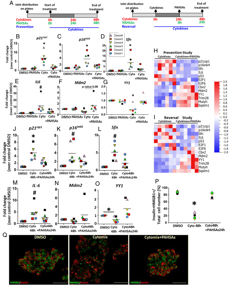
Senescence in pancreatic beta cells plays a major role in beta cell dysfunction, which leads to impaired glucose homeostasis and diabetes. Therefore, prevention of beta cell senescence could reduce the risk of diabetes. Treatment of nonobese diabetic (NOD) mice, a model of type 1 autoimmune diabetes (T1D), with palmitic acid hydroxy stearic acids (PAHSAs), a novel class of endogenous lipids with antidiabetic and antiinflammatory effects, delays the onset and reduces the incidence of T1D from 82% with vehicle treatment to 35% with PAHSAs. Here, we show that a major mechanism by which PAHSAs protect islets of the NOD mice is by directly preventing and reversing the initial steps of metabolic stress-induced senescence. In vitro PAHSAs increased Mdm2 expression, which decreases the stability of p53, a key inducer of senescence-related genes. In addition, PAHSAs enhanced expression of protective genes, such as those regulating DNA repair and glutathione metabolism and promoting autophagy. We demonstrate the translational relevance by showing that PAHSAs prevent and reverse early stages of senescence in metabolically stressed human islets by the same Mdm2 mechanism. Thus, a major mechanism for the dramatic effect of PAHSAs in reducing the incidence of type 1 diabetes in NOD mice is decreasing cellular senescence; PAHSAs may have a similar benefit in humans.

Keywords: cellular senescence; diabetes; lipids; metabolic stress; pancreatic islets.


Conflict of interest statement

Competing interest statement: I.S. and B.B.K. are inventors on patents related to the FAHFAs.

Figures

Fig. 1.
Fig. 1.
PAHSA treatment of NOD mice preserves beta cell phenotype and protects beta cells from senescence. (A) NOD mice were treated with either PAHSAs (5- and 9-PAHSA) or vehicle by oral gavage for 6 wks from 4 wks of age until 10 wks of age before hyperglycemia occurred. (BD) RNAseq data analysis from NOD mouse islets revealed PAHSA effects on the preservation of the expression of genes responsible for maintenance of islet identity and hormones (B), down-regulation of disallowed genes (C), and apoptosis (D). (E) Quantification of TUNEL assay in the beta cells of pancreas sections from NOD mice treated with a combination of 5- and 9-PAHSA or vehicle for 6 wk (n = 5 to 7; *P < 0.05 by two-tailed Student’s t test). (FH) PAHSA treatment of NOD mice also prevented up-regulation of aging-related (F), senescence-related (G), and SASP-related (H) genes. (I and J). Genes involved in DNA repair (I) and GSH metabolism (J) were maintained with PAHSA treatment. (K) Immunohistochemistry and quantification of pancreas sections from the same NOD mice as in A showing P21Cip1 and P16Ink4a (green), insulin (red), and DAPI (blue). Quantification of p21Cip1 or p16Ink4 was also performed in alpha cells; n = 5 to 7 mice/group. (Scale bars, 50 μm.) Data are shown as means ± SEM; *P < 0.001 versus vehicle p21cip1 or p16ink4. Data were analyzed by two-tailed Student’s t test. (L and M) Immunohistochemistry and quantification of pancreas sections from prehyperglycemic NOD mice (11 wk of age) and hyperglycemic (for vehicle-treated mice) NOD mice (15 wks of age) treated for 6 or 11 wks with either PAHSAs or vehicle. (L) Nuclear HMGB1 (green), insulin (magenta), and DAPI images are shown as well as quantification of HMGB1 at 11 wks of age in alpha cells. (M) γH2Ax (green), insulin (magenta), and DAPI images are shown as well as image quantification; n = 4 to 6 (HMGB1) mice/group. (Scale bar, 50 μm.) Data are shown as means ± SEM (*P < 0.001 versus vehicle HMGB1 [L] or γH2Ax [M]) and were analyzed by two-tailed Student’s t test.
Fig. 2.
Fig. 2.
PAHSAs prevent and reverse the effects of metabolic stress in beta cells in vitro. (AC) Schemas of studies to prevent the initiation of senescence in MIN6 cells under different metabolic stress conditions: glucotoxicity (A), cytokine stress (B), and ER stress induced by thapsigargin (C). (DR) PAHSA treatment prevented some changes in gene expression in MIN6 cells exposed to metabolic stress conditions: high glucose (D, G, J, M, and P) cytokine stress (E, H, K, N, and Q) or ER stress (F, I, L, O, and R). Expression of senescence-related genes p21Cip1 (DF), p16Ink4a (GI), and IL-6 (JL), beta cell maturity marker Ucn3 (MO), and DNA damage gene H2Ax (PR) is shown; n = 4 independent experiments. Data are shown as means ± SEM (*P < 0.05, **P < 0.005, and ***P < 0.001 versus control) and were analyzed by one-way ANOVA with Bonferroni multiple comparisons test; Cyto, cytokines; Thap, thapsigargin. (SU) Schema for reversal studies in MIN6 cells under different metabolic stress conditions: glucotoxicity (S), cytokine stress (T), and ER stress induced by thapsigargin (U). The stressors were present until the end of the experiment. (V) HMGB1 immunohistochemistry showing a reversal experiment in MIN6 cells. Separate channels are shown in SI Appendix, Fig. S5. Quantification (WY) of loss of nuclear HMGB1 in MIN6 cells under three different metabolic stress conditions: glucotoxicity (W), cytokine stress (X), and ER stress induced by thapsigargin (Y) (Scale bars, 100 μm, left three columns; Scale bars, 32 μm, right three columns). Data are shown as means ± SEM (**P < 0.005 and ***P < 0.001 versus control) and were analyzed by one-way ANOVA with a Bonferroni multiple comparisons test.
Fig. 3.
Fig. 3.
Mechanism by which PAHSAs protect beta cells under metabolic stress conditions. (A) Proposed model for the mechanism by which PAHSAs reduce stress-induced senescence in beta cells: reduction of p53 by preserving or increasing expression of its negative regulator Mdm2. (B) PAHSA treatment preserved the expression of genes related to Mdm2 and p53 regulation in NOD mouse pancreatic islets (6 wk of treatment, 10 wk of age). (C) PAHSAs reversed cytokine stress–induced changes of senescence and SASP-related gene expression in MIN6 cells transfected with siRNA for Mdm2 (gray bars) or control siRNA (white bars); n = 4 independent experiments, each with three replicates per condition. Data show means ± SEM; *P < 0.05 versus DMSO and PAHSA siRNA control; #P < 0.05 versus cytokines siRNA control; &P < 0.05 different from all other conditions; $P < 0.05 different from all conditions except DMSO and PAHSA controls; P < 0.05 different from both DMSO and PAHSA conditions. (D) PAHSAs reversed ER stress–induced senescence and SASP-related gene expression in MIN6 cells transfected with siRNA for Mdm2 (gray bars) or control siRNA (white bars). Data show means ± SEM; *P < 0.05 versus DMSO and PAHSA siRNA control; #P < 0.05 different from DMSO and PAHSA controls and thapsigargin siRNA control; &P < 0.05 different from all other conditions; $P < 0.05 different from all except DMSO and PAHSA controls; @P < 0.05 different from all except thapsigargin siRNA control; P < 0.05 different from both DMSO and PAHSA conditions. Data in C and D were analyzed by one-way ANOVA with Bonferroni multiple comparisons test. (E and F) Western blotting analysis of p53, phospho-p53Ser15, MDM2, YY1, and vinculin in MIN6 cells with reversal by PAHSAs of cytokine-stress (E) and ER stress (F) induced by thapsigargin. Cells were transfected with either siRNA for Mdm2 or control siRNA (n = 3 replicates per condition). Data are representative of three different Western blots. (G) Quantification of the data in E showing means ± SEM; $P < 0.05 different from all except DMSO siRNA control conditions; *P < 0.05 versus DMSO siRNA control and cytokines 48 h + PAHSAs 24 h siRNA control; P < 0.05 versus all conditions; @P < 0.05 versus all conditions except DMSO siRNA Mdm2. (H) Quantification of F; &P < 0.05 versus all conditions; #P < 0.05 versus DMSO siRNA control conditions and thapsigargin + PAHSAs 3 h siRNA control; @P < 0.05 versus all conditions except DMSO siRNA for Mdm2. Data were analyzed by one-way ANOVA with a Bonferroni multiple comparisons test. (I) PAHSAs preserved and reversed GSH protein levels, which decreased with cytokine exposure in MIN6 cells. Quantification of total GSH, GSSH, and GSH (reduced GSH). White bars show controls containing DMSO, and gray bars show PAHSAs (dissolved in DMSO). Data are shown as means ± SEM; *P < 0.05 versus all conditions; #P < 0.05 versus cytokines without PAHSAs. Data were analyzed by one-way ANOVA with a Bonferroni multiple comparisons test; Cyto, cytokines, Thap, Thapsigargin. (J) Model illustrating the mechanism by which PAHSAs exert their beneficial effects on pancreatic beta cells under metabolic stress conditions. PAHSAs act through Mdm2 and its cofactors to destabilize p53. PAHSAs also alter DNA repair mechanisms and autophagy to preserve the beta cell population. In addition, PAHSAs regulate p53 through the transcription factors E2F1 and E2F6. The effect of PAHSAs on p53 results in decreased expression of downstream genes related to senescence, SASP, and apoptosis. In addition, PAHSAs increase the expression of genes related to GSH metabolism to protect the beta cell from metabolic stress.
Fig. 4.
Fig. 4.
PAHSAs prevent and reverse the initiation of senescence induced by metabolic stress in human islets. (A) Schemas of prevention and reversal experiments of PAHSA treatment during cytokine-induced stress in human islets from six different donors. (BG) PAHSAs prevented up-regulated expression of senescence and SASP genes islets from all human donors (n = 6). Data are shown as means ± SEM; *P < 0.05 versus all conditions; #P < 0.05 versus control DMSO. Data were analyzed by one-way ANOVA with a Tukey’s multiple comparisons test. (H) Heat map of PCR-determined expression of genes related to the mechanism by which PAHSAs prevent senescence in beta cells represented in I. Data are from human islets exposed to cytokines in the prevention study. (I) Heat maps of PCR-determined expression of genes related to the mechanism by which PAHSAs reverse senescence in beta cells represented in Fig. 3J. Data are from human islets exposed to cytokines in the reversal study. (JO) PAHSAs reversed the decreased expression of senescence and SASP-related genes to levels similar to control conditions. PAHSAs maintained or increased MDM2 and YY1 expression. Data are shown as means ± SEM; *P < 0.05 versus all conditions; #P < 0.05 versus control DMSO. Data were analyzed by one-way ANOVA with a Tukey’s multiple comparisons test. (P and Q) Quantification (P) and representative pictures (Q) of nuclear HMGB1 in beta cells from human islets. Cytomix (Cyto, combination of recombinant human IL1β, Tnfα and IFNγ. (Scale bar, 50 μm.) Data are shown as means ± SEM; *P < 0.05 versus all conditions. Data were analyzed by one-way ANOVA with a Tukey’s multiple comparisons test.

References

    1. Atkinson M. A., et al. , How does type 1 diabetes develop?: The notion of homicide or β-cell suicide revisited. Diabetes 60, 1370–1379 (2011). - PMC - PubMed
    1. Cnop M., Toivonen S., Igoillo-Esteve M., Salpea P., Endoplasmic reticulum stress and eIF2α phosphorylation: The Achilles heel of pancreatic β cells. Mol. Metab. 6, 1024–1039 (2017). - PMC - PubMed
    1. Donath M. Y., et al. , Mechanisms of beta-cell death in type 2 diabetes. Diabetes 54 (suppl. 2), S108–S113 (2005). - PubMed
    1. Hudish L. I., Reusch J. E., Sussel L., β cell dysfunction during progression of metabolic syndrome to type 2 diabetes. J. Clin. Invest. 129, 4001–4008 (2019). - PMC - PubMed
    1. Weir G. C., Bonner-Weir S., Five stages of evolving beta-cell dysfunction during progression to diabetes. Diabetes 53 (suppl. 3), S16–S21 (2004). - PubMed

Publication types