. 2023 Mar 30;141(13):1584-1596.
doi: 10.1182/blood.2022016934.

NRX-0492 degrades wild-type and C481 mutant BTK and demonstrates in vivo activity in CLL patient-derived xenografts

Affiliations

NRX-0492 degrades wild-type and C481 mutant BTK and demonstrates in vivo activity in CLL patient-derived xenografts

Deyi Zhang et al. Blood. .

Abstract

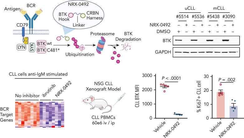
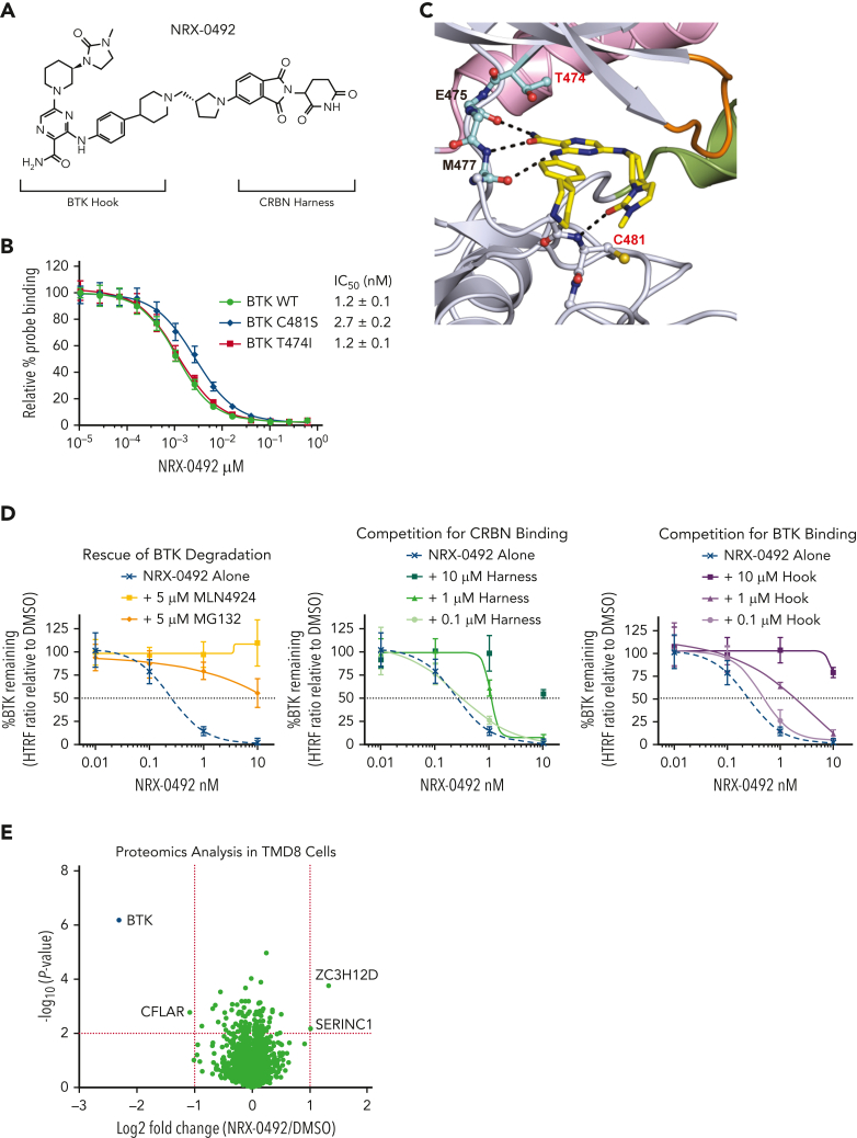
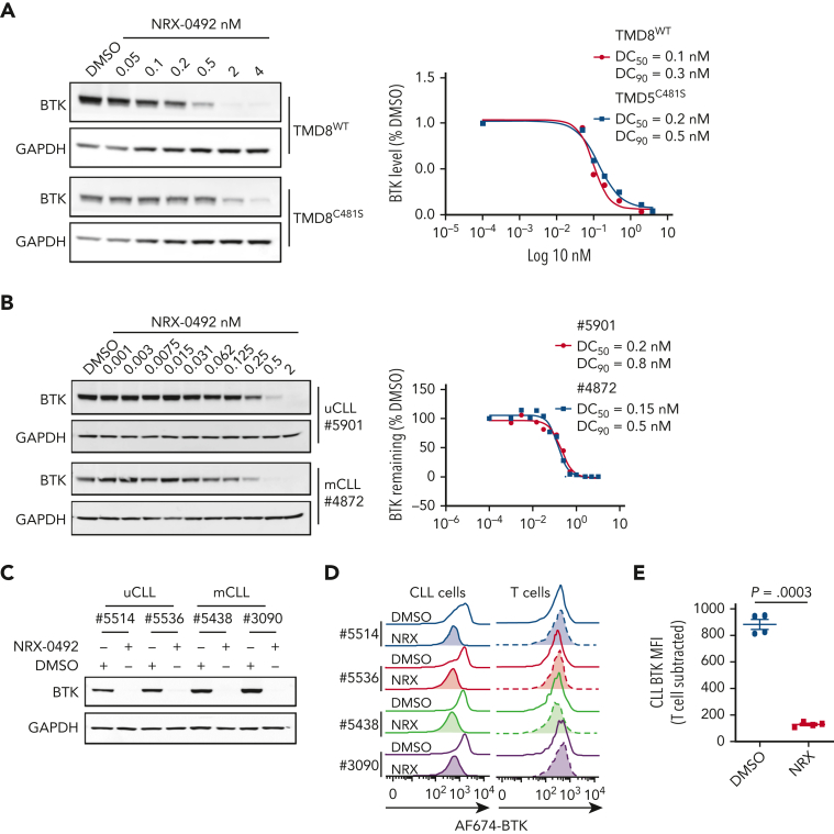
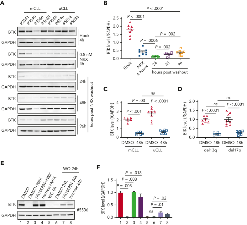
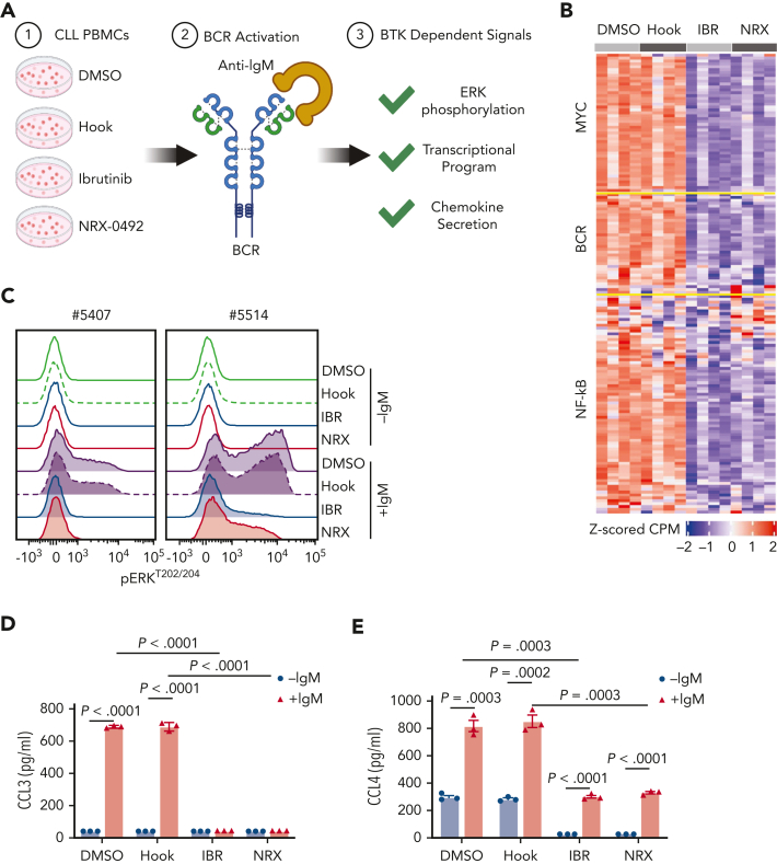
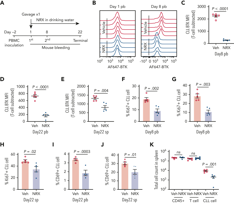
Bruton tyrosine kinase (BTK) is essential for B-cell receptor (BCR) signaling, a driver of chronic lymphocytic leukemia (CLL). Covalent inhibitors bind C481 in the active site of BTK and have become a preferred CLL therapy. Disease progression on covalent BTK inhibitors is commonly associated with C481 mutations. Here, we investigated a targeted protein degrader, NRX-0492, that links a noncovalent BTK-binding domain to cereblon, an adaptor protein of the E3 ubiquitin ligase complex. NRX-0492 selectively catalyzes ubiquitylation and proteasomal degradation of BTK. In primary CLL cells, NRX-0492 induced rapid and sustained degradation of both wild-type and C481 mutant BTK at half maximal degradation concentration (DC50) of ≤0.2 nM and DC90 of ≤0.5 nM, respectively. Sustained degrader activity was maintained for at least 24 hours after washout and was equally observed in high-risk (deletion 17p) and standard-risk (deletion 13q only) CLL subtypes. In in vitro testing against treatment-naïve CLL samples, NRX-0492 was as effective as ibrutinib at inhibiting BCR-mediated signaling, transcriptional programs, and chemokine secretion. In patient-derived xenografts, orally administered NRX-0492 induced BTK degradation and inhibited activation and proliferation of CLL cells in blood and spleen and remained efficacious against primary C481S mutant CLL cells collected from a patient progressing on ibrutinib. Oral bioavailability, >90% degradation of BTK at subnanomolar concentrations, and sustained pharmacodynamic effects after drug clearance make this class of targeted protein degraders uniquely suitable for clinical translation, in particular as a strategy to overcome BTK inhibitor resistance. Clinical studies testing this approach have been initiated (NCT04830137, NCT05131022).


Conflict of interest statement

Conflict-of-interest disclosure: A.K., J.M., A.T.-M., E.A., Y.S.T., H.L., S.G., M.C.C., S.Y., D.W.R., and M.N. were or currently are full-time employees and shareholders of Nurix Therapeutics. A.W. received research funding from Pharmacyclics, Acerta Pharma, Merck, Verastem, Genmab, and Nurix Therapeutics. C.S. received research funding from Genmab. The remaining authors declare no competing financial interests.

Figures

None
Graphical abstract
Figure 1.
Figure 1.
NRX-0492 selectively degrades BTK via a cereblon (CRBN)- and proteasome-dependent mechanism. (A) Chemical structure of NRX-0492. (B) Binding of NRX-0492 to BTK WT, C481S, and T474I mutant BTK measured in a FRET competition assay. Data represent average values and standard deviations from 3 experiments. (C) Crystal structure of kinase domain of WT BTK in gray cartoon bound to the “hook” of NRX-0492 in yellow sticks. Functional kinase motives are colored for orientation: c-Helix in pink, P-loop in orange, activation loop in green, and gate/hinge in cyan. Mainchain atoms that engage in hydrogen bonding interaction are shown in sticks and the corresponding hinge region residues are labeled in black. The hydrogen bonding interactions are indicated with dashed black lines. Sidechains of mutational hotspots T474 and C481 are shown in sticks and labeled in red. (D) TMD8 cells were pretreated with MLN4924, MG132, harness, or hook at the indicated concentrations for 1 hour. Cells were then treated with NRX-0492 for an additional 4 hours, and BTK levels were quantified by total-BTK homologous time–resolved fluorescence. Each experiment was performed in triplicate. Graphs represent mean ± SD. (E) TMD8 cells were treated for 6 hours with 50 nM NRX-0492 or DMSO in triplicate. Samples were analyzed with tandem mass tag mass spectrometry. Results are graphed in a volcano plot with a statistical significance threshold of P < .01 indicated by the horizontal dotted line.
Figure 2.
Figure 2.
NRX-0492 induces degradation of WT and C481S mutant BTK. Degradation of BTK by NRX-0492 was assessed by Western blot in TMD8 (A) and primary CLL cells (B) and DC50 and DC90 were calculated based on immunoblot quantification. Comparative analysis of BTK degradation in PBMCs from 4 patients exposed to 2nM NRX-0492 for 4 hours using (C) immunoblotting and (D) flow cytometry. NRX-0492 reduced BTK staining intensity in CLL cells but not T cells. (E) Mean fluorescent intensity of anti-BTK AF647 in T cells was used as background control; comparison by paired t test. MFI, mean fluorescent intensity.
Figure 3.
Figure 3.
BTK degradation by NRX-0492 in CLL cells is sustained and equally achieved in high and standard-risk disease. (A) Immunoblots of CLL PBMCs that were treated with 0.5 nM hook or 0.5 nM NRX-0492 for 4 hours and then cultured for the indicated times after drug washout, with (B) quantification of BTK relative to loading control (GAPDH) over time. (C, D) Mean (± SEM) BTK levels in CLL PBMCs 48 hours after drug washout of vehicle (DMSO) or NRX-0492 with samples divided by (C) IGHV status into mutated CLL and unmutated CLL or (D) cytogenetic risk group, del13q (low risk), del17p (high risk). Comparisons over time by paired t test, comparisons between treatment subgroups by unpaired t test; ns, not significant. (E) Representative immunoblot showing BTK in CLL PBMCs treated as indicated: lane 1 DMSO only; lanes 2 to 4, cells were treated with DMSO, MLN4924, or harness for 1 hour followed by 2 nM NRX-0492 treatment for 4 hours. Lanes 5 to 8, cells were treated with NRX-0492 for 4 hours followed by drug washout (WO). 24 hours post washout, DMSO, MLN4924, or harness were added for an additional 24 hours. (F) Summary of experiments using cells from 3 different patients, statistics by paired t test.
Figure 4.
Figure 4.
BTK inhibition and BTK degradation have comparable effects on CLL biology. (A) In vitro assays using CLL PBMCs to investigate the impact of different drugs on BTK-dependent signaling in response to BCR engagement. (B) CLL cell transcriptome assessed by RNA sequencing of CLL cells from 4 patients treated with DMSO, hook, 1 μM ibrutinib (IBR), or 2 nM NRX-0492, DMSO for 18 hours followed by 20 μg/mL anti-IgM stimulation for 6 hours. Median centered heatmap scaled as indicated depicts changes in defined gene signatures representing MYC, BCR, and NF-κB regulated genes. Each row represents a gene and each column represents a sample. supplemental Table 2 lists the genes in the same sequence as shown in the heat map together with the expression value across the different conditions. (C) Histogram depicting flow cytometry staining of p-ERK1/2Thr202/Tyr204 in CLL cells from 2 representative patients, drug treatment as in B, anti-IgM stimulation for 15 minutes. (D) CCL3 and (E) CCL4 concentrations by enzyme-linked immunosorbent assay in supernatant of CLL cells treated as in B, cell culture supernatants collected 48 hours after stimulation. ERK, extracellular signal-regulated kinase.
Figure 5.
Figure 5.
BTK degradation and anti-CLL activity of NRX-0492 in PDX model. In vivo activity was tested in the PDX model of CLL. Ten NSG mice were inoculated with PBMCs from patient #5539 with CLL. (A) Schematic display of the experimental schedule. (B) Representative histogram of AF647-BTK staining in CLL cells from peripheral blood (pb) on day1 before drug treatment (left) and after 7 days of treatment with NRX-0492 (day 8, right). (C-E) Mean (± SEM) CLL AF647-BTK MFI in peripheral blood on day 8 (C), day 22 (D), and spleen on day 22 (E). Each symbol represents 1 mouse. (F, G) Mean (± SEM) percentage of Ki67 expression in CLL cells in peripheral blood on day 8 (F), day 22 (G), and spleen on day 22 (H). (I, J) The percentage of CD69 positive CLL cells in peripheral blood (I) and spleen (J) on day 22. (K) Mean total cell counts (± SEM) of human cells (CD45+), T cells, and CLL cells in spleen on day 22. Comparisons are by unpaired student t test; ns, not significant. Veh, vehicle; NRX, NRX-0492; pb, peripheral blood; sp, spleen.
Figure 6.
Figure 6.
Summary of NRX-0492 testing in CLL PDX model. Experimental design as in Figure 5. PBMCs from 4 different patients (represented by unique symbols) were inoculated into NSG mice that were divided to vehicle control (Veh) and NRX-0492 (NRX) treatment groups. Each symbol represents 1 mouse. (A) Mean (± SEM) MFI of AF647-BTK in CLL cells in peripheral blood of mice after 7 days of NRX-0492 treatment. (B) Mean (± SEM) MFI of AF647-BTK in CLL cells from spleens on day 22. (C) Mean (± SEM) percentage of CLL cells expressing Ki67 in peripheral blood before treatment start (Day 1) and after 1 week of treatment and in cells from spleens on day 22. (D) Mean (± SEM) percentage of live CLL cells among CD45+ cells in the spleens on day 22. Statistical analysis by unpaired student t test; ns, not significant.
Figure 7.
Figure 7.
Activity of NRX-0492 against IBR-resistant CLL with BTK C481S mutation. (A) PBMCs from 2 patients with CLL with clinical progression on IBR and BTK C481S mutations were treated with NRX-0492 for 24 hours at the indicated concentrations and analyzed for BTK expression by Western blotting. (B) Estimates of DC50 and DC90 concentrations based on data shown in A. The fraction of residual BTK and the cancer cell fractions of C481S mutations for the samples tested is indicated (C-F) PBMCs from patient with CLL #12803 who progressed on IBR with BTK C481S were used to establish the PDX model. (C) BTK and (D) Ki67 expression by flow cytometry in peripheral blood CLL cells of vehicle (Veh) and NRX-0492 treated mice on day 8; (E) Western blotting for BTK in CLL cells, selected by CD3 negative selection, from spleens on day 20.

Comment in

References

    1. Burger JA. Treatment of chronic lymphocytic leukemia. N Engl J Med. 2020;383(5):460–473. - PubMed
    1. Herishanu Y, Perez-Galan P, Liu D, et al. The lymph node microenvironment promotes B-cell receptor signaling, NF-κB activation, and tumor proliferation in chronic lymphocytic leukemia. Blood. 2011;117(2):563–574. - PMC - PubMed
    1. Wiestner A. The role of B-cell receptor inhibitors in the treatment of patients with chronic lymphocytic leukemia. Haematologica. 2015;100(12):1495–1507. - PMC - PubMed
    1. Stevenson FK, Krysov S, Davies AJ, Steele AJ, Packham G. B-cell receptor signaling in chronic lymphocytic leukemia. Blood. 2011;118(16):4313–4320. - PubMed
    1. Treon SP, Tripsas CK, Meid K, et al. IBR in previously treated Waldenstrom's macroglobulinemia. N Engl J Med. 2015;372(15):1430–1440. - PubMed

MeSH terms

Substances

Associated data