. 2022 Dec:86:104351.
doi: 10.1016/j.ebiom.2022.104351. Epub 2022 Nov 11.

Fulminant lung fibrosis in non-resolvable COVID-19 requiring transplantation

Affiliations

Fulminant lung fibrosis in non-resolvable COVID-19 requiring transplantation

Soma S K Jyothula et al. EBioMedicine. 2022 Dec.

Abstract

Background: Coronavirus Disease 2019 (COVID-19) can lead to the development of acute respiratory distress syndrome (ARDS). In some patients with non-resolvable (NR) COVID-19, lung injury can progress rapidly to the point that lung transplantation is the only viable option for survival. This fatal progression of lung injury involves a rapid fibroproliferative response and takes on average 15 weeks from initial symptom presentation. Little is known about the mechanisms that lead to this fulminant lung fibrosis (FLF) in NR-COVID-19.

Methods: Using a pre-designed unbiased PCR array for fibrotic markers, we analyzed the fibrotic signature in a subset of NR-COVID-19 lungs. We compared the expression profile against control lungs (donor lungs discarded for transplantation), and explanted tissue from patients with idiopathic pulmonary fibrosis (IPF). Subsequently, RT-qPCR, Western blots and immunohistochemistry were conducted to validate and localize selected pro-fibrotic targets. A total of 23 NR-COVID-19 lungs were used for RT-qPCR validation.

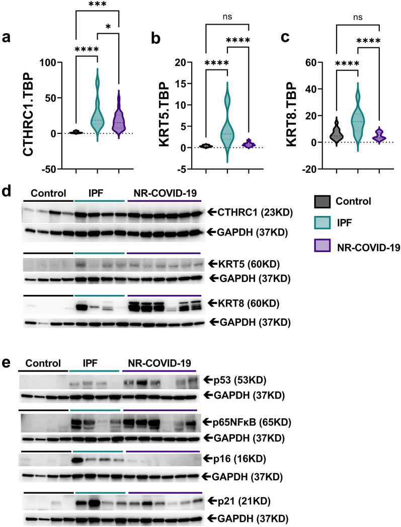
Findings: We revealed a unique fibrotic gene signature in NR-COVID-19 that is dominated by a hyper-expression of pro-fibrotic genes, including collagens and periostin. Our results also show a significantly increased expression of Collagen Triple Helix Repeat Containing 1(CTHRC1) which co-localized in areas rich in alpha smooth muscle expression, denoting myofibroblasts. We also show a significant increase in cytokeratin (KRT) 5 and 8 expressing cells adjacent to fibroblastic areas and in areas of apparent epithelial bronchiolization.

Interpretation: Our studies may provide insights into potential cellular mechanisms that lead to a fulminant presentation of lung fibrosis in NR-COVID-19.

Funding: National Institute of Health (NIH) Grants R01HL154720, R01DK122796, R01DK109574, R01HL133900, and Department of Defense (DoD) Grant W81XWH2110032 to H.K.E. NIH Grants: R01HL138510 and R01HL157100, DoD Grant W81XWH-19-1-0007, and American Heart Association Grant: 18IPA34170220 to H.K.-Q. American Heart Association: 19CDA34660279, American Lung Association: CA-622265, Parker B. Francis Fellowship, 1UL1TR003167-01 and The Center for Clinical and Translational Sciences, McGovern Medical School to X.Y.

Keywords: CTHRC1; ECMO; Extracellular matrix; Extracorporeal life support; KRT5; KRT8; Periostin (POSTN); Post-acute SARS-CoV-2 sequelae (PASC).


Conflict of interest statement

Declaration of interests All authors declare no conflict of interest except for HJH who served as speaker and advisory panel member for Boehringer Ingelheim for Nintedanib.

Figures

Fig. 1
Fig. 1
Macroscopic, radiologic, and histological findings in COVID-19 patients that require lung transplantation.a) Macroscopic picture of a donor lung discarded for lung transplantation (control CTRL2), a COVID-19 lung explant (TXC7, TXC4) with non-resolvable (NR) COVID-19. b) High resolution CT (HRCT) image from two patients with NR-COVID-19. c) Masson's Trichome stained section showing the entire slide and a magnified window from a control (upper panel, CTRL2), and a NR-COVID-19 patient (bottom panel, TXC1). d) Frequency distribution histogram depicting the age of NR-COVID-19 patients who received a lung transplantation. e) Frequency distribution plot depicting the days between the first positive SARS-CoV-2 test and time of transplantation, the red line depicts the mean of 119 days. f) Pie-chart representing the different ethnicities and gender that make up our NR-COVID-19 transplant population percentages are shown in brackets. Blue scale bars in panel a represent 3 cm, green scale bars in panel b represent 10 cm, red scale bars in panel c represent 500 μm and black scale bars represent 100 μm.
Fig. 2
Fig. 2
Unbiased PCR Array findings. Clustergram, for all samples, run using the Bio-Rad pulmonary fibrosis PCR Array. Control samples are in black font and NR-COVID-19 in red font. The gene names in red were significantly changed between control and NR-COVID-19. The colors indicate normalized expression using GAPDH and TBP as control genes. Green represents reduced expression whereas red represents increased expression levels.
Fig. 3
Fig. 3
Individual expression of PCR Array findings. Expression levels for profibrotic genes: collagen (COL)1A2 (a), COL3A1 (b), Periostin (POSTN, c), Tenascin C (TNC, d), Versican (VCAN), osteopontin (SPP1, f), Gremlin 1 (GREM1, g) and SMAD2 (h) derived from the PCR Array data where TBP, GAPDH, ANGPT1, LRP5, and LRP6 were used as reference genes. Individual plots for each lung sample (upper or lower lobes) for each patient are plotted independently. Significance levels ∗P < 0.05 refers to comparisons between Control (dark grey, N = 10 from 5 individual lungs) and NR-COVID-19 (light grey, N = 13 from 7 individual lungs). Dashed lines represent the median and the dotted lines represent the upper and lower quartiles.
Fig. 4
Fig. 4
Confirmatory RT-PCR for extracellular matrix (ECM) constituents. Transcript levels for collagen 1 A 2 (COL1A2, a), COL2A1 (b), COL3A1(c), periostin (POSTN, d). tenascin C (TNC, e), and versican (VCAN, f) using TBP as a reference gene. Western blot for POSTN and VCAN using GAPDH as a reference gene for control, IPF, and NR-COVID-19 samples (g and h). Transcript levels for Gremlin 1 (GREM1, i), SMAD2 (j) using TBP as a reference gene. Western blot for pSMAD 2,3; and SMAD2,3; using GAPDH as a reference gene for control, IPF, and NR-COVID-19 (k). Western blots for pSMAD 1,5; SMAD1; SMAD5 and ID1 using GAPDH as a reference gene for control, IPF, and NR-COVID-19 samples (l). Significance levels: ∗P < 0.05, ∗∗P < 0.01, ∗∗∗P < 0.001, ∗∗∗∗P < 0.0001, refers to One-way ANOVAs with the Benjamini, Krieger and Yekutieli post-hoc test between Control (grey, N = 13), IPF (teal, N = 11) and NR-COVID-19 (purple, N = 23). Dashed lines represent the median and the dotted lines represent the upper and lower quartiles.
Fig. 5
Fig. 5
Elevated CTHRC1 and KRT5/KRT8 expression in NR-COVID-19. Transcript levels for Collagen Triple Helix Repeat Containing 1(CTHRC1, a), cytokeratin 5 (KRT5, b), and KRT8 (c) using TBP as a reference gene. Western blots (d) for CTHRC1, KRT5, and KRT8 using GAPDH as a reference gene for control, IPF, and NR-COVID-19 samples. Western blots (e) for p53, p65 NFκB, p16, and p21 using GAPDH as a reference gene for control, IPF, and NR-COVID-19 samples. Significance levels: ∗P < 0.05, and ∗∗∗∗P < 0.0001, refers to One-way ANOVAs with the Benjamini, Krieger, and Yekutieli post-hoc test between Control (grey, N = 13), IPF (teal, N = 11) and NR-COVID-19 (purple, N = 23). Dashed lines represent the median and the dotted lines represent the upper and lower quartiles.
Fig. 6
Fig. 6
Localization of CTHRC1, KRT5, and KRT8 signals in NR-COVID-19. Double immunohistochemistry for CTHRC1, KRT5, or KRT8 and smooth muscle actin (SMA). a) Representative histological images from dual CTHRC1 and SMA signals from 3 distinct NR-COVID-19 patients denoting CTHRC1 adjacent to SMA expressing cells. Representative histological images from dual KRT5 and SMA signals (b) or dual KRT8/SMA (c) from 3 distinct NR-COVID-19 patients denoting KRT5 cells adjacent to SMA expressing cells and in the bronchiolization lung epithelium. Scale bar represents 50 μm.
Fig. 7
Fig. 7
Localization of CTHRC1, KRT5, and KRT8 signals in IPF patients. Double immunohistochemistry for CTHRC1, KRT5, or KRT8 and smooth muscle actin (SMA). a) Representative histological images from dual CTHRC1 and SMA signals from 3 distinct IPF patients denoting CTHRC1 adjacent to SMA expressing cells. Representative histological images from dual KRT5 and SMA signals (b) or dual KRT8/SMA (c) from 3 distinct IPF patients denoting KRT5 cells adjacent to SMA expressing cells. Scale bar represents 50 μm.

References

    1. Grasselli G., Tonetti T., Protti A., et al. Pathophysiology of COVID-19-associated acute respiratory distress syndrome: a multicentre prospective observational study. Lancet Respir Med. 2020;8(12):1201–1208. - PMC - PubMed
    1. Wu C., Chen X., Cai Y., et al. Risk factors associated with acute respiratory distress syndrome and death in patients with coronavirus disease 2019 pneumonia in Wuhan, China. JAMA Intern Med. 2020;180(7):934–943. - PMC - PubMed
    1. Richardson S., Hirsch J.S., Narasimhan M., et al. Presenting characteristics, comorbidities, and outcomes among 5700 patients hospitalized with COVID-19 in the New York City area. JAMA. 2020;323(20):2052–2059. - PMC - PubMed
    1. Williams G.W., Berg N.K., Reskallah A., Yuan X., Eltzschig H.K. Acute respiratory distress syndrome: contemporary management and novel approaches during COVID-19. Anesthesiology. 2020;134(2):270–282. - PMC - PubMed
    1. CDC . 2020. COVID-19 pandemic planning scenarios.