Review
. 2023 May 2;15(5):a041232.
doi: 10.1101/cshperspect.a041232.

Plasticity of Epithelial Cells during Skin Wound Healing

Affiliations
Review

Plasticity of Epithelial Cells during Skin Wound Healing

Xiaoyan Sun et al. Cold Spring Harb Perspect Biol. .

Abstract

Epithelial tissues line the outer surfaces of the mammalian body and protect from external harm. In skin, the epithelium is maintained by distinct stem cell populations residing in the interfollicular epidermis and various niches of the hair follicle. These stem cells give rise to the stratified epidermal layers and the protective hair coat, while being confined to their respective niches. Upon injury, however, all stem cell progenies can leave their niche and collectively contribute to a central wound healing process, called reepithelialization, for restoring the skin's barrier function. This review explores how epithelial cells from distinct niches respond and adapt during acute wound repair. We discuss when and where cells sense and react to damage, how cellular identity is regulated at the molecular and behavioral level, and how cells memorize past experiences and their origin. This collective knowledge highlights cellular plasticity as a brilliant feature of epithelial tissues to heal.


Figures

Figure 1.
Figure 1.
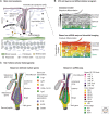
Heterogeneity of epithelial cells in homeostatic mouse skin. (A) Microanatomy of telogen mouse skin. The epidermis consists of interfollicular epidermis (IFE) and hair follicles (HFs) (with associated sebaceous glands), which are embedded in a cell-type-rich dermis and hypodermis. (B) IFE cell layers and differentiation program. Classically, the IFE has been divided into basal, spinous, granular, and cornified layers that undergo a stepwise differentiation program. Recent scRNA-seq and intravital imaging revealed the IFE differentiation as a gradual process (Lin et al. 2020; Cockburn et al. 2021). (C) Cellular heterogeneity of the telogen HF based on previously described marker genes (left panel) or based on unbiased scRNA-seq analysis (right panel, classification based on Joost et al. 2016, 2020).
Figure 2.
Figure 2.
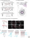
Epithelial cell contribution and cell behavior during wound repair. (A) Schematic of the multistep reepithelialization process, involving the activation of keratinocytes in early stage, proliferation and migration in intermediate stage, and complete wound closure and remodeling of epidermal barrier in late stage. (B) Illustration of the wound repairing epithelium. The wound-surrounding epithelial tissue is spatially divided into a migratory leading edge closest to the wound, a proliferative zone, and a “mixed” region between the two zones. Top panel shows a sagittal view. Lower panel shows a top view. (C) Contribution of lineage-traced Lgr5 hair follicle (HF) or Lgr6 interfollicular epidermis (IFE) stem cells during wound reepithelialization. Representative images of wounds taken 1, 3, 5, or 12 days post-wounding (dpw), show the temporospatial distribution of Lgr5- and Lgr6-progeny that have been labeled before wounding. (The right side in panel C is reprinted from Joost et al. 2018 under the terms of Creative Commons BY-NC-ND 4.0 license.) Dotted lines: approximate location of the basement membrane (thin) and wound front (thick). Filled lines: approximate location of the skin surface. (WF) Wound front. Scale bars, 100 μm. (D) Schematic overview of different injury types and corresponding major cellular contribution.
Figure 3.
Figure 3.
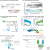
Molecular deconstruction of the epidermal wound healing process. (A) Cellular behavior and metabolic changes based on molecular signatures and functional enrichment. The wound cells’ transcriptome during an active reepithelialization process is a spatially coordinated program of proliferation, migration, and metabolism (Aragona et al. 2017; Haensel et al. 2020). (B) Illustration of how a cell's identity is shaped. Every wound (epithelial) cell's transcriptome is the sum of all influences that a cell experiences at any given moment: the active wound signature (early → mid → late) and the original and/or new niche (hair follicle [HF] → interfollicular epidermis [IFE]) (Joost et al. 2018). (C) Schematic showing transient lineage infidelity and transcriptional convergence of IFE and adjacent HF (including damaged HF) cells during wound reepithelialization revealed by recent scRNA-seq and ATAC-seq (Ge et al. 2017; Joost et al. 2018; Gonzales et al. 2021). (D) Transcriptional adaptation of individual cells revealed through combined lineage tracing and RNA sequencing. Lgr5-traced HF stem cell progeny (Lgr5TOM) gradually lose their typical bulge identity and acquire wound IFE-like identity during reepithelialization. (Panel D is modified from Joost et al. 2018 under the terms of Creative Commons BY-NC-ND 4.0 license.) (EF) Wound-induced common chromatin accessibility of HF and IFE-derived cells. Dual expression of both lineage transcription factors (TFs) (e.g., IFE-TF KLF5 and HF-TF SOX9) is transiently present in the mobilized cells from both HF and IFE niches (Ge et al. 2017; Adam et al. 2020). Superenhancer: group of enhancers densely occupied by TFs and mediators. Epicenter: enhancer elements where (lineage-specific) TF clustering occurs.
Figure 4.
Figure 4.
Early adaptations of hair follicle (HF) stem cells and their cross talk with the wound environment. (A) Adaptation of bulge cells starts within 1 day post-wounding (dpw). Distribution of Lgr5-traced cells (shown as violin plots) along the bulge → interfollicular epidermis (IFE) pseudotime (see Fig. 3D). Note that 1-dpw cells already span the entire spectrum of bulge → IFE transcriptional identity. (B) Illustration of the signaling cross talk between HF stem cells and the early wound environment. Arrow indicates that the signals secreted from the wound stroma are interacting with up-regulated receptors in the stem cell niche. (C) Detection of Itgb1 and Thbs1 by mRNA fluorescence in situ hybridization (RNA-FISH) in wound sections at 1 dpw. Dashed lines: approximate location of the basement membrane (thin) and wound front (thick). Filled lines: approximate location of the skin surface. (WF) Wound front, (WGA) wheat germ agglutinin (stains cell membranes). Scale bars, 100 μm (panoramas), 50 μm (zoom-ins). (D) Expression of selected receptors in Lgr5- and Lgr6-traced control (unwounded) and early wound cells (which has been defined as wound state 1B in Joost et al. 2018) matching their wound-derived ligand of 1-dpw samples. (Panels AD reprinted from Joost et al. 2018 under the terms of Creative Commons BY-NC-ND 4.0 license.) (E) Whole-mount top view of lineage-traced wounds from Lgr5creERT2;R26Tomato mice. Adult mice were treated with tamoxifen at indicated times relative to wounding (experimental scheme). Wounds were collected after 12 d and examined for labeling on the wound epidermal side (n ≥ 2 mice per time point) (X. Sun, original data). Dashed lines: outline approximate wound areas. Scale bars, 500 μm.

References

    1. Adam RC, Yang H, Ge Y, Infarinato NR, Gur-Cohen S, Miao Y, Wang P, Zhao Y, Lu CP, Kim JE, et al. 2020. NFI transcription factors provide chromatin access to maintain stem cell identity while preventing unintended lineage fate choices. Nat Cell Biol 22: 640–650. 10.1038/s41556-020-0513-0 - DOI - PMC - PubMed
    1. Alonso L, Fuchs E. 2006. The hair cycle. J Cell Sci 119: 391–393. 10.1242/jcs.02793 - DOI - PubMed
    1. Aragona M, Dekoninck S, Rulands S, Lenglez S, Mascré G, Simons BD, Blanpain C. 2017. Defining stem cell dynamics and migration during wound healing in mouse skin epidermis. Nat Commun 8: 14684. 10.1038/ncomms14684 - DOI - PMC - PubMed
    1. Arwert EN, Hoste E, Watt FM. 2012. Epithelial stem cells, wound healing and cancer. Nat Rev Cancer 12: 170–180. 10.1038/nrc3217 - DOI - PubMed
    1. Belokhvostova D, Berzanskyte I, Cujba A-M, Jowett G, Marshall L, Prueller J, Watt FM. 2018. Homeostasis, regeneration and tumour formation in the mammalian epidermis. Int J Dev Biol 62: 571–582. 10.1387/ijdb.170341fw - DOI - PMC - PubMed

Publication types

LinkOut - more resources