Review
. 2022 Nov 14;9(6):ENEURO.0092-22.2022.
doi: 10.1523/ENEURO.0092-22.2022. Print 2022 Nov-Dec.

P2X7 Receptor and Purinergic Signaling: Orchestrating Mitochondrial Dysfunction in Neurodegenerative Diseases

Affiliations
Review

P2X7 Receptor and Purinergic Signaling: Orchestrating Mitochondrial Dysfunction in Neurodegenerative Diseases

Alexsandra S Zelentsova et al. eNeuro. .

Abstract

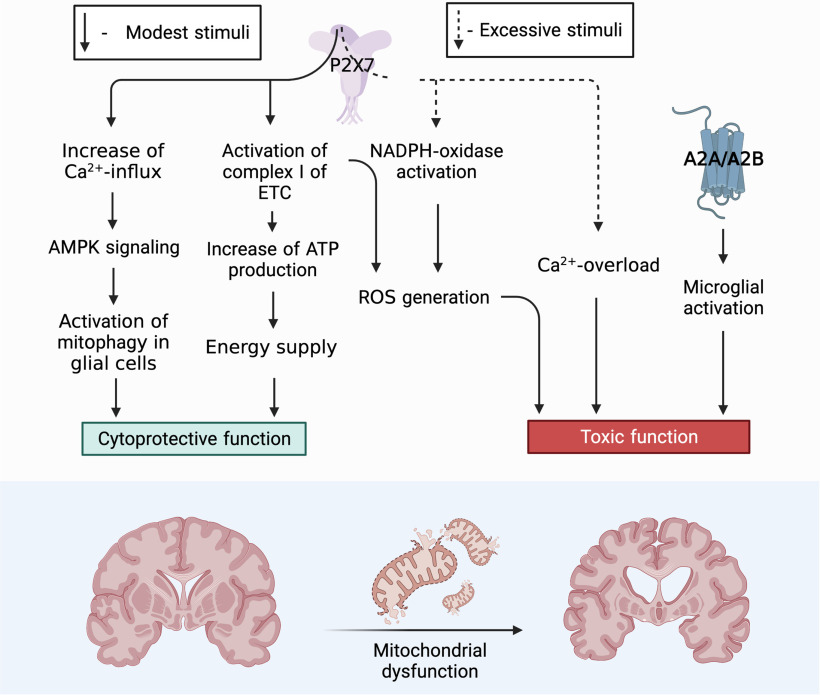
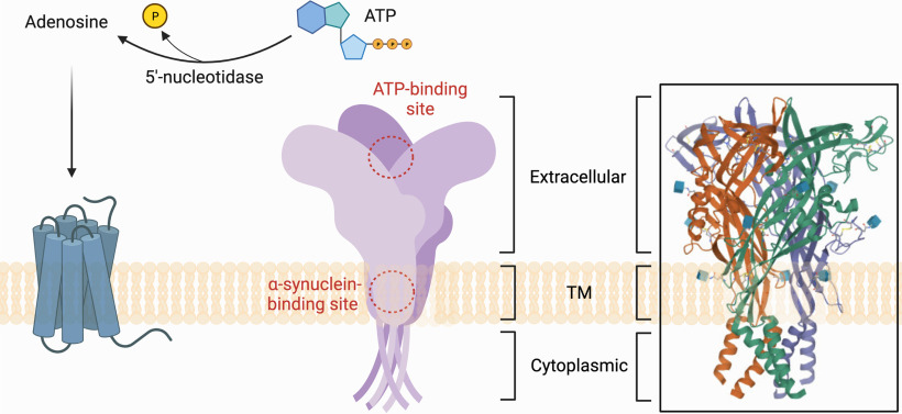
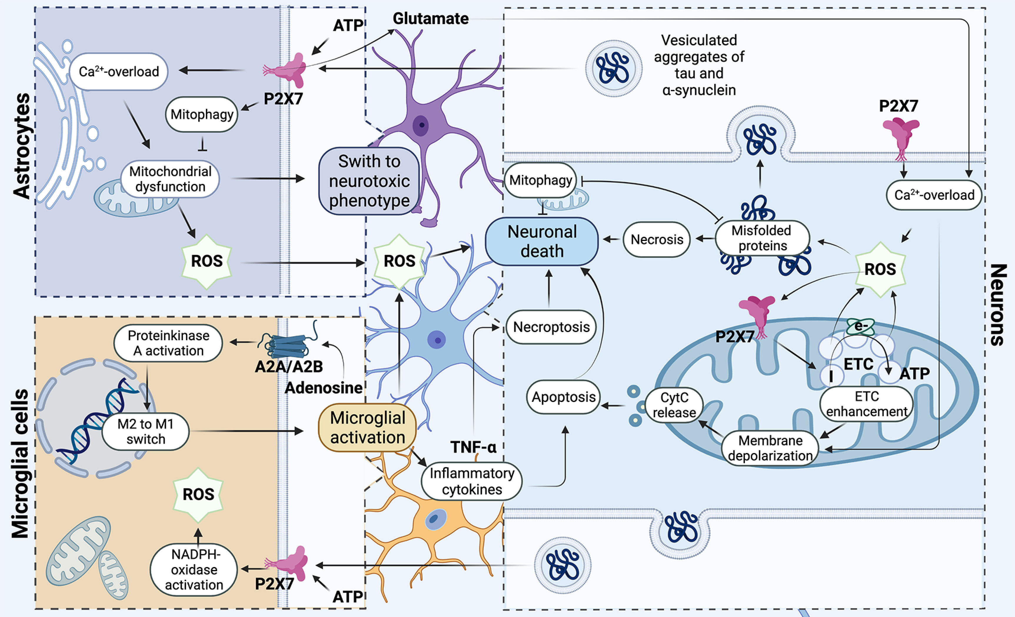
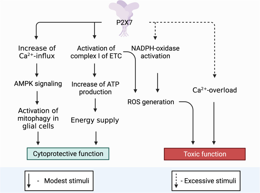
Mitochondrial dysfunction is one of the basic hallmarks of cellular pathology in neurodegenerative diseases. Since the metabolic activity of neurons is highly dependent on energy supply, nerve cells are especially vulnerable to impaired mitochondrial function. Besides providing oxidative phosphorylation, mitochondria are also involved in controlling levels of second messengers such as Ca2+ ions and reactive oxygen species (ROS). Interestingly, the critical role of mitochondria as producers of ROS is closely related to P2XR purinergic receptors, the activity of which is modulated by free radicals. Here, we review the relationships between the purinergic signaling system and affected mitochondrial function. Purinergic signaling regulates numerous vital biological processes in the CNS. The two main purines, ATP and adenosine, act as excitatory and inhibitory neurotransmitters, respectively. Current evidence suggests that purinergic signaling best explains how neuronal activity is related to neuronal electrical activity and energy homeostasis, especially in the development of Alzheimer's and Parkinson's diseases. In this review, we focus on the mechanisms underlying the involvement of the P2RX7 purinoreceptor in triggering mitochondrial dysfunction during the development of neurodegenerative disorders. We also summarize various avenues by which the purine signaling pathway may trigger metabolic dysfunction contributing to neuronal death and the inflammatory activation of glial cells. Finally, we discuss the potential role of the purinergic system in the search for new therapeutic approaches to treat neurodegenerative diseases.

Keywords: exosomes; mitochondrion; oxidative stress; purinergic metabolome; tau protein; α-synuclein.


Conflict of interest statement

The authors declare no competing financial interests.

Figures

None
Graphical abstract
Figure 1.
Figure 1.
Schematic models of P2X7 receptor. Extracellular ATP is a P2RX7 receptor agonist, as well as a substrate for 5′-ectonucleotidase, which hydrolyzes ATP to adenosine and temporarily generates ADP, which is a P2Y receptor agonist. Adenosine, the product of hydrolysis of adenine nucleotides, activates adenosine receptors or P1 receptors. P2X receptors are ionotropic, and their activation opens the cation channel, which leads to cell hyperpolarization because of the outflow of K+ and the influx of extracellular Na+ and Ca2+. Figure Contributions: Alexsandra S. Zelentsova prepared the structure of P2X7 receptors and several sites for ligands. Plamena R. Angelova developed the ATP-binding site and activation of P2X7 receptor by ATP molecule.
Figure 2.
Figure 2.
Participation of the P2X7 receptor in the development of mitochondrial degeneration in neurons. The P2X7 purinergic receptor is involved in modulating the redox potential, triggering the production of hydrogen peroxide through the mechanism of Ca2+ release from intracellular stores from the primary microglia. At the same time, in neurons, mitochondria are the primary source of ROS and generate superoxide at the initial site in the respiratory chain under conditions of ischemia and hypoxia. ROS initiates increase of P2X7 expression on the mitochondrial membrane and activity of Complex I, hence increasing mitochondrial polarization, which leads to an elevation of a cascade of unfavorable processes: depolarization of the mitochondrial membrane, calcium overload, and release of cytochrome c, which ultimately leads to cell death by apoptosis way. Under conditions of excessive accumulation of ROS, the process of incorrect aggregation of α-synuclein and tau protein triggered. Oligomeric proteins are involved in coordinating neuroinflammation via the direct interaction of extracellular oligomers with the P2X7 receptor of microglia, which triggers NADPH oxidase activation. Oligomeric proteins trigger the development of mitochondrial dysfunction, localizing in mitochondria and reducing their oxygen consumption and significantly increasing extracellular production of hydrogen peroxide by astrocytes. Purinergic receptors play a key role in the process of neuroinflammation and can trigger activation of microglia. In activated microglial cells, P2X7 is directly involved in triggering the production of TNFα cytokine that can be involved in the process of neuronal death both through apoptosis and through necroptosis; in addition, this receptor stimulates ROS production and triggers mitochondrial dysfunction. Adenosine receptors are involved in the regulation of microglial polarization. Activation of microglia closely connected with purine receptors P1 associated with G-proteins. Activation of adenosine receptor (AR) regulates the activity of protein kinase A (PKA) and participates in the switching of phenotype M1/M2 in the microglial cells. Figure Contributions: Alexsandra S. Zelentsova prepared the mechanism developing of neuronal death. Alexei V. Deykin developed the involvement of oligomeric proteins in the organization of neuroinflammation. Vladislav O. Soldatov performed microglial activation. Anastasia A. Ulezko developed forming ROS by mitochondrion. Alina Y. Borisova developed the pathogenetic mechanisms between microglia, astrocytes, and neuron. Veronika S. Belyaeva performed the formation of unregular folding oligomeric proteins. Marina Y. Skorkina performed the mechanisms of organization of neuroinflammation. Plamena R. Angelova developed the activation of the microglial neuron and the role of P2X7 receptor in neuroinflammation and death of neuron ny necroptosis and apoptosis.
Figure 3.
Figure 3.
Role of P2X7 receptor in the functional activity of neurons. Toxic function: P2X7 triggers an intracellular signaling pathway for the activation of free radical oxidation processes and the accumulation of ROS, as a result, mitochondria are actively involved in the process of ROS production. ROS via AMPK signaling pathway can trigger mitophagy, thereby controlling the presence of defective mitochondria in the neuron removing them and ensuring cell viability. However, with excessive accumulation of ROS and a decrease in the activity of the antioxidant systems of the cell, the correct functioning of the PARKIN protein is blocked, which leads to inhibition of the mitophagy process. Misfolding of proteins, defective mitochondria accumulate in the cell. One of the defective proteins is α-synuclein that in turn has binding sites for the P2X7 receptor stimulating it and thereby increasing the Ca2+ concentration in the cell. An elevated level of Ca2+ blocks AMPK activity, after which the death of neurons can follow the path of necrosis. Cytoprotective function: P2X7 triggers mitophagy throught the Ca2+ increases which activation AMPK signaling pathway. It eliminates defective mitochondria thereby contributing to the survival of neuroglia and maintaining its viability in conditions of short-term stimulation. P2X7, localized on the outer mitochondrial membrane, captures the concentration of ATP in the cell and, with a decrease in its production, enhances the work of Complex I (NADH-DH) of the electron transport chain, as a result of which oxidative phosphorylation is stimulated and ATP production in the cell increases. Figure Contributions: Alexei V. Deykin described the cytoprotective function of P2X7 through activating AMPK-signaling pathway. Vladislav O. Soldatov performed the cytoprotective function by activation Complex I of ETC. Marina Y. Skorkina described the toxic function through ROS. Plamena R. Angelova developed the toxic function through Ca2+ overload.

References

    1. Abramov AY, Berezhnov V, Fedotova RI, Zinchenko VP, Dolgacheva LP (2017) Interaction of misfolded proteins and mitochondria in neurodegenerative disorders. Biochem Soc Trans 45:1025–1033. 10.1042/BST20170024 - DOI - PubMed
    1. Agosta F, Dalla Libera D, Spinelli EG, Finardi A, Canu E, Bergami A, Bocchio Chiavetto L, Baronio M, Comi G, Martino G, Matteoli M, Magnani G, Verderio C, Furlan R (2014) Myeloid microvesicles in cerebrospinal fluid are associated with myelin damage and neuronal loss in mild cognitive impairment and Alzheimer disease. Ann Neurol 76:813–825. 10.1002/ana.24235 - DOI - PubMed
    1. Alnouri MW, Jepards S, Casari A, Schiedel AC, Hinz S, Müller CE (2015) Selectivity is species-dependent: characterization of standard agonists and antagonists at human, rat, and mouse adenosine receptors. Purinergic Signal 11:389–407. 10.1007/s11302-015-9460-9 - DOI - PMC - PubMed
    1. An S, Kumar R, Sheets ED, Benkovic SJ (2008) Reversible compartmentalization of de novo purine biosynthetic complexes in living cells. Science 320:103–106. 10.1126/science.1152241 - DOI - PubMed
    1. Anderson CM, Nedergaard M (2006) Emerging challenges of assigning P2X7 receptor function and immunoreactivity in neurons. Trends Neurosci 29:257–262. 10.1016/j.tins.2006.03.003 - DOI - PubMed

Publication types

MeSH terms

Substances