. 2022 Dec 12;40(52):7676-7692.
doi: 10.1016/j.vaccine.2022.11.002. Epub 2022 Nov 12.

Immunization with a tri-antigen syphilis vaccine significantly attenuates chancre development, reduces bacterial load, and inhibits dissemination of Treponema pallidum

Affiliations

Immunization with a tri-antigen syphilis vaccine significantly attenuates chancre development, reduces bacterial load, and inhibits dissemination of Treponema pallidum

Sheila A Lukehart et al. Vaccine. .

Abstract

Syphilis continues to be a significant public health concern worldwide. The disease is endemic in many low- and middle-income countries, and rates have risen sharply in high-income countries over the last decade. The continued prevalence of infectious and congenital syphilis worldwide highlights the need for the development of an effective syphilis vaccine to complement public health measures for syphilis control. The complex, multi-stage course of syphilis infection necessitates a holistic approach to the development of an effective vaccine, in which immunization prevents both the localized stage of infection (typified by the highly infectious chancre) and the disseminated stages of infection (typified by the secondary rash, neurosyphilis, and destructive tertiary lesions, as well as congenital syphilis). Inhibiting development of the infectious chancre would reduce transmission thus providing community- level protection, while preventing dissemination would provide individual-level protection by reducing serious sequelae and may also provide community level protection by reducing shedding during secondary syphilis. In the current study we build upon prior investigations which demonstrated that immunizations with individual, well characterized T. pallidum TprK, TprC, and Tp0751 peptides elicits partial protection against infection in the animal model. Specifically, we show here that immunization with a TprC/TprK/Tp0751 tri-antigen cocktail protects animals from progressive syphilis lesions and substantially inhibits dissemination of the infection.

Keywords: Bacterium; Syphilis; Treponema pallidum; Vaccine.


Conflict of interest statement

Declaration of Competing Interest The authors declare the following financial interests/personal relationships which may be considered as potential competing interests: Caroline Cameron has patent issued to University of Victoria.

Figures

Fig. 1.
Fig. 1.. Protection experiment pipeline.
Shown is a schematic for the immunization and challenge experiments (left) and subsequent rabbit infectivity test analysis (right), for testing the protective capacity of the tri-antigen cocktail vaccine. Unique aspects of the experiments conducted at the different locations are indicated by different colored boxes: University of Washington, purple boxes; University of Victoria, red boxes.
Fig 2.
Fig 2.
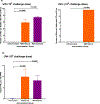
Immunization with the tri-antigen cocktail induces both a delayed type hypersensitivity (Panel a) and an interferon-γ (IFN-γ, Panel b) response upon challenge with virulent T. pallidum. a. Following immunization, rabbits were challenged intradermally on their backs at 10 sites per rabbit. The challenge doses at UW and UVic were 105 and 106 T. pallidum Nichols strain per site, respectively. Forty-eight hours post-challenge, both immunized groups (RIBI Natural, orange, n = 8 rabbits UW, n = 7 UVic; RIBI Synthetic, purple, n = 7 rabbits UW) had evidence of clinical DTH (erythema and induration) at the majority (Fig. 2a left panel) or all (Fig. 2a right panel) of the challenge sites, while no erythema/induration was seen at any site in the unimmunized control rabbits (blue; n = 7 rabbits UW, n = 4 UVic). b. At UW, biopsies of two challenge sites per rabbit were examined by qPCR for interferon-γ message (n = 16 lesions for RIBI Natural, and 14 lesions each for RIBI Synthetic and Unimmunized). IFNγ was detected in the immunized, but not the unimmunized control, rabbits. Results are presented as mean +/− SEM. An unpaired t test with Welch’s correction (Fig. 2a, left panel), a Chi-squared test (Fig. 2a, right panel), and an unpaired t test (Fig. 2b) were used to analyze the differences between the immunized and unimmunized groups.
Fig. 3.
Fig. 3.
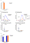
Immunization with the tri-antigen cocktail decreases both lesion volume (Panel a) and ulceration (Panel b). a. At both UW and UVic, immunization with the tri-antigen cocktail decreased lesion volume compared to unimmunized control rabbits, regardless of the Nichols challenge dose (RIBI Natural-immunized rabbits, orange circles, n = 8 UW, n = 7 UVic; RIBI Synthetic-immunized rabbits, purple squares, n = 7 UW; unimmunized control rabbits, blue triangles, n = 7 UW, n = 4 UVic). Lesion volume was greater in immunized vs control rabbits on days 2 and 5 (Fig. 3a left panel, P < 0.0001) due to clinical DTH, while lesion volume was smaller in immunized vs control rabbits on days 10–25 (105 dose, Fig. 3a left panel, P < 0.0001) and days 7–14 (106 dose, Fig. 3a right panel, P = 0.03) due to attenuation of chancre development in immunized rabbits. b. Lesions in immunized rabbits challenged with either 106 or 105 T. pallidum per site exhibited less ulceration. At days 14 (106 dose, Fig. 3b right panel), 19 and 30 (105 dose, Fig. 3b left panel), the proportion of lesions ulcerating was significantly lower in RIBI Natural-immunized animals compared to unimmunized control rabbits (P = 0.009 [d.14], P = 0.001 [d.19] and P = 0.0003 [d.30]). At both time points (days 19 and 30, Fig. 3b left panel), there was a non-significant trend toward a lower proportion of lesions ulcerating in RIBI Synthetic-immunized animals, compared to unimmunized animals (P = 0.11 and P = 0.17, respectively). Data shown are mean +/− SEM. Comparisons were conducted by unpaired t test (Fig. 3a and Fig. 3b left panels), unpaired t test with Welch’s correction (Fig. 3b right panel), and two-way ANOVA (Fig. 3a right panel).
Fig. 4.
Fig. 4.
Presence of T. pallidum in lesion aspirates at Days 10 and 19 post challenge (pc). Presence of T. pallidum Nichols strain were determined in aspirates of 8 to 10 lesions per rabbit from each individual rabbit. a. At days 10 (106 dose, Fig. 4a right panel) and 19 (105 dose, Fig. 4a left panel) pc, lesions were examined for the presence of viable T. pallidum using darkfield microscopy. All immunized groups had significantly lower T. pallidum burden in lesions compared to the unimmunized group, regardless of the challenge dose (P = 0.001 and P = 0.04 for Natural and Synthetic, respectively, compared to unimmunized, Fig. 4a left panel and P = 0.005, Fig. 4a right panel). b. Total number of T. pallidum seen in darkfield microscopic examination of 100 fields per lesion aspirate and the number of copies of tp0574 (single copy gene) in pooled lesion aspirates of 8 lesions per rabbit for unimmunized (blue, n = 7 rabbits); RIBI Natural (orange, n = 8 rabbits); and RIBI Synthetic (purple, n = 7 rabbits) groups. The number of T. pallidum seen by darkfield microscopy was significantly lower in both immunized groups than in unimmunized controls, (P = 0.001 and P = 0.004 for Natural and Synthetic, respectively, compared to unimmunized, Fig. 4b left panel). The RIBI-Natural group had fewer T. pallidum detected by qPCR than the unimmunized rabbits (P = 0.008, Fig. 4b right panel). The T. pallidum burden in the RIBI-Synthetic group was not significantly different from the unimmunized rabbits as measured by qPCR. Results are presented as mean and 95 % CI. Unpaired t test (Fig. 4a right panel and Fig. 4b) and an unpaired t test with Welch’s correction (Fig. 4a left panel) were used to analyze the differences between the immunized and unimmunized groups; P < 0.05 was considered to be a significant difference.
Fig. 5.
Fig. 5.. Antibody titers in pre-challenge sera.
ELISA assay was used to determine IgG (Fig. 5a left panel and 5b) and IgM (Fig. 5a right panel) titers in pre-challenge sera. At UVic, IgG titers were determined only for the Tp0751 antigen. Values shown are mean +/− SEM of the reciprocal dilutions yielding ODs 2-fold higher than the pre-immune serum for that rabbit. Note that the Y-axis for IgG is given in thousands (e.g. 5120 K = 5,120,000) while the IgM titer is the actual dilution. Except for the IgG titers against Tp0751 (P = 0.0001), there were no significant differences in pre-challenge titers between the two adjuvant groups. Titers were compared using unpaired t test; P < 0.05 was considered to be significant. Different secondary antibodies and a longer primary antibody incubation were used at UW compared to UVic, which could explain the different apparent IgG titer for the Tp0751 antigen.
Fig 6.
Fig 6.. Cytokine message in 48-hour biopsies demonstrates a Th1/Th17 immune response was generated by the tri-antigen immunization.
Erythema and induration were observed at all challenge sites at 48 h, consistent with a delayed type hypersensitivity (DTH) response. The cytokines generally associated with this response are those seen in Th1-skewed profiles. In biopsies of the 48-hr 105 T. pallidum Nichols strain challenge sites, IL-2, Interferon-γ (IFN γ), and TNFα message levels were significantly higher in both immunized groups compared to controls, indicating a Th1 response. IL-17 A and IL-17F levels were also significantly higher in immunized versus control rabbits, consistent with a Th17 profile. In contrast, levels of the anti-inflammatory cytokines TGF-β and IL-10 were significantly higher in the unimmunized vs the immunized rabbits at 48 h post challenge. No differences in cytokine levels were seen between the two immunized groups. Data shown are mean +/− SEM values from 14 to 16 biopsies per group, and comparisons were analyzed using an unpaired t test with Holm-Sidak adjustment for multiple comparisons. P < 0.05 was considered to be a significant difference: (* <0.05; ** <0.02; *** <0.005; ****<0.0001).
Fig. 7.
Fig. 7.. Rabbit infectivity test (RIT) and number of viable T. pallidum in popliteal lymph nodes post challenge.
RIT was used to determine the approximate number of viable T. pallidum Nichols strain in the popliteal lymph nodes (LN) of challenged rabbits, as time to development of orchitis following injection of LN suspensions into naïve recipient rabbits is inversely related to the number of viable T. pallidum inoculated. a. Time (days) to orchitis for the unimmunized control, and tri-antigen-immunized RIBI Natural and RIBI Synthetic groups. Means and 95 % CI are shown. The RIBI Natural group had a significantly longer time to orchitis, indicating fewer treponemes in their LN, compared to both the unimmunized and RIBI Synthetic groups. This demonstrates a significant reduction in T. pallidum dissemination in the RIBI Natural-immunized rabbits. Comparisons were analyzed using an unpaired t test. P < 0.05 was considered to be a significant difference. b. Time to orchitis with the Nichols strain of T. pallidum is inversely correlated with the number of infectious T. pallidum inoculated. Using historical [42] and contemporary unpublished data from our laboratory, we plotted the time to orchitis (days) for inocula of 200, 500, 100,000 and 25,000,000 T. pallidum Nichols strain, and used the resulting best-fit line (Y = −0.2718x + 9.6013) to estimate the number of infectious treponemes present in the LN from the unimmunized and immunized rabbits. Immunized rabbits had 88 % (RIBI Natural) and 38 % (RIBI Synthetic) reductions in the number of T. pallidum disseminating to the popliteal LN, compare to unimmunized rabbits. c. In the experiment conducted at UVic, for RIT animals that did develop orchitis, the RIBI Natural group had a longer time to orchitis compared to the unimmunized group, indicating fewer treponemes in their LN, although the trend did not achieve statistical significance, perhaps due to the small sample size in the unimmunized group. Mean +/− SEM are shown, and comparisons were analyzed using an unpaired t test. In this experiment, RIT animals receiving LN from two unimmunized rabbits and one tri-antigen-immunized RIBI Natural rabbit did not develop orchitis but seroconverted at day 42 post-LN transfer and were euthanized, while one RIT rabbit receiving LN from the tri-antigen-immunized RIBI Natural group failed to seroconvert or develop orchitis by day 90 post-LN transfer. d. Estimation of the number of infectious treponemes present in the LN from the unimmunized and immunized rabbits that did develop orchitis using the best-fit line (Fig. 7b). Immunized rabbits had an approximately 85 % reduction in the number of T. pallidum disseminating to the popliteal LN compared to unimmunized rabbits; an approximation is provided as the time to orchitis for the immunized rabbits fell just outside of the best-fit line.
Fig. 8.
Fig. 8.. Antibody titers in pre-challenge sera.
Pre-challenge IgG and IgM antibody titers were determined at UW for experiment Tri II as described for Fig. 5. There were no significant differences in pre-challenge titer between the 105 and 103 challenge groups.
Fig. 9.
Fig. 9.. Lesion attenuation in immunized animals challenged with different doses.
Following immunization, rabbits were challenged intradermally on their backs at 10 sites per rabbit with 105 T. pallidum Nichols strain per site (n = 4) or 103 T. pallidum per site (n = 4). At 48 h, evidence of DTH (erythema and induration) was recorded. Unimmunized rabbits showed no evidence of DTH at 48 h. While immunized rabbits challenged with 105 T. pallidum per site developed DTH at nearly all sites by 48 h, rabbits challenged with 103 T. pallidum per site failed to show evidence of DTH at 48 h. The lesions in immunized rabbits were significantly smaller (Fig. 9b) and had fewer T. pallidum detected by qPCR (Fig. 9c) than in unimmunized rabbits, regardless of the challenge dose. Inset in Fig. 9b shows early lesion volumes in rabbits challenged with 103 T. pallidum per site; immunized rabbits demonstrated DTH (erythema and induration) at Day 10 post challenge following the lower dose challenge. Lesions in immunized rabbits challenged with 105 T. pallidum per site had a lower proportion of lesions ulcerating, compared to unimmunized rabbits. In contrast, high proportions of lesions progressed to ulceration in rabbits challenged with 103 T. pallidum per site, regardless of immunization status (Fig. 9d). Data reflect the proportion of challenge sites ulcerating by the end of the observation period: day 41 for 105 dose, and day 48 for 103 dose. Data are shown as mean +/− SEM (Fig. 9 a, b and d) and mean and 95 % CI (Fig. 9c). An unpaired t test was used to analyze the difference between the immunized and unimmunized groups in panels a, c, and d; two-way ANOVA was used in panel b. P < 0.05 was considered to be a significant difference.
Fig. 10.
Fig. 10.. Heterologous protection of the tri-antigen cocktail (UW).
a. Immunization with the tri-antigen cocktail resulted in decreased lesion volume in rabbits challenged with the heterologous strains T. pallidum subsp. pallidum Chicago (Fig. 10a, left panel) and T. pallidum subsp. pallidum Sea 81–4 (Fig. 10a, right panel). Means +/− SEM are shown. Differences between immunized and unimmunized animals in each group were analyzed by an unpaired t test with Welch’s correction at each time point. P < 0.05 was considered to be significant and is shown by *. b. Number of copies of tp0574 (single copy gene) in pooled (by rabbit) lesion aspirates for unimmunized and immunized rabbits challenged with T. pallidum subsp. pallidum Chicago (Fig. 10b, left panel) and pooled (by rabbit) biopsies for animal groups challenged with T. pallidum subsp. pallidum Sea 81–4 (Fig. 10b, right panel). Lesion aspirates were taken on day 22 (Chicago) and biopsies were collected on day 25 (Sea 81–4) post-challenge; timing of the sampling was determined by the appearance of the lesions (large but not yet ulcerating) in the unimmunized animals for each strain c. Time (days) to orchitis or qPCR quantitation for the RIT rabbits receiving LN from the unimmunized control and tri-antigen-immunized rabbit groups challenged with T. pallidum subsp. pallidum Chicago (Fig. 10c, left panel) or T. pallidum subsp. pallidum Sea 81–4 (Fig. 10c, right panel). Animals receiving LN from immunized rabbits exhibited a slight trend towards delayed orchitis, although statistical significance was not achieved. For the Sea 81–4 challenged rabbits, because the Sea 81–4 strain produces virtually no lesions or orchitis following infection, time to development of orchitis in recipient animals could not be determined, nor could lesion aspirates be collected to assess T. pallidum burden. Instead dissemination in the Sea 81–4 RIT recipient animals was determined by extracting the testes from RIT recipient rabbits into a defined volume of saline, followed by qPCR to determine the relative amount of T. pallidum dissemination in the challenged rabbits. There was a significant difference between the immunized and unimmunized groups, although the absolute tp0574 copies were very low. Data shown are mean and 95 % CI and differences between immunized and unimmunized animals in each group were analyzed by the Mann-Whitney test. P < 0.05 was considered significant.

References

    1. O’Byrne P, MacPherson P. Syphilis. BMJ 2019;365:. 10.1136/bmj.l4159l4159. - DOI - PMC - PubMed
    1. Choudhri Y, Miller J, Sandhu J, Leon A, Aho J. Infectious and congenital syphilis in Canada, 2010–2015. Can Commun Dis Rep 2018;44(2):43–8. 10.14745/ccdr.v44i02a02. - DOI - PMC - PubMed
    1. Spiteri G, Unemo M, Mardh O, Amato-Gauci AJ. The resurgence of syphilis in high-income countries in the 2000s: a focus on Europe. Epidemiol Infect 2019;147:e143. 10.1017/S0950268819000281. - DOI - PMC - PubMed
    1. Arrieta AC, Singh J. Congenital Syphilis. N Engl J Med 2019;381(22):2157. 10.1056/NEJMicm1904420. - DOI - PubMed
    1. Lancet (ed). Congenital syphilis in the USA. Lancet 2018;392(10154):1168. 10.1016/S0140-6736(18)32360-2. - DOI - PubMed

Publication types

Substances