. 2022 Nov 14;12(1):19467.
doi: 10.1038/s41598-022-21817-w.

A flexible adhesive surface electrode array capable of cervical electroneurography during a sequential autonomic stress challenge

Affiliations

A flexible adhesive surface electrode array capable of cervical electroneurography during a sequential autonomic stress challenge

Yifeng Bu et al. Sci Rep. .

Abstract

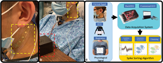
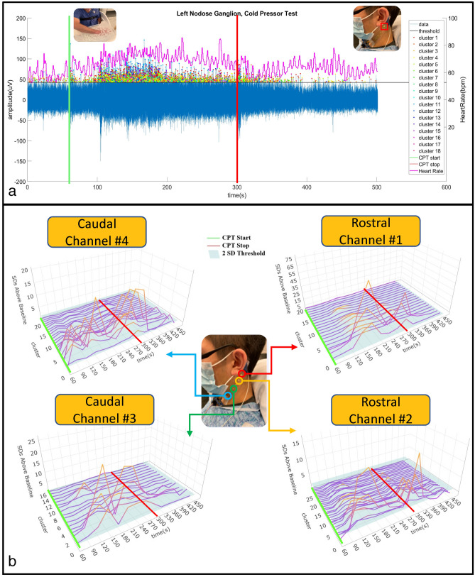
This study introduces a flexible, adhesive-integrated electrode array that was developed to enable non-invasive monitoring of cervical nerve activity. The device uses silver-silver chloride as the electrode material of choice and combines it with an electrode array consisting of a customized biopotential data acquisition unit and integrated graphical user interface (GUI) for visualization of real-time monitoring. Preliminary testing demonstrated this electrode design can achieve a high signal to noise ratio during cervical neural recordings. To demonstrate the capability of the surface electrodes to detect changes in cervical neuronal activity, the cold-pressor test (CPT) and a timed respiratory challenge were employed as stressors to the autonomic nervous system. This sensor system recording, a new technique, was termed Cervical Electroneurography (CEN). By applying a custom spike sorting algorithm to the electrode measurements, neural activity was classified in two ways: (1) pre-to-post CPT, and (2) during a timed respiratory challenge. Unique to this work: (1) rostral to caudal channel position-specific (cephalad to caudal) firing patterns and (2) cross challenge biotype-specific change in average CEN firing, were observed with both CPT and the timed respiratory challenge. Future work is planned to develop an ambulatory CEN recording device that could provide immediate notification of autonomic nervous system activity changes that might indicate autonomic dysregulation in healthy subjects and clinical disease states.


Conflict of interest statement

The authors declare no competing interests.

Figures

Figure 1
Figure 1
Custom surface electrode array for non-invasive testing of cervical neuronal activity. (a) Device cross section (5 mm sensor diameter and 500 μm interconnect width). (b) Cleanroom post-processed wafer ready to be printed with Ag/AgCl ink. (c) Ag/AgCl ink screen-printed wafer on the active electrode region. (d) Device transfer-printed onto a self-adhering flexible silicone substrate (Ecoflex/Silbione) on PET backing via water soluble tape.
Figure 2
Figure 2
The custom design allows for free movement without distorting the adhesiveness and robustness of the physical structure of the electrode array. (a) The custom surface electrode array is adhesive integrated, flexible, and non-invasively attached to the subject's anterior cervical neck, placed lateral to the trachea and medial to the sternocleidomastoid. A 3 M RedDot electrode was used as a reference electrode. The four channels were aligned rostral to caudal, so that each channel was positioned over an anatomical target: (1) channel #1 over the upper nodose ganglion, (2) channel #2 over the lower nodose ganglion, (3) Channel #3 over the upper carotid artery, and (4) channel #4 over the lower carotid artery. (b) Data collection was carried out with flexible multi-electrodes attached to the wireless biopotential data acquisition board, i.e., the HackEEG (red dashed outline), and 3 M RedDot deployed as ground/reference electrodes. (c) Subject recording pipeline and workflow. Cervical electroneurography, electrocardiography, and respiration were recorded; signals recorded to the data acquisition system underwent post processing algorithms.
Figure 3
Figure 3
Cervical neural firing coordinately increases with onset of CPT. (a) Cervical Electroneurography (CEN) rostral Channel #1 recording post-spike sorting analysis for the cold-pressor test (CPT). The green vertical line indicates CPT start i.e., the subject immersed their right hand into the ice-water bucket, while the red vertical line indicates CPT cessation i.e., the subjects removed their hand from the ice water bucket. Channel #1 (blue line = neural firing) captures the amplitude of the recorded neural action potential data. The peak of each detected spike (blue line) that exceeds the threshold value sorted each to 18 different clusters identified by different color. Heart rate in beats per minute is plotted in magenta using the right y-axis. (b) Exemplar subject cervical electroneurography (CEN) neural firing change during the cold pressor test. Top left Responsive clusters at the Caudal Channel #4 (located over the carotid artery, vagus nerve, glossopharyngeal nerve, sympathetic chain, and sensory C2/C3 dermatomal nerves) immediately increase in firing frequency with cold pressor test (CPT) onset (i.e., hand placed in ice water bath). Top right Responsive clusters from the rostral Channel #1 (located over nodose ganglion and in close approximation to the auriculotemporal nerve) demonstrate delayed firing onset (at 120–150 s). Bottom panels Both Caudal Channel #3 (overlying the carotid artery, vagus nerve, glossopharyngeal nerve, sympathetic chain, and sensory C2/C3 dermatomal nerves) and Rostral Channel #2 (located over nodose ganglion and in close approximation to the auriculotemporal nerve) increase in firing frequency at 90–120 s post recording initiation. In all panels, the transition from purple to orange lines indicates increases in cervical neural firing greater than 2 SD above baseline (i.e., prior to CPT start), and the green line denotes initiation of CPT, while cessation is denoted by the red line.
Figure 4
Figure 4
All subject channel activity across CPT indicated as % time intervals: (a) 0–20% of CPT; (b) 20–40% of CPT; (c) 40–60% of CPT; (d) 60–80% of CPT, and (e) 80–100% of CPT duration. Panel A: For the first interval of 0–20%, Channels #3 and #4 demonstrate highly significant increases in firing frequency (**p < 0.001), while Channels #1 and #2 also increased in firing frequency, but to a lesser extent (*p < 0.05) when compared to baseline. Channel #4 (lower carotid artery) demonstrated significantly greater (connected segment) firing than Channels #1 and #2 (p < 0.05). Panel (b) All channels remained at relatively increased activity (p < 0.05) when compared to baseline. An increase in firing in Channel #2 was observed when compared to Channel #4 (p < 0.05). Panels (ce) All channels do not show increases in activity when compared to baseline. Channel 2 continued to demonstrate significantly greater firing than Channel 4 for the remaining CPT duration (p < 0.05). Channel #1 = overlying upper nodose ganglion; Channel #2 = overlying lower nodose ganglion and trigeminal auriculotemporal branch; Channel #3 = overlying upper carotid artery; Channel #4 = overlying lower carotid artery. X Axis: Channel 1–4; Y Axis: Percentage Increase in neural firing frequency with respect to baseline activity.
Figure 5
Figure 5
Neuronal firing comparison between CPTd and CPTi groups during the respiratory challenge. (a, b) The green vertical line indicates initiation of the respiratory challenge, while the red vertical line indicates cessation of the respiratory challenge. The blue line captures the amplitude of the recorded electrode data for electrode positioned over the upper nodose ganglion (Channel #1). The peak of each detected spike (exceeding the threshold value)  is indicated by spike coloraction in the positive y-axis, representing the different clusters. The respiration belt voltage is plotted in cyan using the right y-axis. (a) Exemplar CPTd CEN neural firing change during deep respiratory challenge. (b) Exemplar CPTi CEN neural firing change during deep respiratory challenge. (c) Average firing frequencies comparison between pre- and post-challenges for CPTd and CPTi groups across four channels. All channels in the CPTi group have significant neuronal activity increase after deep respiratory challenge (p < 0.05), whereas only Channel #4 in the CPTd group has significant decreased activity. We observed significant between group (CPTd vs CPTi) differences (p < 0.01)  in neuronal firing change across all channels.

References

    1. McCorry LK. Physiology of the autonomic nervous system. Am. J. Pharm. Educ. 2007;71:4. - PMC - PubMed
    1. Zygmunt A, Stanczyk J. Methods of evaluation of autonomic nervous system function. Arch. Med. Sci. 2010;6:11. - PMC - PubMed
    1. Marques AH, Silverman MN, Sternberg EM. Evaluation of stress systems by applying noninvasive methodologies: Measurements of neuroimmune biomarkers in the sweat, heart rate variability and salivary cortisol. NeuroImmun. Modul. 2010;17:205–208. - PMC - PubMed
    1. Jänig W, McLachlan EM. Characteristics of function-specific pathways in the sympathetic nervous system. Trends Neurosci. 1992;15:475–481. - PubMed
    1. Levy MN, Koeppen BM, Stanton BA. Berne & Levy Principles of Physiology E-Book. Elsevier Health Sciences; 2005.

Publication types