

Electron microscopy study on the transport of lead oxide nanoparticles into brain structures following their subchronic intranasal administration in rats
- PMID: 36376368
- PMCID: PMC9663722
- DOI: 10.1038/s41598-022-24018-7
Electron microscopy study on the transport of lead oxide nanoparticles into brain structures following their subchronic intranasal administration in rats
Abstract
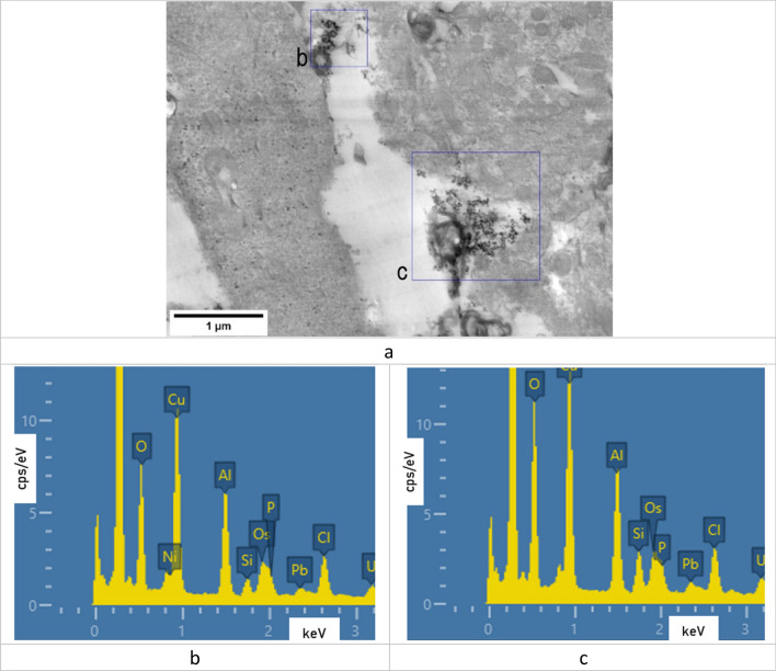
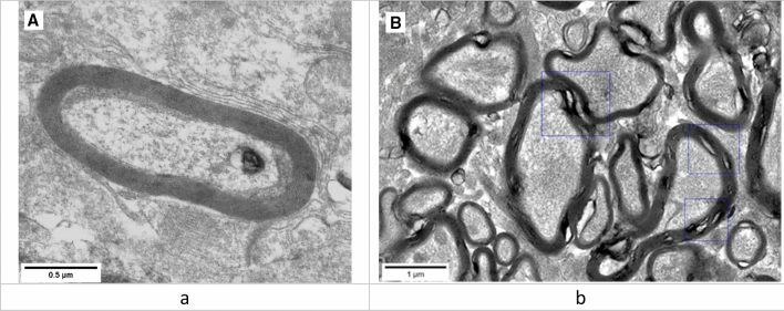
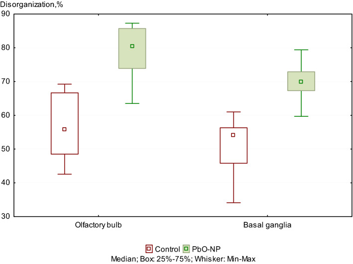
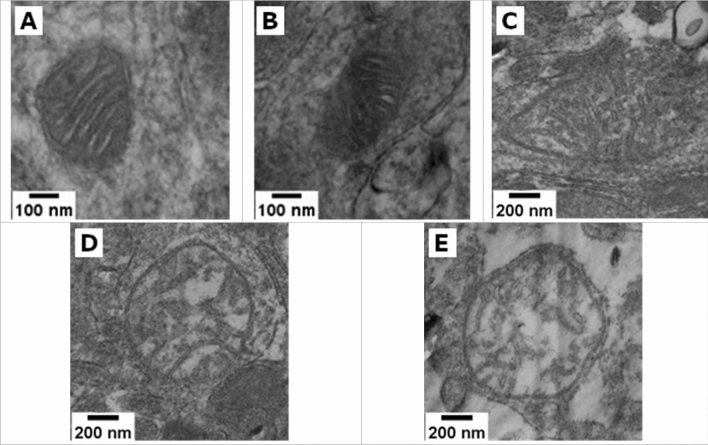
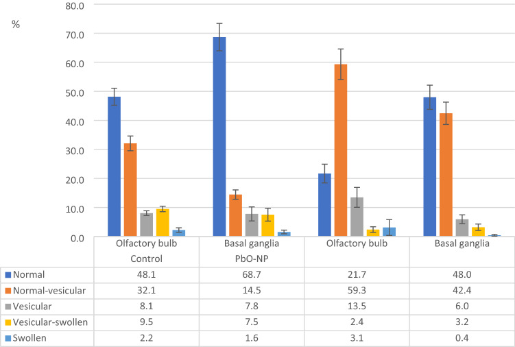
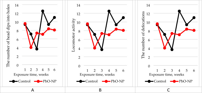
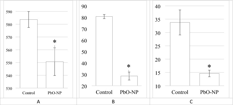
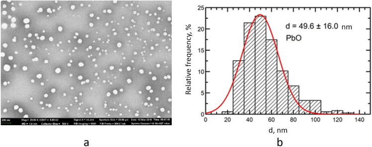
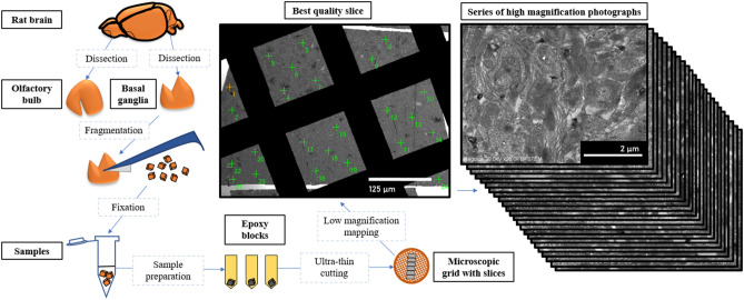
White outbred female rats were exposed intranasally to 50-µL of suspension of lead oxide nanoparticles (PbO NPs) at a concentration of 0.5 mg/mL thrice a week during six weeks. A control group of rats was administered deionized water in similar volumes and conditions. The developed intoxication was manifested by altered biochemical and cytochemical parameters, as well as behavioral reactions of animals. Using electron microscopy and energy-dispersive X-ray spectroscopy techniques, we revealed deposition of PbO NPs in the olfactory bulb, but not in basal ganglia, and an increase in the number of axons with damage to the myelin sheath in the tissues of olfactory bulb and basal ganglia, changes in the ultrastructure of mitochondria of neurons in the tissues of olfactory bulb and basal ganglia of the brain, and differences in the mitochondrial profile of neurons in different regions of the rat brain. Our results collectively suggest that the central nervous system may be a target of low-level toxicity of lead oxide nanoparticles.
© 2022. The Author(s).
Conflict of interest statement
The authors declare no competing interests.
Figures

Similar articles
-
Analysis of Experimental Data on Changes in Various Structures and Functions of the Rat Brain following Intranasal Administration of Fe2O3 Nanoparticles.Int J Mol Sci. 2023 Feb 10;24(4):3572. doi: 10.3390/ijms24043572. Int J Mol Sci. 2023. PMID: 36834983 Free PMC article.
-
Effect of Intranasally Administered Stem Cell-Derived Exosomes on Rat's Olfactory Bulb Histological Structure After Lead-Oxide Nanoparticle Administration.Microsc Microanal. 2025 Mar 17;31(2):ozaf008. doi: 10.1093/mam/ozaf008. Microsc Microanal. 2025. PMID: 40112249
-
Effects of intranasal instillation of nanoparticulate matter in the olfactory bulb.Sci Rep. 2021 Aug 20;11(1):16997. doi: 10.1038/s41598-021-96593-0. Sci Rep. 2021. PMID: 34417533 Free PMC article.
-
Acute toxicity of zinc oxide nanoparticles to the rat olfactory system after intranasal instillation.J Appl Toxicol. 2013 Oct;33(10):1079-88. doi: 10.1002/jat.2842. Epub 2013 Jan 11. J Appl Toxicol. 2013. PMID: 23315988
-
[Characteristics of olfactory epithelium and manipulations of neural functions in the brain by the intranasal administration].Yakugaku Zasshi. 2012;132(11):1247-53. doi: 10.1248/yakushi.12-00229-1. Yakugaku Zasshi. 2012. PMID: 23123715 Review. Japanese.
Cited by
-
Nanoparticles-induced potential toxicity on human health: Applications, toxicity mechanisms, and evaluation models.MedComm (2020). 2023 Jul 14;4(4):e327. doi: 10.1002/mco2.327. eCollection 2023 Aug. MedComm (2020). 2023. PMID: 37457660 Free PMC article. Review.
-
Analysis of Experimental Data on Changes in Various Structures and Functions of the Rat Brain following Intranasal Administration of Fe2O3 Nanoparticles.Int J Mol Sci. 2023 Feb 10;24(4):3572. doi: 10.3390/ijms24043572. Int J Mol Sci. 2023. PMID: 36834983 Free PMC article.
-
On the Mechanisms of the Cardiotoxic Effect of Lead Oxide Nanoparticles.Cardiovasc Toxicol. 2024 Jan;24(1):49-61. doi: 10.1007/s12012-023-09814-5. Epub 2023 Dec 18. Cardiovasc Toxicol. 2024. PMID: 38108959 Free PMC article.
References
Publication types
MeSH terms
Substances
LinkOut - more resources
Full Text Sources
