. 2023 Feb;37(2):326-338.
doi: 10.1038/s41375-022-01758-z. Epub 2022 Nov 14.

VIP152 is a selective CDK9 inhibitor with pre-clinical in vitro and in vivo efficacy in chronic lymphocytic leukemia

Affiliations

VIP152 is a selective CDK9 inhibitor with pre-clinical in vitro and in vivo efficacy in chronic lymphocytic leukemia

Steven Sher et al. Leukemia. 2023 Feb.

Abstract

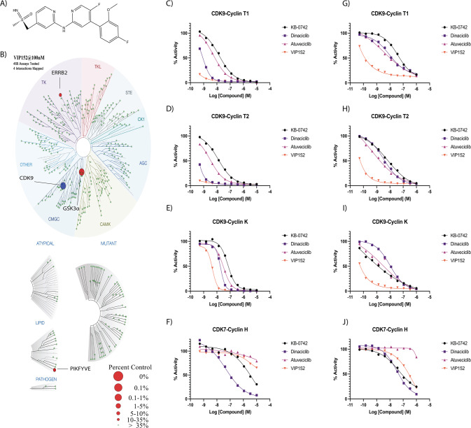
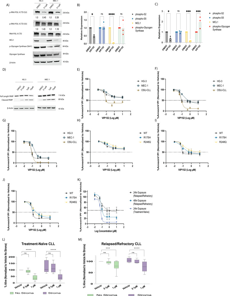
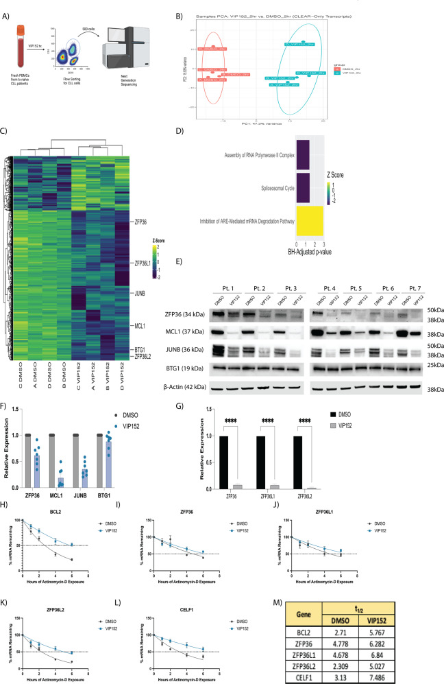
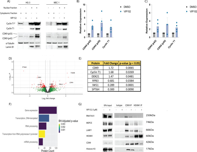
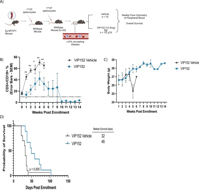
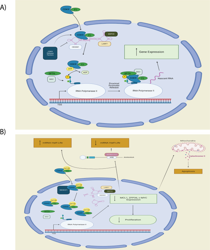
Chronic lymphocytic leukemia (CLL) is effectively treated with targeted therapies including Bruton tyrosine kinase inhibitors and BCL2 antagonists. When these become ineffective, treatment options are limited. Positive transcription elongation factor complex (P-TEFb), a heterodimeric protein complex composed of cyclin dependent kinase 9 (CDK9) and cyclin T1, functions to regulate short half-life transcripts by phosphorylation of RNA Polymerase II (POLII). These transcripts are frequently dysregulated in hematologic malignancies; however, therapies targeting inhibition of P-TEFb have not yet achieved approval for cancer treatment. VIP152 kinome profiling revealed CDK9 as the main enzyme inhibited at 100 nM, with over a 10-fold increase in potency compared with other inhibitors currently in development for this target. VIP152 induced cell death in CLL cell lines and primary patient samples. Transcriptome analysis revealed inhibition of RNA degradation through the AU-Rich Element (ARE) dysregulation. Mechanistically, VIP152 inhibits the assembly of P-TEFb onto the transcription machinery and disturbs binding partners. Finally, immune competent mice engrafted with CLL-like cells of Eµ-MTCP1 over-expressing mice and treated with VIP152 demonstrated reduced disease burden and improvement in overall survival compared to vehicle-treated mice. These data suggest that VIP152 is a highly selective inhibitor of CDK9 that represents an attractive new therapy for CLL.


Conflict of interest statement

AJ, MF, JG, AH, and RI are currently employed by Vincerx Pharma Inc. JG, AH, RI, and JCB are current equity holders in Vincerx Pharma Inc (a publicly traded company) or hold membership on the entity’s Board of Directors or advisory committees. RL is on the scientific advisory board of Vincerx Pharma Inc. JCB holds membership on the Board of Directors or advisory committees of Novartis and Newave and holds Consultancy, Honoraria on Novartis, Trillium Astellas, Astellas, AstraZeneca, Pharmacyclics, and Syndax. J.W. is a consultant for AbbVie Inc, ArQule Inc, AstraZeneca Pharmaceuticals LP, Janssen Biotech Inc, Pharmacyclics LLC, an AbbView Company, is on the advisory committee of AbbVie Inc, ArQule Inc, Janssen Biotech Inc, AstraZeneca, and Beigene. JW has received research funding from AbbVie Inc, and Loxo Oncology Inc. JB holds consultancy and honoraria from AbbVie Inc. AstraZeneca, Kite, and Innate. All other authors declare no conflicts of interest.

Figures

Fig. 1
Fig. 1. VIP152 is a selective CDK9 inhibitor with improved activity over other inhibitors.
A Chemical structure of VIP152. B Inhibitory activity of VIP152 was plotted in a representation of the human kinome with results from the DiscoverX Kinome Scan, (CF) Dose-response curves of VIP152, dinaciclib, KB-0742, and atuveciclib for CDK9 in complex with Cyclin T1/T2/K, CDK7 in complex with Cyclin H, in the radiometric acellular HotSpot Assay. GJ Dose-response curves of VIP152, dinaciclib, KB-0742, and atuveciclib for CDK9 in complex with Cyclin T1/T2/K, CDK7 in complex with Cyclin H, in the proximity-based cellular NanoBRET assay.
Fig. 2
Fig. 2. VIP152 inhibits proliferation and induces apoptosis in CLL cells.
A Representative western blot of 8 h treatment of HG-3 and MEC-1 cell lines with DMSO or 0.1 µM VIP152. Densitometry analyses below bands indicate phosphorylation relative to total protein. B, C Densitometry analysis of three independent immunoblot experiments examining phospho-Serine 2 and phoshpo-Serine 5 residues of RNA Polymerase II, MCL1, and phospho-Glycogen Synthase upon VIP152 treatment in HG-3 (B) and MEC-1 (C). Results are shown as mean ± SEM. (*, ***, **** = p < 0.05, 0.001, and 0.0001 respectively). D Representative western blot of apoptosis induction in HG-3 and MEC-1 treated with VIP152 for six hours. Western blot is a representative image of three biological replicates. EG Annexin V/propidium iodide flow cytometry dose-response curves of VIP152 treated HG-3, MEC-1, and OSU-CLL at 24 h (E), 48 h (F), and 72 h (G). Results are averaged of three independent biological replicates with results shown as mean ± SEM. HJ Annexin V/propidium iodide flow cytometry dose-response curves of VIP152 treated HG-3 TP53WT, HG-3 TP53R175H, and HG-3 TP53R248Q at 24 h (H), 48 h (I), and 72 h (J). Results are averaged of three independent biological replicates with results shown as mean ± SEM. K Annexin V/propidium iodide flow cytometry dose-response curves of VIP152 treated treatment-naïve or relapsed/refractory patient samples for the listed timepoints. L, M Results of four-hour treatment with washout of 10 treatment-naïve (L) and 8 BTKi/venetoclax relapse/refractory (M) primary CLL patient samples plated with or without the human stromal cell line, HS5. (***, **** = p < 0.0005 and p < 0.0001 respectively).
Fig. 3
Fig. 3. VIP152 induces rapid transcriptomic changes.
A Schematic cartoon of limiting-cell RNA-seq experiment. B Principal component analysis of RNA sequencing data. Each dot represents a unique patient sample treated with either DMSO (red) or 1 µM VIP152 (teal). C Heat map of most differentially expressed genes. Genes selected by overall DEG’s with a LFC > 1 or LFC < −1 adj. p-value < 0.05. D Plot of pathways from Ingenuity Pathway Analysis with p-value < 0.05 and z-score > 0 or z-score < 0. EG 7 CLL patient samples were treated for 8 h with 1 µM VIP152 and then analyzed for protein expression by western blot (E, F) and ZFP36, ZFP36L1, and ZFP36L2 expression by qRT-PCR (G). HL 5 CLL patient samples were treated for 8 h with 1 µM VIP152 and then supplemented with 500 nM Actinomycin-D. RNA was taken at varying timepoints and analyzed via qRT-PCR. M mRNA half-lives of analyzed transcripts in (HL) as determined by nonlinear regression using a one-phase decay method.
Fig. 4
Fig. 4. VIP152 disrupts P-TEFb canonical binding partners.
AC Cytoplasmic and nuclear extracts of HG-3 and MEC-1 treated with 1 µM VIP152 for two hours with quantification (B, C). D, E Proteomic characterization of nuclear immunoprecipitation of VIP152 treated HG-3 cells. Gray dots indicate non-differentially associated proteins. Green dots indicate transcripts with a LFC > 0.5 or LFC < −0.5 but p-value > 0.05. Blue dots indicate proteins with a p-value < 0.05 but −0.5 < LFC < 0.5. Red dots indicate proteins with a p-value < 0.05 and a LFC > 0.5 or LFC < −0.5. D Volcano plot of differentially associated proteins. E Table of P-TEFb and POLII-associated proteins. F Plot of pathways from Ingenuity Pathway Analysis with p-value < 0.05 and z-score > 0 or z-score < 0 (G) Nuclear IP of CDK9 and HEXIM1 of VIP152 treated HG-3 cells probing for components of the 7SK-RNA complex.
Fig. 5
Fig. 5. VIP152 improves survival in the Eµ-MTCP1 CLL model.
20 C57BL/6 J mice were engrafted via tail vein with 106 splenocytes from leukemic Eµ-MTCP1 mice. A Schematic cartoon of in vivo adoptive transfer study. BD Measurement of peripheral disease as characterized by CD45+/CD5+/CD19+ % in peripheral blood (B), weight monitoring (C), and survival (D) of mice following enrollment upon either VIP152 (blue) or vehicle treatment (red). 10 mice were enrolled into each treatment arm. (*,** = p < 0.01 and p < 0.005 respectively). Error bars indicate mean of surviving mice ± SEM.
Fig. 6
Fig. 6. VIP152 mechanistic illustration.
A Normal signaling pathway for P-TEFb. B Proposed signaling pathway for VIP152 inhibition of P-TEFb. VIP152 bound to P-TEFb leads to intranuclear accumulation with dissociation from 7SK RNA complex from DDX21 cleavage. Decreased binding to POLII leads to decreased transcriptional activity with subsequent loss of proliferative signaling, increased apoptosis with loss of MCL1 expression, and increased mRNA half-life from decreased AU-Rich element mediated mRNA degradation subsequent to loss of ZFP36 family expression.

References

    1. Landgren O, Ma W, Abbasi F, Hayes RB, Ghia P, Marti GE, et al. B-cell clones as early markers for chronic lymphocytic leukemia. N. Engl J Med. 2009;360:659–67. - PMC - PubMed
    1. Cheson BD, Meyer RM. Chronic lymphocytic leukemia. N. Engl J Med. 2005;352:804–15. - PubMed
    1. Byrd JC, Brown JR, O’brien S, Barrientos JC, Kay NE, Reddy NM, et al. Ibrutinib versus ofatumumab in previously treated chronic lymphoid leukemia ABSTRACT. N. Engl J Med. 2014;371:213–36.. - PMC - PubMed
    1. Byrd JC, Furman RR, Coutre SE, Flinn IW, Burger JA, Blum KA, et al. Targeting BTK with Ibrutinib in Relapsed Chronic Lymphocytic Leukemia ABSTRACT. N. Engl J Med. 2013;369:32–42. - PMC - PubMed
    1. Burger JA, Tedeschi A, Barr PM, Robak T, Owen C, Ghia P, et al. Ibrutinib as initial therapy for patients with chronic lymphocytic leukemia. N. Engl J Med. 2015;373:2425–37. - PMC - PubMed

Publication types

MeSH terms

Substances