. 2023 Feb;30(2):341-355.
doi: 10.1038/s41418-022-01087-9. Epub 2022 Nov 14.

SNORD88C guided 2'-O-methylation of 28S rRNA regulates SCD1 translation to inhibit autophagy and promote growth and metastasis in non-small cell lung cancer

Affiliations

SNORD88C guided 2'-O-methylation of 28S rRNA regulates SCD1 translation to inhibit autophagy and promote growth and metastasis in non-small cell lung cancer

Kangyu Wang et al. Cell Death Differ. 2023 Feb.

Abstract

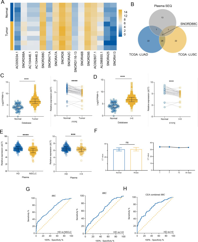
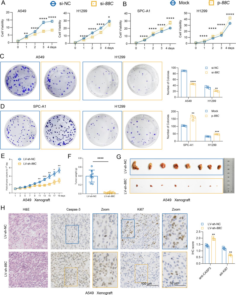
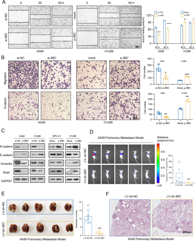
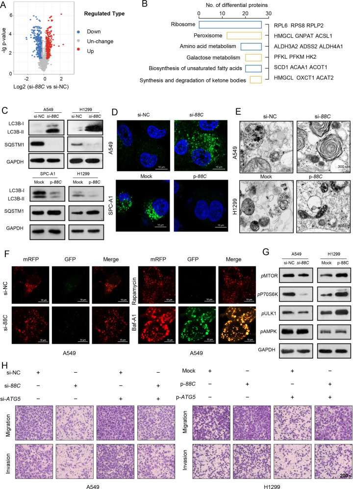
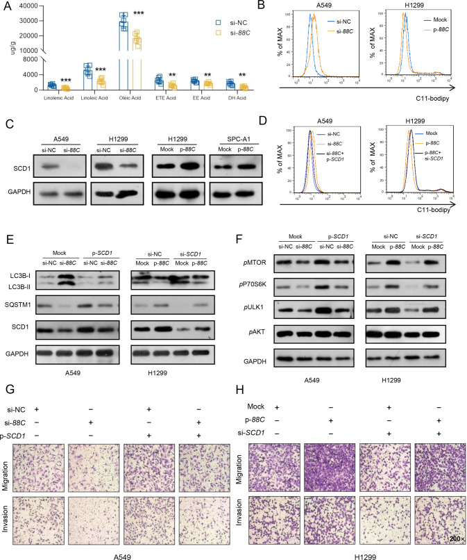
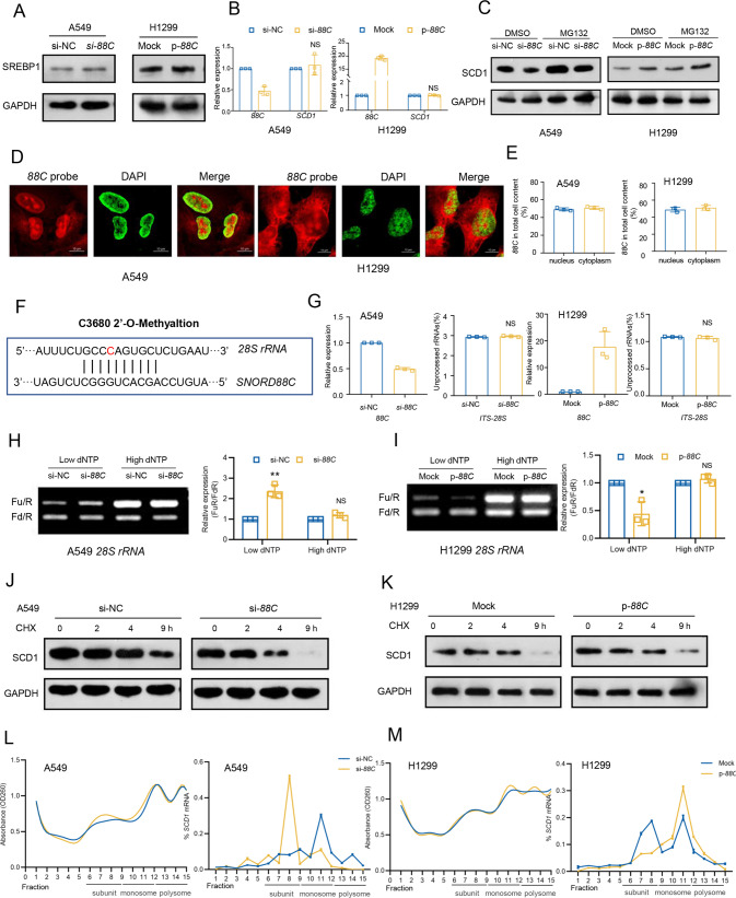
Small nucleolar RNAs (snoRNAs) have been shown to play critical regulatory roles in cancer development. SNORD88C, which located at the intronic region of C19orf48 in chromosome 19q.33 with a 97-nt length was screened through database and snoRNA-sequencing. We firstly verified this snoRNA was up-regulated in tissue and plasma and served as a non-invasive diagnostic biomarker; then confirmed that SNORD88C promoted proliferation and metastasis of NSCLC in vitro and in vivo. Mechanistically, SNORD88C promoted 2'-O-methylation modification at the C3680 site on 28S rRNA and in turn enhanced downstream SCD1 translation, a central lipogenic enzyme for the synthesis of MUFA that can inhibit autophagy by regulating lipid peroxidation and mTOR, providing the novel insight into the regulation of SNORD88C in NSCLC.


Conflict of interest statement

The authors declare no competing interests.

Figures

Fig. 1
Fig. 1. SNORD88C is up-regulated in NSCLC and serves as a non-invasive diagnostic biomarker.
A Though the snoRNAs-sequencing in plasma from NSCLC patients and donors, the differential snoRNAs was shown by heatmap. B The top 50 snoRNAs with the largest fold change in the LUAD and LUSC data sets from TCGA and the statistically differential snoRNAs in the sequencing results were analyzed using the VENN diagram, in which only SNORD88C was overlapped. C, D SNORD88C was significantly increased in NSCLC and early NSCLC tissues from TCGA database and FFPE samples, compared with para-tumor tissues (E) SNORD88C was significantly increased in the plasma of NSCLC and early NSCLC patients, compared with the plasma of donors. F SNORD88C was stably expressed in plasma treated with RNase (left) or stored at −80 °C at time-points of 1, 7, 15, 30 days (right). G The diagnostic efficacy of plasma SNORD88C for NSCLC (left) and early-NSCLC (right). H The diagnostic efficacy of SNORD88C combined with CEA for early-NSCLC. Two-tailed paired t-test, two-tailed unpaired t-test, Mann-Whitney test, one-way analysis of variance (ANOVA) or Kruskal-Wallis test; ns, no significance; *P < 0.05, **P < 0.01, ***P < 0.001, ****P < 0.0001.
Fig. 2
Fig. 2. SNORD88C promotes proliferation of NSCLC in vitro and in vivo.
CCK8 (A, B) and clone formation (C, D) assays showed that knockdown of SNORD88C inhibited proliferation (A) and clonogenicity (C) in A549 and H1299 cells, while overexpression of SNORD88C in SPC-A1 and H1299 cells showed the opposite effect (B, D). In the A549 tumor transplantation model, the growth rate (E) of xenograft was slower, and the weight (F) and volume (G) decreased in LV-sh-SNORD88C compared with the control. H Immunohistochemical staining showed that the expression of Caspas-3 increased and the expression of Ki67 decreased in the tumor tissue of LV-sh-SNORD88C group compared with the control. Scale bar indicates 50 μm and 100 μm.; Two-tailed unpaired t-test, Mann-Whitney test, one-way analysis of variance (ANOVA) or Kruskal-Wallis test; *P < 0.05, **P < 0.01, ***P < 0.001, ****P < 0.0001.
Fig. 3
Fig. 3. SNORD88C promotes NSCLC migration and invasion in vitro and in vivo.
AB Wound healing, migration and invasion assays showed that knockdown of SNORD88C in A549 cells inhibited the motility, migration and invasion of NSCLC, while overexpression of SNORD88C in H1299 cells showed the opposite effect. Original magnification for wound healing, ×40; Original magnification for migration and invasion, ×200. C Western blot showed that SNORD88C knockdown decreased the expression of N-cadherin, Vimentin, and Snail and increased E-cadherin expression in A549 and H1299, while overexpression of SNORD88C showed the opposite expression for these proteins in SPC-A1 and H1299 cells. D, E In A549 pulmonary metastasis model the luciferase signal intensities in the LV-sh-SNORD88C group was remarkably lower than that in the control. F H&E staining showed that metastatic nodules in the lung tissue invaded the pulmonary capsule in the LV-sh-NC group, but not in the LV-sh-SNORD88C. Scale bar indicates 200 μm; Two-tailed unpaired t-test, *P < 0.05, **P < 0.01, ***P < 0.001, ****P < 0.0001.
Fig. 4
Fig. 4. SNORD88C-attenuated autophagy is required for its promotion of migration and invasion.
A The volcano map showed that the differential proteins analyzed by LC-MS/MS-based TMT-labeled quantitative proteomic in SNORD88C-knockdown H1299 cells and the control group. (p ≤ 0.05 and FC ≥ 1.2) B The differential proteins enrichment pathways. CE WB, immunofluorescence labeled by anti-LC3 antibody as well as transmission electron microscopy showed that autophagy was activated in SNORD88C-knockdown NSCLC cells, but inhibited in SNORD88C-overexpression NSCLC cells. Scale bar indicates 10 μm and 300 μm. F A549 cells expressing mRFP-GFP-LC3 were transfected with si-NC or si-88C, or treated with 1 nM rapamycin or 1 nM Baf-A1 for 24 h and imaged by confocal microscopy. Scale bars: 10 μm. G WB assay showed that the phosphorylation levels of MTOR, P70S6K, and ULK1 were decreased in SNORD88C-knockdown A549 cells, while the phosphorylation levels of these proteins were enhanced in SNORD88C-overexpression H1299 cells. The AMPK phosphorylation level was not affected by SNORD88C knockdown or overexpression. H ATG5 silencing could reverse the inhibitory effect of SNORD88C knockdown on the migration and invasion of A549 cells (left). ATG5 overexpression could abolish the promotion of SNORD88C overexpression on the migration and invasion of H1299 cells (right). Original magnification, ×200. Two-tailed unpaired t-test, **P < 0.01.
Fig. 5
Fig. 5. SNORD88C inhibits autophagy via upregulating SCD1 protein expression.
A GC-MS-based metabolomics targeting fatty acids showed many kinds of unsaturated fatty acids were decreased in SNORD88C-knockdown H1299 cells compared to the control group. B Flow cytometry showed that through C11-bodipy staining, lipid peroxidation was promoted in SNORD88C-knockdown A549 cells, while inhibited in SNORD88C-overexpression H1299 cells. C WB showed that SCD1 expression was decreased in SNORD88C-knockdown A549 and H1299 cells, and increased in SNORD88C-overexpression SPC-A1 and H1299 cells. D The promotion of lipid peroxidation by SNROD88C knockdown in A549 cells and the inhibition of lipid peroxidation by SNROD88C overexpression in H1299 cells could be reversed by SCD1 overexpression and knockdown. E In A549, the overexpression of SCD1 reversed the increase of LC3B and the decrease of SQSTM1 caused by SNORD88C silencing. In H1299, knocking down SCD1 abolished the decrease of LC3B and the increase of SQSTM1 induced by SNORD88C overexpression. F SCD1 overexpression reversed the decreased phosphorylation of MTOR, P70S6K, and ULK1 caused by SNORD88C knockdown in A549. SCD1 knockdown abolished the increased phosphorylation of these proteins induced by SNORD88C overexpression in H1299. G In A549, SCD1 overexpression could reverse the inhibitory effect of SNORD88C knockdown on migration and invasion. H In H1299, SCD1-knockdown could abolish the promoting effect of SNORD88C overexpression on the migration and invasion. Original magnification, ×200. Two-tailed unpaired t-test, **P < 0.01, ***P < 0.001.
Fig. 6
Fig. 6. SNORD88C regulates the translation activity of SCD1 though guiding 2′-O-me of 28S rRNA.
A Western blot showed that SREBP1 expression was not affected by SNORD88C knockdown or overexpression. B q-PCR showed that SCD1 mRNA level was not affected by SNORD88C knockdown or overexpression. C Western blot showed MG132 treatment clearly increased SCD1 protein levels, but failed to reverse the regulation of SNORD88C knockdown or overexpression on SCD1. D, E Fluorescent in situ hybridization (FISH) using Cy3-labeled probes targeting SNORD88C and q-PCR showed SNORD88C was mainly located in the nucleolus and cytoplasm. Red represented SNORD88C, green (artifact) represented nucleus. Scale bar indicates 10 μm. F The snoPY database predicted the complementary sequence and 2′-O-me site of rRNA targeted by SNORD88C, and red represented the methylation site. G q-PCR assay to detect ITS-28S showed that SNORD88C knockdown (left) or overexpression (right) did not affect the processing of 28S rRNA. The value of the vertical coordinate represented the average of primer pairs d/c (unprocessed) over b/a (total) and primer pairs f/e (unprocessed) over b/a (total) for 28S rRNA, respectively. Primers were shown in Table S4. H, I The 2′-O-me activity of 28S rRNA at C3680 site detected by RTL-P assay was decreased after SNORD88C knockdown in A549, while increased after SNORD88C overexpression in H1299. J, K The translation activity of SCD1 was reduced by SNORD88C knockdown treated by the CHX in A549 cells, while increased by SNORD88C overexpression in H1299 cells. Two-tailed unpaired t test; ns, no significance; *P < 0.05, **P < 0.01. L, M Ribosomal components in cytoplasmic extracts of si-NC and si-88C A549 cells (L) or mock and p-88C H1299 cells (M) were fractionated through sucrose gradients, and the relative levels of SCD1 mRNA were analyzed by qRT-PCR in the gradient fractions.

Similar articles

Cited by

References

    1. Bray F, Ferlay J, Soerjomataram I, Siegel RL, Torre LA, Jemal A. Global cancer statistics 2018: GLOBOCAN estimates of incidence and mortality worldwide for 36 cancers in 185 countries. CA Cancer J Clin. 2018;68:394–424. doi: 10.3322/caac.21492. - DOI - PubMed
    1. Herbst RS, Morgensztern D, Boshoff C. The biology and management of non-small cell lung cancer. Nature. 2018;553:446–54.. doi: 10.1038/nature25183. - DOI - PubMed
    1. Mannoor K, Liao J, Jiang F. Small nucleolar RNAs in cancer. Biochim Biophys Acta. 2012;1826:121–8. - PMC - PubMed
    1. Liang J, Wen J, Huang Z, Chen XP, Zhang BX, Chu L. Small nucleolar RNAs: Insight into their function in cancer. Front Oncol. 2019;9:587. doi: 10.3389/fonc.2019.00587. - DOI - PMC - PubMed
    1. Williams GT, Farzaneh F. Are snoRNAs and snoRNA host genes new players in cancer? Nat Rev Cancer. 2012;12:84–8. doi: 10.1038/nrc3195. - DOI - PubMed

Publication types